Anti-TMS1/ASC 抗体 [EPR28201-61] - BSA and Azide free
Anti-TMS1/ASC antibody [EPR28201-61] - BSA and Azide free
- BOND RX™ Validated
- RabMAb
- Recombinant
- 詳細を見る
Be the first to review this product! Submit a review
|
(0 Publication)
Rabbit Recombinant Monoclonal TMS1/ASC antibody. Carrier free. Suitable for IP, Flow Cyt (Intra), ICC/IF, IHC-P, WB and reacts with Mouse, Rat samples.
別名を表示する
Asc, Pycard, Apoptosis-associated speck-like protein containing a CARD, mASC, PYD and CARD domain-containing protein
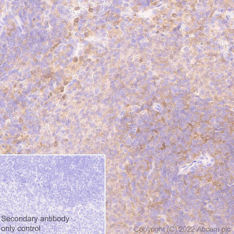
- IHC-P
Supplier Data
Immunohistochemistry (Formalin/PFA-fixed paraffin-embedded sections) - Anti-TMS1/ASC antibody [EPR28201-61] - BSA and Azide free (AB307561)
This data was developed using ab307560, the same antibody clone in a different buffer formulation. Immunohistochemical analysis of paraffin-embedded Rat spleen tissue labeling TMS1/ASC with ab307560 at 1/1000 (0.484 ug/ml) followed by a ready to use LeicaDS9800 (Bond™ Polymer Refine Detection) was used. Positive staining on rat spleen.The section was incubated with ab307560 for 30 mins at room temperature.The immunostaining was performed on a Leica Biosystems BOND® RX instrument Counterstained with Hematoxylin. Secondary antibody only control : Secondary antibody is a ready to use LeicaDS9800 (Bond™ Polymer Refine Detection) was used. Heat mediated antigen retrieval was performed with Tris-EDTA buffer (pH 9.0, Epitope Retrieval Solution2) for 20 mins
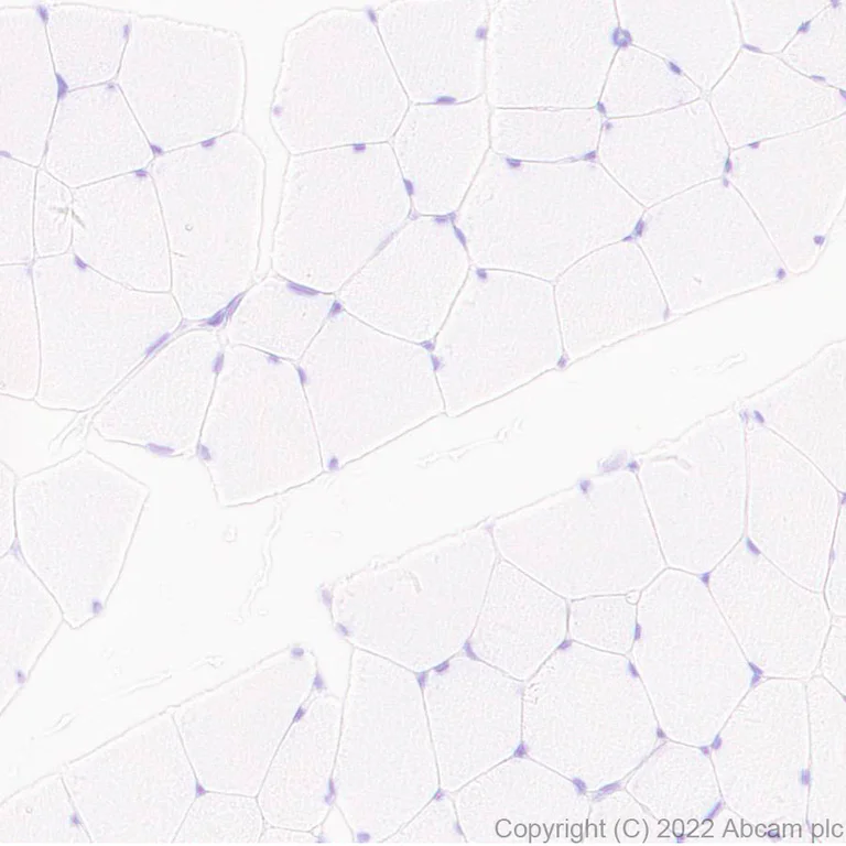
- IHC-P
Supplier Data
Immunohistochemistry (Formalin/PFA-fixed paraffin-embedded sections) - Anti-TMS1/ASC antibody [EPR28201-61] - BSA and Azide free (AB307561)
This data was developed using ab307560, the same antibody clone in a different buffer formulation. Immunohistochemical analysis of paraffin-embedded Rat skeletal muscle tissue labeling TMS1/ASC with ab307560 at 1/1000 (0.484 ug/ml) followed by a ready to use LeicaDS9800 (Bond™ Polymer Refine Detection) was used. Low expression tissue : no staining on rat skeletal muscle.The section was incubated with ab307560 for 30 mins at room temperature.The immunostaining was performed on a Leica Biosystems BOND® RX instrument Counterstained with Hematoxylin. Secondary antibody only control : Secondary antibody is a ready to use LeicaDS9800 (Bond™ Polymer Refine Detection) was used. Heat mediated antigen retrieval was performed with Tris-EDTA buffer (pH 9.0, Epitope Retrieval Solution2) for 20 mins
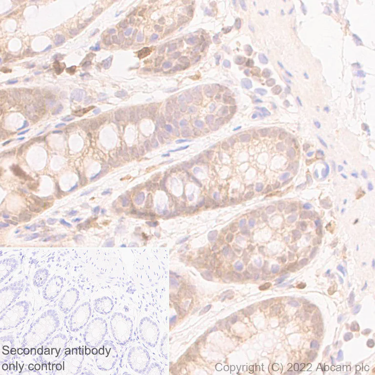
- IHC-P
Supplier Data
Immunohistochemistry (Formalin/PFA-fixed paraffin-embedded sections) - Anti-TMS1/ASC antibody [EPR28201-61] - BSA and Azide free (AB307561)
This data was developed using ab307560, the same antibody clone in a different buffer formulation. Immunohistochemical analysis of paraffin-embedded Rat colon tissue labeling TMS1/ASC with ab307560 at 1/1000 (0.484 ug/ml) followed by a ready to use LeicaDS9800 (Bond™ Polymer Refine Detection) was used. Positive staining on rat colon.The section was incubated with ab307560 for 30 mins at room temperature.The immunostaining was performed on a Leica Biosystems BOND® RX instrument Counterstained with Hematoxylin. Secondary antibody only control : Secondary antibody is a ready to use LeicaDS9800 (Bond™ Polymer Refine Detection) was used. Heat mediated antigen retrieval was performed with Tris-EDTA buffer (pH 9.0, Epitope Retrieval Solution2) for 20 mins
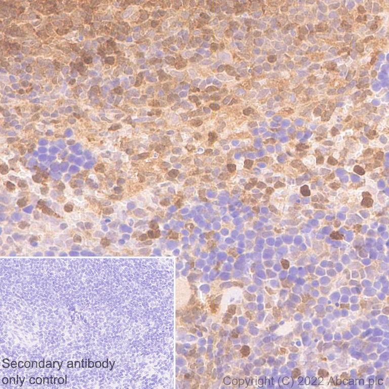
- IHC-P
Supplier Data
Immunohistochemistry (Formalin/PFA-fixed paraffin-embedded sections) - Anti-TMS1/ASC antibody [EPR28201-61] - BSA and Azide free (AB307561)
This data was developed using ab307560, the same antibody clone in a different buffer formulation. Immunohistochemical analysis of paraffin-embedded Mouse spleen tissue labeling TMS1/ASC with ab307560 at 1/1000 (0.484 ug/ml) followed by a ready to use LeicaDS9800 (Bond™ Polymer Refine Detection) was used. Positive staining on mouse spleen.The section was incubated with ab307560 for 30 mins at room temperature.The immunostaining was performed on a Leica Biosystems BOND® RX instrument Counterstained with Hematoxylin. Secondary antibody only control : Secondary antibody is a ready to use LeicaDS9800 (Bond™ Polymer Refine Detection) was used. Heat mediated antigen retrieval was performed with Tris-EDTA buffer (pH 9.0, Epitope Retrieval Solution2) for 20 mins
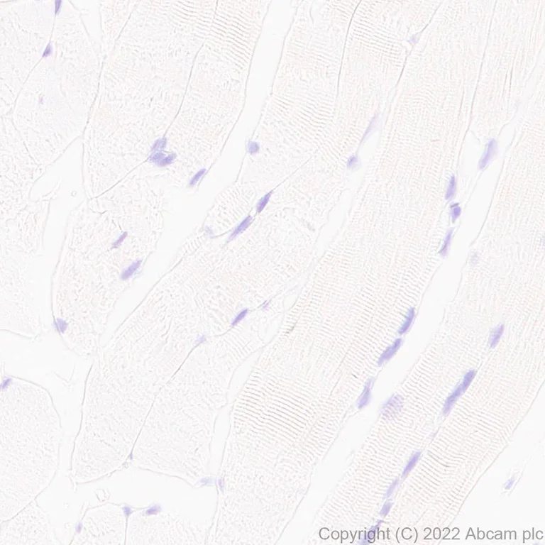
- IHC-P
Supplier Data
Immunohistochemistry (Formalin/PFA-fixed paraffin-embedded sections) - Anti-TMS1/ASC antibody [EPR28201-61] - BSA and Azide free (AB307561)
This data was developed using ab307560, the same antibody clone in a different buffer formulation. Immunohistochemical analysis of paraffin-embedded Mouse skeletal muscl tissue labeling TMS1/ASC with ab307560 at 1/1000 (0.484 ug/ml) followed by a ready to use LeicaDS9800 (Bond™ Polymer Refine Detection) was used. Low expression tissue : no staining on mouse skeletal muscle.The section was incubated with ab307560 for 30 mins at room temperature.The immunostaining was performed on a Leica Biosystems BOND® RX instrument Counterstained with Hematoxylin. Secondary antibody only control : Secondary antibody is a ready to use LeicaDS9800 (Bond™ Polymer Refine Detection) was used. Heat mediated antigen retrieval was performed with Tris-EDTA buffer (pH 9.0, Epitope Retrieval Solution2) for 20 mins
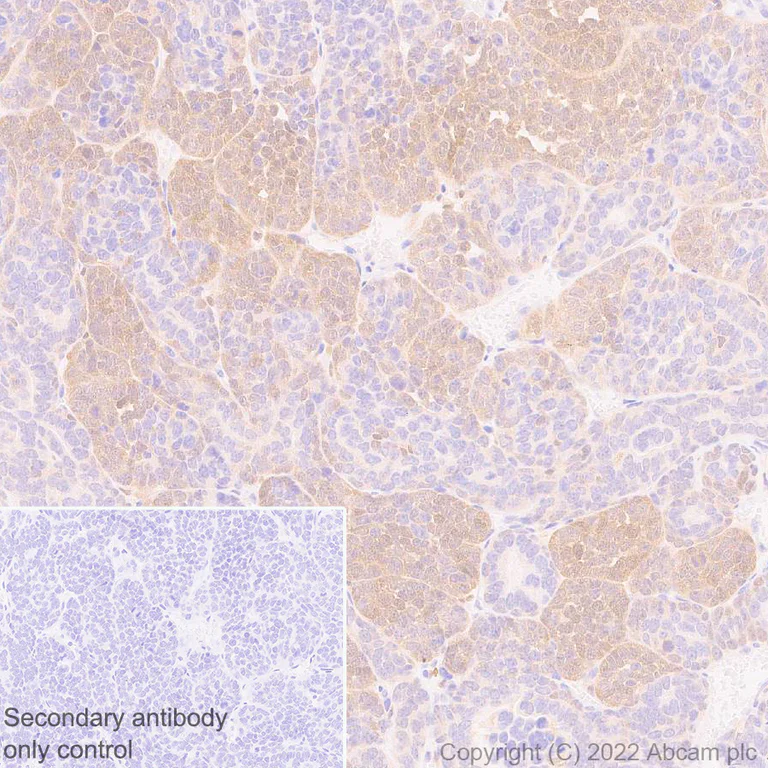
- IHC-P
Supplier Data
Immunohistochemistry (Formalin/PFA-fixed paraffin-embedded sections) - Anti-TMS1/ASC antibody [EPR28201-61] - BSA and Azide free (AB307561)
This data was developed using ab307560, the same antibody clone in a different buffer formulation. Immunohistochemical analysis of paraffin-embedded Mouse head and neck tissue labeling TMS1/ASC with ab307560 at 1/1000 (0.484 ug/ml) followed by a ready to use LeicaDS9800 (Bond™ Polymer Refine Detection) was used. Positive staining on mouse head and neck squamous cell carcinoma.The section was incubated with ab307560 for 30 mins at room temperature.The immunostaining was performed on a Leica Biosystems BOND® RX instrument Counterstained with Hematoxylin. Secondary antibody only control : Secondary antibody is a ready to use LeicaDS9800 (Bond™ Polymer Refine Detection) was used. Heat mediated antigen retrieval was performed with Tris-EDTA buffer (pH 9.0, Epitope Retrieval Solution2) for 20 mins
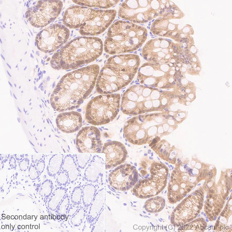
- IHC-P
Supplier Data
Immunohistochemistry (Formalin/PFA-fixed paraffin-embedded sections) - Anti-TMS1/ASC antibody [EPR28201-61] - BSA and Azide free (AB307561)
This data was developed using ab307560, the same antibody clone in a different buffer formulation. Immunohistochemical analysis of paraffin-embedded Mouse colon tissue labeling TMS1/ASC with ab307560 at 1/1000 (0.484 ug/ml) followed by a ready to use LeicaDS9800 (Bond™ Polymer Refine Detection) was used. Positive staining on mouse colon.The section was incubated with ab307560 for 30 mins at room temperature.The immunostaining was performed on a Leica Biosystems BOND® RX instrument Counterstained with Hematoxylin. Secondary antibody only control : Secondary antibody is a ready to use LeicaDS9800 (Bond™ Polymer Refine Detection) was used. Heat mediated antigen retrieval was performed with Tris-EDTA buffer (pH 9.0, Epitope Retrieval Solution2) for 20 mins
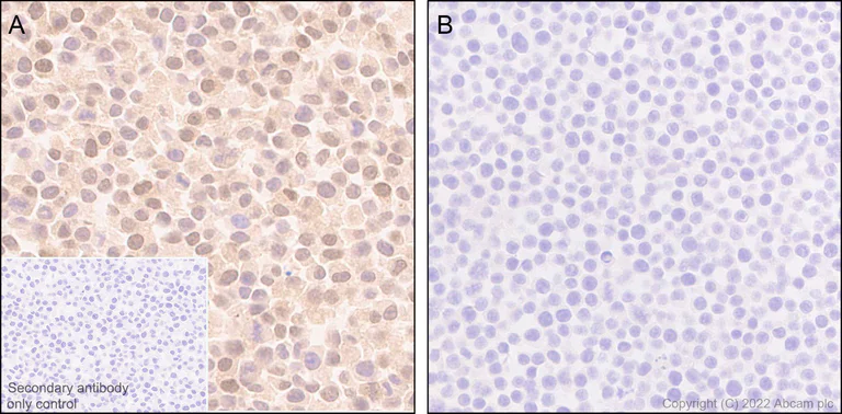
- IHC-P
Supplier Data
Immunohistochemistry (Formalin/PFA-fixed paraffin-embedded sections) - Anti-TMS1/ASC antibody [EPR28201-61] - BSA and Azide free (AB307561)
This data was developed using ab307560, the same antibody clone in a different buffer formulation. Immunohistochemical analysis of paraffin-embedded (A) J774A.1 (mouse r tissue labeling TMS1/ASC with ab307560 at 1/1000 (0.484 ug/ml) followed by a ready to use LeicaDS9800 (Bond™ Polymer Refine Detection) was used. Positive staining on (A) J774A.1 (mouse reticulum cell sarcoma monocyte/macrophage) cell pellet, no staining on (B) RAW 264.7 (Mouse Abelson murine leukemia virus-induced tumor macrophage) cell pellet.The section was incubated with ab307560 for 30 mins at room temperature.The immunostaining was performed on a Leica Biosystems BOND® RX instrument Counterstained with Hematoxylin. Secondary antibody only control : Secondary antibody is a ready to use LeicaDS9800 (Bond™ Polymer Refine Detection) was used. Heat mediated antigen retrieval was performed with Tris-EDTA buffer (pH 9.0, Epitope Retrieval Solution2) for 20 mins
- ICC/IF
Supplier Data
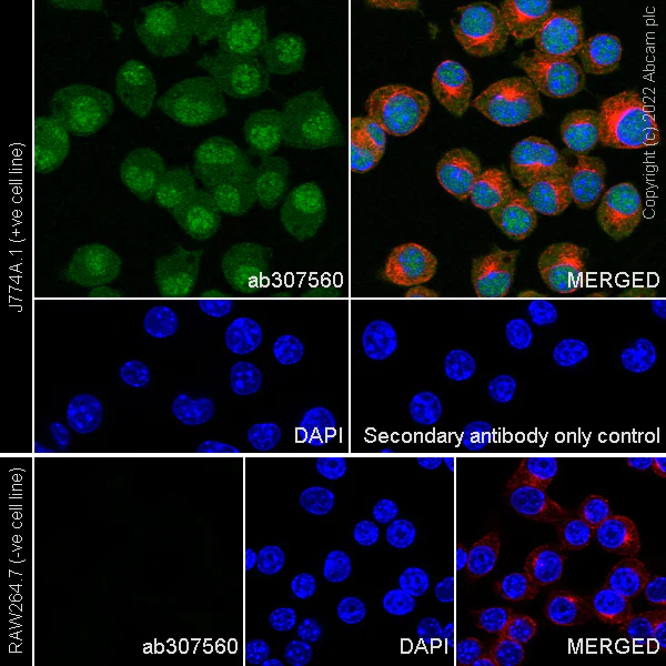
Immunocytochemistry/ Immunofluorescence - Anti-TMS1/ASC antibody [EPR28201-61] - BSA and Azide free (AB307561)
This data was developed using ab307560, the same antibody clone in a different buffer formulation. Immunofluorescent analysis of 4% Paraformaldehyde-fixed, 0.1% TritonX-100 permeabilized J774A.1 (mouse reticulum cell sarcoma monocyte/macrophage) cells labelling TMS1/ASC with ab307560 at 1/500 (0.968 ug/ml) dilution, followed by ab150081 Goat Anti-Rabbit IgG H&L (Alexa Fluor® 488) preadsorbed antibody at 1/1000 2 ug/ml dilution (Green). Confocal image showing nuclear and cytoplasmic staining in J774A.1 cell line. Negative control : RAW 264.7 (PMID : 28766178Ј©
Image was taken with a confocal microscope(Leica-Microsystems, TCS SP8). ab195889 Anti-alpha Tubulin mouse monoclonal antibody - Microtubule Marker (Alexa Fluor® 594) was used to counterstain tubulin at 1/200 2.5ug/ml dilution (Red). The Nuclear counterstain was DAPI (Blue). Secondary antibody only control : Secondary antibody is ab150081 Goat Anti-Rabbit IgG H&L (Alexa Fluor® 488) preadsorbed at 1/1000 2 ug/ml dilution.
- Flow Cyt (Intra)
Supplier Data
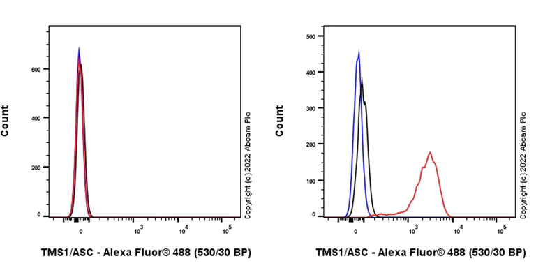
Flow Cytometry (Intracellular) - Anti-TMS1/ASC antibody [EPR28201-61] - BSA and Azide free (AB307561)
This data was developed using ab307560, the same antibody clone in a different buffer formulation.
Flow cytometric analysis of 4% paraformaldehyde fixed 90% methanol permeabilized RAW264.7 (mouse abelson murine leukemia virus-induced tumor macrophage, Left) / J774A.1 (mouse reticulum cell sarcoma monocyte macrophage, Right) cells labelling TMS1/ASC with ab307560 at 1/500 dilution (0.1ug) (Red) compared with a Rabbit monoclonal IgG (ab172730) (Black) isotype control and an unlabelled control (cells without incubation with primary antibody and secondary antibody) (Blue). A Goat Anti-Rabbit IgG (Alexa Fluor® 488, ab150081) at 1/2000 dilution was used as the secondary antibody. Negative control : RAW 264.7 (PMID : 28766178).
- IP
Supplier Data
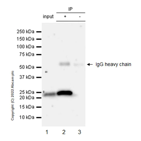
Immunoprecipitation - Anti-TMS1/ASC antibody [EPR28201-61] - BSA and Azide free (AB307561)
This data was developed using ab307560, the same antibody clone in a different buffer formulation.
TMS1/ASC was immunoprecipitated from 0.35 mg J774A.1 (mouse reticulum cell sarcoma monocyte/macrophage ) whole cell lysate 10 ug with ab307560 at 1/30 dilution (2ug in 0.35mg lysates). Western blot was performed on the immunoprecipitate using ab307560 at 1/1000 dilution. VeriBlot for IP secondary antibody (HRP) (ab131366) was used at 1/5000 dilution.
Lane 1 : J774A.1 (mouse reticulum cell sarcoma monocyte/macrophage ) whole cell lysate 10 ug
Lane 2 : ab307560 IP in J774A.1 whole cell lysate
Lane 3 : Rabbit monoclonal IgG (ab172730) instead of ab307560 in J774A.1 whole cell lysate
Blocking and dilution buffer and concentration : 5% NFDM/TBST.
Exposure time : 50 seconds
All lanes:
Immunoprecipitation - Anti-TMS1/ASC antibody [EPR28201-61] (<a href='/products/primary-antibodies/tms1-asc-antibody-epr28201-61-ab307560'>ab307560</a>) at 1/1000 dilution
Lane 1:
J774A.1 (mouse reticulum cell sarcoma monocyte/macrophage ) whole cell lysate 10 μg
Lane 2:
J774A.1 whole cell lysate
Secondary
All lanes:
Immunoprecipitation - VeriBlot for IP Detection Reagent (HRP) (<a href='/products/reagents/veriblot-for-ip-detection-reagent-hrp-ab131366'>ab131366</a>) at 1/5000 dilution
Observed band size: 21 kDa
false
Exposure time: 50s
- WB
Supplier Data
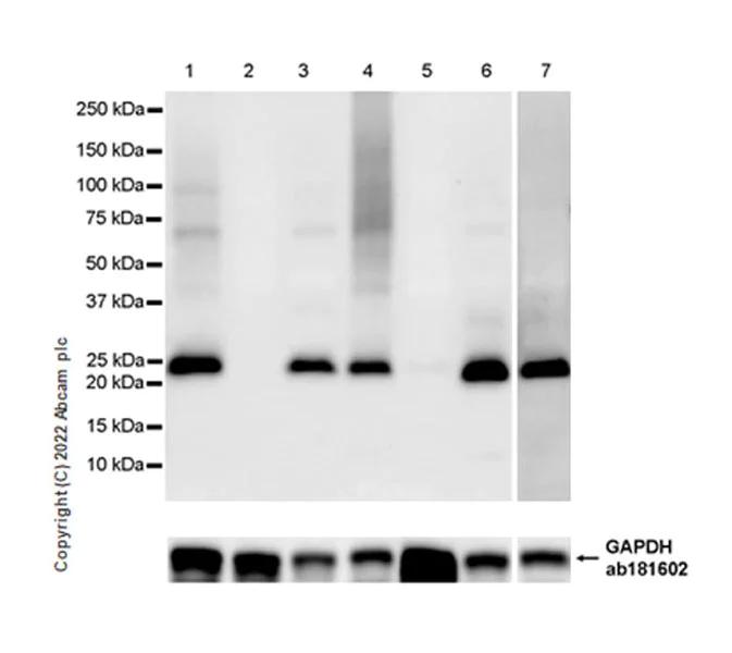
Western blot - Anti-TMS1/ASC antibody [EPR28201-61] - BSA and Azide free (AB307561)
This data was developed using 307560, the same antibody clone in a different buffer formulation. Blocking and diluting buffer and concentration : Negative control : RAW 264.7 (PMID : 28766178); Low expression tissue : skeletal muscle (PMID : 32659370). Lanes 1-6 : 15 seconds; Lane 7 : 125 seconds. Exposure time :
All lanes:
Western blot - Anti-TMS1/ASC antibody [EPR28201-61] (<a href='/products/primary-antibodies/tms1-asc-antibody-epr28201-61-ab307560'>ab307560</a>) at 1/1000 dilution
Lane 1:
J774A.1 (mouse reticum cell sarcoma monocyte/macrophage ) whole cell lysate 20 μg
Lane 2:
RAW 264.7 (mouse elson murine leukemia virus-induced tumor macrophage) whole cell lysate 20 μg
Lane 3:
Mouse spleen tissue lysate 20 μg
Lane 4:
Mouse colon tissue lysate 20 μg
Lane 5:
Mouse skeletal muscle tissue lysate 20 μg
Lane 6:
Rat spleen tissue lysate 20 μg
Lane 7:
Rat colon tissue lysate 20 μg
Secondary
All lanes:
Western blot - Goat Anti-Rabbit IgG H&L (HRP) (<a href='/products/secondary-antibodies/goat-rabbit-igg-h-l-hrp-ab97051'>ab97051</a>) at 1/20000 dilution
Observed band size: 21 kDa
false
Exposure time: 15s
関連する標識済み抗体及び組成の異なる製品 (3)
-
Anti-TMS1/ASC antibody [EPR28201-61]
-
519 Alexa Fluor® 488
Alexa Fluor® 488 Anti-TMS1/ASC antibody [EPR28201-61]
-
665 Alexa Fluor® 647
Alexa Fluor® 647 Anti-TMS1/ASC antibody [EPR28201-61]
組合せ製品

Primary Antibodies
AB263899
Anti-NLRP3 antibody [EPR23094-1]
KO Validated|RabMAb|Recombinant|20ul selling size
- 4
- 2

Primary Antibodies
AB179515
Anti-pro Caspase-1 + p10 + p12 antibody [EPR16883]
RabMAb|Recombinant|20ul selling size

- 3
- 3
Primary Antibodies
AB219800
Anti-GSDMD antibody [EPR20859]
BOND RX™ Validated|RabMAb|Recombinant|KO Validated|20ul selling size

- 5
- 2
Reactivity data
製品の詳細
Patented technology
Our RabMAb® technology is a patented hybridoma-based technology for making rabbit monoclonal antibodies. For details on our patents, please refer to RabMAb® patents.
What are the advantages of a recombinant monoclonal antibody?
This product is a recombinant monoclonal antibody, which offers several advantages including:
- - High batch-to-batch consistency and reproducibility
- - Improved sensitivity and specificity
- - Long-term security of supply
- - Animal-free batch production
For more information, read more on recombinant antibodies.
Conjugation ready
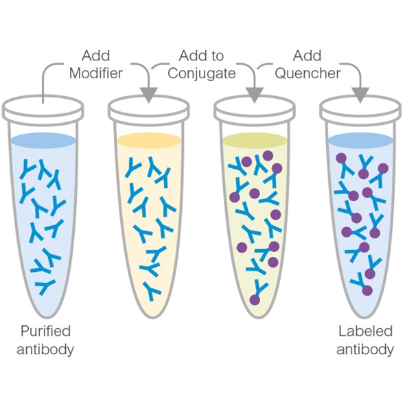
Our carrier-free antibodies are typically supplied in a PBS-only formulation, purified and free of BSA, sodium azide and glycerol. This conjugation-ready format is designed for use with fluorochromes, metal isotopes, oligonucleotides, and enzymes, which makes them ideal for antibody labelling, functional and cell-based assays, flow-based assays (e.g. mass cytometry) and Multiplex Imaging applications.
Use our conjugation kits for antibody conjugates that are ready-to-use in as little as 20 minutes with 1 minute hands-on-time and 100% antibody recovery: available for fluorescent dyes, HRP, biotin and gold.
Compatibility
This product is compatible with the Maxpar® Antibody Labeling Kit from Fluidigm, without the need for antibody preparation. Maxpar® is a trademark of Fluidigm Canada Inc.
出荷温度及び保存条件
製品の状態
精製方法
バッファー組成
出荷温度
短期保存温度
長期保存温度
製品プロトコール
- Visit the General protocols
- Visit the Troubleshooting
ターゲットの情報
Abcam product promise
Please note: All products are 'FOR RESEARCH USE ONLY. NOT FOR USE IN DIAGNOSTIC OR THERAPEUTIC PROCEDURES'.
For licensing inquiries, please contact partnerships@abcam.com