Anti-RIP 抗体 [EPR4689-100]
Anti-RIP antibody [EPR4689-100]
- 20ul selling size
- KO Validated
- RabMAb
- Recombinant
- 詳細を見る
Be the first to review this product! Submit a review
|
(2 Publications)
Knockout Tested Rabbit Recombinant Monoclonal RIP antibody. Suitable for WB, Flow Cyt (Intra) and reacts with Human samples. Cited in 2 publications.
別名を表示する
RIP, RIP1, RIPK1, Receptor-interacting serine/threonine-protein kinase 1, Cell death protein RIP, Receptor-interacting protein 1, RIP-1
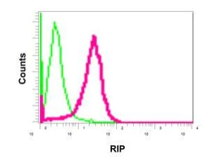
- Flow Cyt (Intra)
Supplier Data
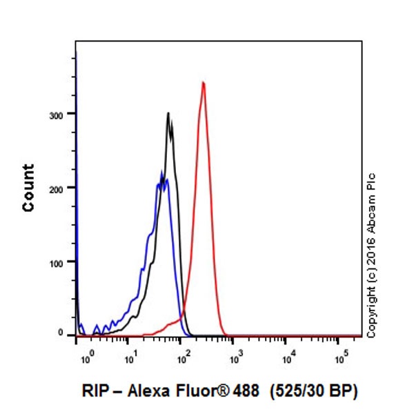
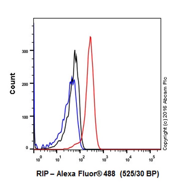
Flow Cytometry (Intracellular) - Anti-RIP antibody [EPR4689-100] (AB178420)
Intracellular flow cytometric analysis of permeabilized HeLa cells labeling RIP with ab178420 at 1/10 dilution (red) compared with a rabbit IgG negative control (green).
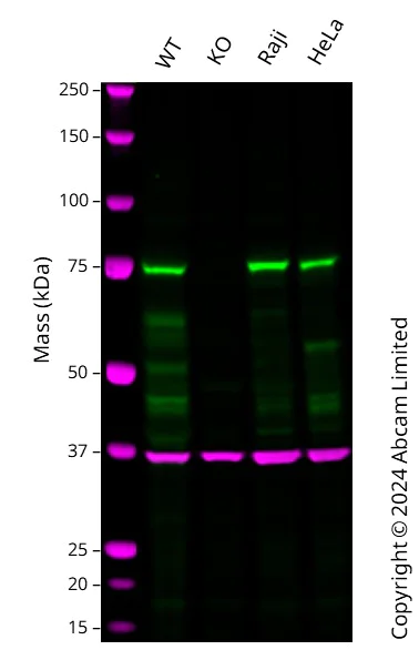
- WB
Lab
Western blot - Anti-RIP antibody [EPR4689-100] (AB178420)
Western blot : Anti-RIPK1 antibody [EPR4689-100] (ab178420) staining at 1/1000 dilution, shown in green; Mouse anti-GAPDH antibody [6C5] (ab8245) loading control staining at 1/20000 dilution, shown in magenta. In Western blot, ab178420 was shown to bind specifically to RIPK1. A band was observed at 75 kDa in wild-type THP-1 cell lysates with no signal observed at this size in RIPK1 knockout cell line. To generate this image, wild-type and RIPK1 knockout THP-1 cell lysates were analysed. First, samples were run on an SDS-PAGE gel then transferred onto a nitrocellulose membrane. Membranes were blocked in 3 % milk in TBS-0.1 % Tween® 20 (TBS-T) before incubation with primary antibodies overnight at 4 °C. Blots were washed four times in TBS-T, incubated with secondary antibodies for 1 h at room temperature, washed again four times then imaged. Secondary antibodies used were Goat anti-Rabbit IgG H&L 800CW and Goat anti-Mouse IgG H&L 680RD at 1/20000 dilution.
All lanes:
Western blot - Anti-RIP antibody [EPR4689-100] (ab178420) at 1/1000 dilution
Lane 1:
Wild-type THP-1 cell lysate at 20 µg
Lane 2:
RIPK1 knockout THP-1 cell lysate at 20 µg
Lane 3:
Raji cell lysate at 20 µg
Lane 4:
HeLa cell lysate at 20 µg
Secondary
All lanes:
Goat anti-Rabbit IgG H&L 800CW and Goat anti-Mouse IgG H&L 680RD at 1/20000 dilution
Predicted band size: 75 kDa
Observed band size: 75 kDa
false
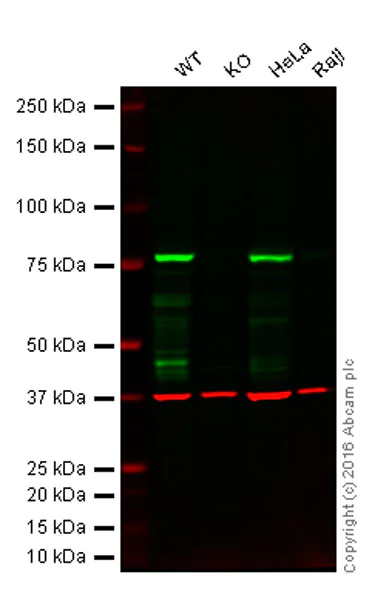
- WB
Unknown
Western blot - Anti-RIP antibody [EPR4689-100] (AB178420)
Lane 1 : Wild-type HAP1 cell lysate (20 μg)
Lane 2 : RIP knockout HAP1 cell lysate (20 μg)
Lane 3 : HeLa cell lysate (20 μg)
Lane 4 : Raji cell lysate (20 μg)
Lanes 1 to 4 : Merged signal (red and green). Green - ab178420 observed at 78 kDa. Red - loading control, ab8245, observed at 37 kDa.
ab178420 was shown to react with RIP in wild-type HAP1 cells along with additional cross-reactive bands. No band was observed when RIP knockout samples were examined. Wild-type and RIP knockout samples were subjected to SDS-PAGE. ab178420 and ab8245 (loading control to GAPDH) were both diluted 1/1000 and 1/10,000 respectively and incubated overnight at 4°C. Blots were developed with Goat anti-Rabbit IgG H&L (IRDye® 800CW) ab216773 and Goat anti-Mouse IgG H&L (IRDye® 680RD) ab216776 secondary antibodies at 1/10,000 dilution for 1 hour at room temperature before imaging.
All lanes:
Western blot - Anti-RIP antibody [EPR4689-100] (ab178420)
Predicted band size: 75 kDa
false
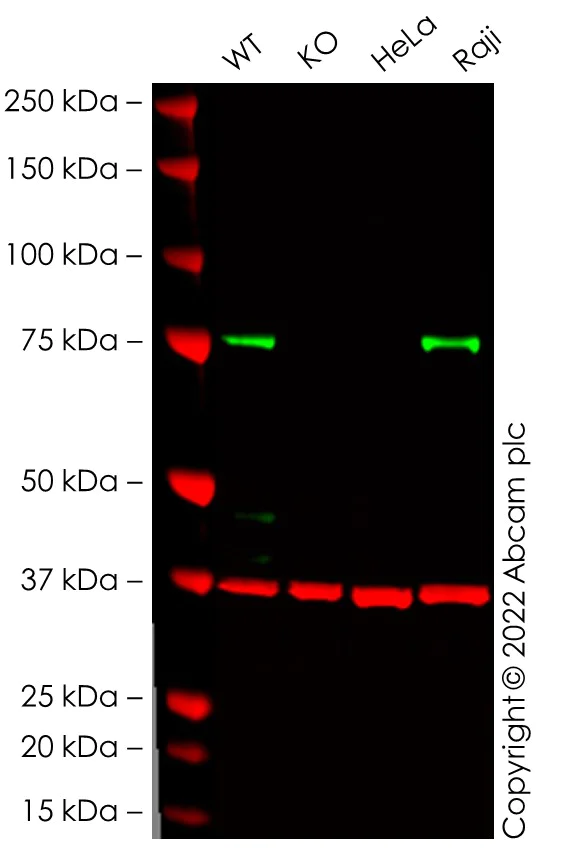
- WB
Lab
Western blot - Anti-RIP antibody [EPR4689-100] (AB178420)
All lanes:
Western blot - Anti-RIP antibody [EPR4689-100] (ab178420) at 1/1000 dilution
Lanes 1 - 4:
Western blot - Human RIPK1 knockout THP-1 cell line (<a href='/products/cell-lines/human-ripk1-knockout-thp-1-cell-line-ab276121'>ab276121</a>)
Lane 1:
Wild-type THP-1 cell lysate at 20 µg
Lane 2:
RIPK1 knockout THP-1 cell lysate at 20 µg
Lane 3:
HeLa cell lysate at 20 µg
Lane 4:
Raji cell lysate at 20 µg
Predicted band size: 75 kDa
Observed band size: 76 kDa
true
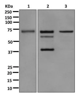
- WB
Supplier Data
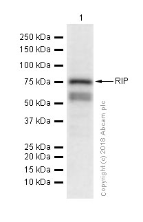
Western blot - Anti-RIP antibody [EPR4689-100] (AB178420)
All lanes:
Western blot - Anti-RIP antibody [EPR4689-100] (ab178420) at 1/1000 dilution
Lane 1:
Raji cell lysate at 10 µg
Lane 2:
HeLa cell lysate with Staurosporine at 10 µg
Lane 3:
HeLa cell lysate at 10 µg
Secondary
All lanes:
Goat anti-rabbit HRP at 1/2000 dilution
Predicted band size: 75 kDa
false
関連する標識済み抗体及び組成の異なる製品 (1)
-
Anti-RIP antibody [EPR4689-100] - BSA and Azide free
代替製品
Primary Antibodies
AB125072
20ul selling size|KO Validated|RabMAb|Recombinant|Trial Size
- 0
- 0
Primary Antibodies
AB238451
Anti-RIP antibody [EPR19697] - BSA and Azide free
RabMAb|Recombinant
- 0
- 0
Primary Antibodies
AB234924
Anti-RIP antibody [EPR4689] - BSA and Azide free
KO Validated|RabMAb|Recombinant
- 0
- 0
組合せ製品
Primary Antibodies
AB187091
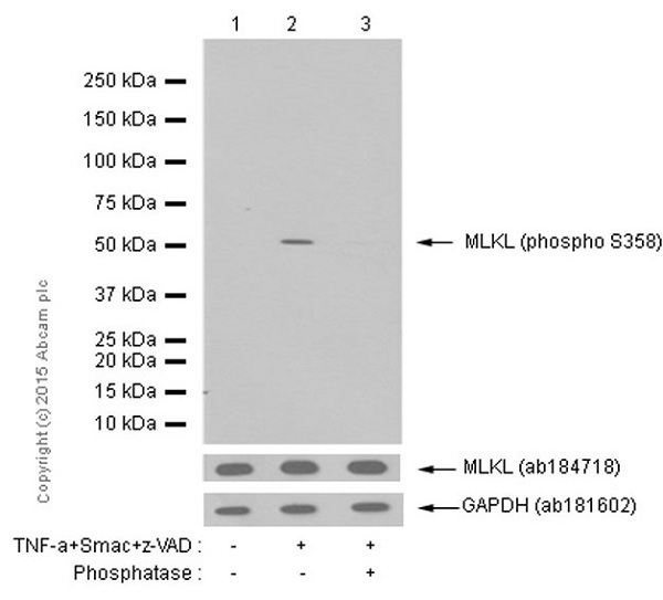
Anti-MLKL (phospho S358) antibody [EPR9514]
RabMAb|Recombinant|20ul selling size
- 4
- 19
Primary Antibodies
AB209384
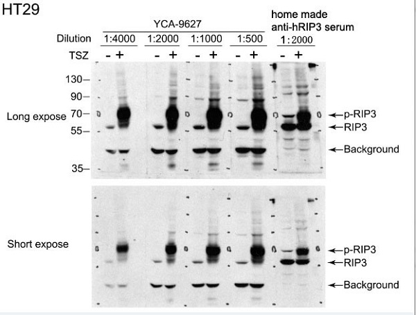
Anti-RIP3 (phospho S227) antibody [EPR9627]
RabMAb|Recombinant|20ul selling size
- 0
- 0
Primary Antibodies
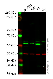
AB184718
RabMAb|Recombinant|KO Validated|20ul selling size
- 5
- 5
Primary Antibodies
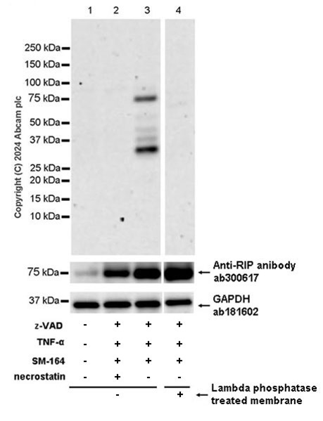
AB316923
Anti-RIP (phospho S166) antibody [EPR25654-166]
20ul selling size|Recombinant|RabMAb|KO Validated
- 0
- 0
Reactivity data
製品の詳細
Species reactivity
Mouse, Rat: We have preliminary internal testing data to indicate this antibody may not react with these species.
Please contact us for more information.
Patented technology
Our RabMAb® technology is a patented hybridoma-based technology for making rabbit monoclonal antibodies. For details on our patents, please refer to RabMAb® patents.
What are the advantages of a recombinant monoclonal antibody?
This product is a recombinant monoclonal antibody, which offers several advantages including:
- - High batch-to-batch consistency and reproducibility
- - Improved sensitivity and specificity
- - Long-term security of supply
- - Animal-free batch production
For more information, read more on recombinant antibodies.
出荷温度及び保存条件
製品の状態
精製方法
バッファー組成
出荷温度
短期保存期間
短期保存温度
長期保存温度
分注に関する情報
保管に関する情報
補足情報
This supplementary information is collated from multiple sources and compiled automatically.
Biological function summary
Receptor-Interacting Protein Kinase 1 (RIPK1) participates in regulating both necroptosis and apoptosis distinguishing itself as an important mediator in cell death mechanisms. As part of the necrosome complex which includes RIPK3 and MLKL RIPK1 functions in necroptosis—a programmed form of necrosis. This characteristic involvement shows its dual role in maintaining cell fate decisions making it an integral part of immune response and inflammation control.
Pathways
RIPK1 strongly associates with the TNF signaling pathway and NF-kB pathway. Its interaction with TNF receptor 1 (TNFR1) and consequent involvement with TRADD and TRAF2 mediates the signal transduction necessary for the activation of NF-kB leading to transcription of genes involved in survival and inflammation. This connection illustrates its capability to switch between promoting cell survival through NF-kB and facilitating cell death via necroptosis or apoptosis depending on cellular context and cues.
製品プロトコール
- Visit the General protocols
- Visit the Troubleshooting
ターゲットの情報
文献 (2)
Recent publications for all applications. Explore the full list and refine your search
Science signaling 13: PubMed32398349
2020
Applications
Unspecified application
Species
Unspecified reactive species
Carcinogenesis 38:402-410 PubMed28334272
2017
Applications
Unspecified application
Species
Unspecified reactive species
Abcam product promise
Please note: All products are 'FOR RESEARCH USE ONLY. NOT FOR USE IN DIAGNOSTIC OR THERAPEUTIC PROCEDURES'.
For licensing inquiries, please contact partnerships@abcam.com