Anti-RanBP2 抗体 [RM2047]
Anti-RanBP2 antibody [RM2047]
- KD Validated
- BOND RX™ Validated
- 20ul selling size
- Recombinant
- RabMAb
- 詳細を見る
Be the first to review this product! Submit a review
|
(0 Publication)
Rabbit Recombinant Multiclonal RanBP2 antibody. Suitable for WB, IHC-P, ICC/IF, IP and reacts with Human, Mouse, Rat samples.
別名を表示する
NUP358, RANBP2, E3 SUMO-protein ligase RanBP2, 358 kDa nucleoporin, Nuclear pore complex protein Nup358, Nucleoporin Nup358, Ran-binding protein 2, p270, RanBP2
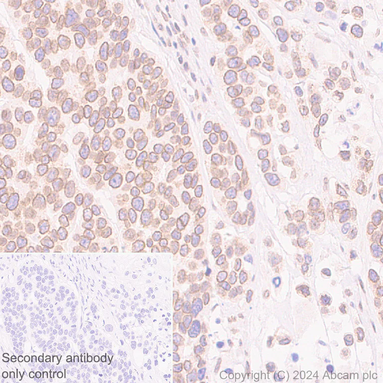
- IHC-P
Supplier Data
Immunohistochemistry (Formalin/PFA-fixed paraffin-embedded sections) - Anti-RanBP2 antibody [RM2047] (AB316336)
Immunohistochemical analysis of paraffin-embedded Human gastric carcinoma tissue labeling RanBP2 with ab316336 at 1/500 (1.002 ug/ml) dilution, followed by a ready to use Rabbit specific IHC polymer detection kit HRP/DAB (ab209101).
Positive staining on human gastric carcinoma. The section was incubated with ab316336 for 30 mins at room temperature.
The immunostaining was performed on a Leica Biosystems BOND® RX instrument
Counterstained with Hematoxylin.
Secondary antibody only control : Secondary antibody is a ready to use Rabbit specific IHC polymer detection kit HRP/DAB (ab209101).
Heat mediated antigen retrieval was performed with Tris-EDTA buffer (pH 9.0, Epitope Retrieval Solution2) for 20 mins
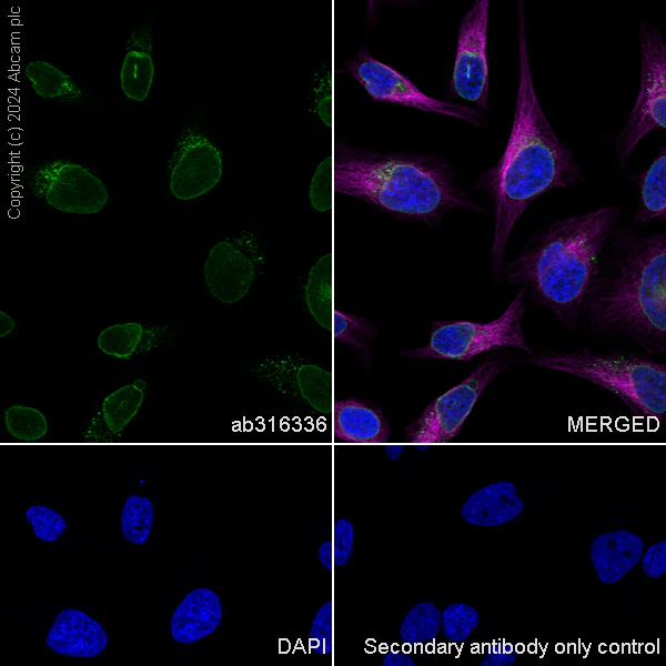
- ICC/IF
Supplier Data
Immunocytochemistry/ Immunofluorescence - Anti-RanBP2 antibody [RM2047] (AB316336)
Immunofluorescent analysis of 4% Paraformaldehyde-fixed, 0.1% TritonX-100 permeabilized HeLa (human cervical adenocarcinoma epithelial cell) cells labelling RanBP2 with ab316336 at 1/500 (1.002 ug/ml) dilution, followed by ab150081 Goat Anti-Rabbit IgG H&L (Alexa Fluor® 488) preadsorbed antibody at 1/1000 (2 ug/ml) dilution (Green).
Confocal image showing nuclear envelope with puncta signal in HeLa cell line.
Image was taken with a confocal microscope(Leica-Microsystems, TCS SP8).
ab195889 Anti-alpha Tubulin mouse monoclonal antibody - Microtubule Marker (Alexa Fluor® 594) was used to counterstain tubulin at 1/200 (2.5ug/ml) dilution (Magenta). The Nuclear counterstain was DAPI (Blue).
Secondary antibody only control : Secondary antibody is ab150081 Goat Anti-Rabbit IgG H&L (Alexa Fluor® 488) preadsorbed at 1/1000 (2 ug/ml) dilution.
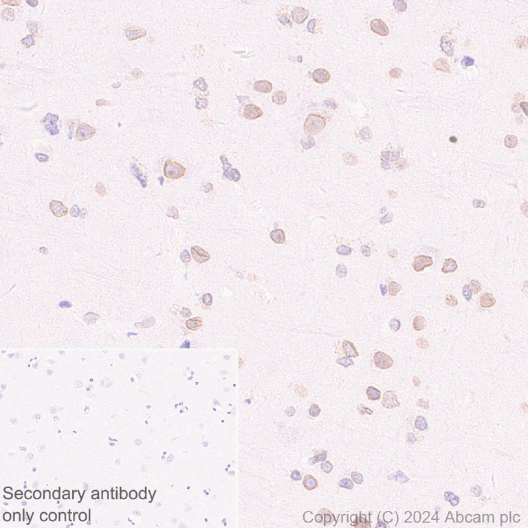
- IHC-P
Supplier Data
Immunohistochemistry (Formalin/PFA-fixed paraffin-embedded sections) - Anti-RanBP2 antibody [RM2047] (AB316336)
Immunohistochemical analysis of paraffin-embedded Human cerebrum tissue labeling RanBP2 with ab316336 at 1/500 (1.002 ug/ml) dilution, followed by a ready to use Rabbit specific IHC polymer detection kit HRP/DAB (ab209101).
Positive staining on human cerebrum. The section was incubated with ab316336 for 30 mins at room temperature.
The immunostaining was performed on a Leica Biosystems BOND® RX instrument
Counterstained with Hematoxylin.
Secondary antibody only control : Secondary antibody is a ready to use Rabbit specific IHC polymer detection kit HRP/DAB (ab209101).
Heat mediated antigen retrieval was performed with Tris-EDTA buffer (pH 9.0, Epitope Retrieval Solution2) for 20 mins
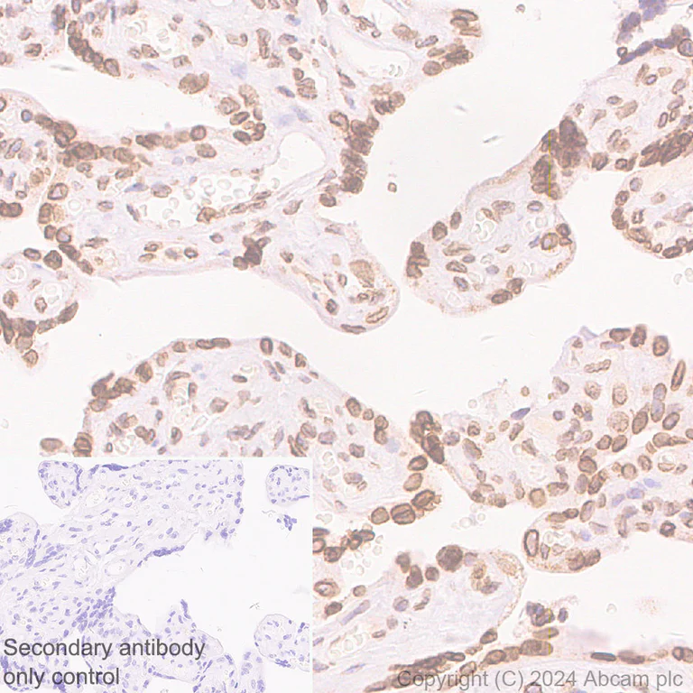
- IHC-P
Supplier Data
Immunohistochemistry (Formalin/PFA-fixed paraffin-embedded sections) - Anti-RanBP2 antibody [RM2047] (AB316336)
Immunohistochemical analysis of paraffin-embedded Human placenta tissue labeling RanBP2 with ab316336 at 1/500 (1.002 ug/ml) dilution, followed by a ready to use Rabbit specific IHC polymer detection kit HRP/DAB (ab209101).
Positive staining on human placenta. The section was incubated with ab316336 for 30 mins at room temperature.
The immunostaining was performed on a Leica Biosystems BOND® RX instrument
Counterstained with Hematoxylin.
Secondary antibody only control : Secondary antibody is a ready to use Rabbit specific IHC polymer detection kit HRP/DAB (ab209101).
Heat mediated antigen retrieval was performed with Tris-EDTA buffer (pH 9.0, Epitope Retrieval Solution2) for 20 mins
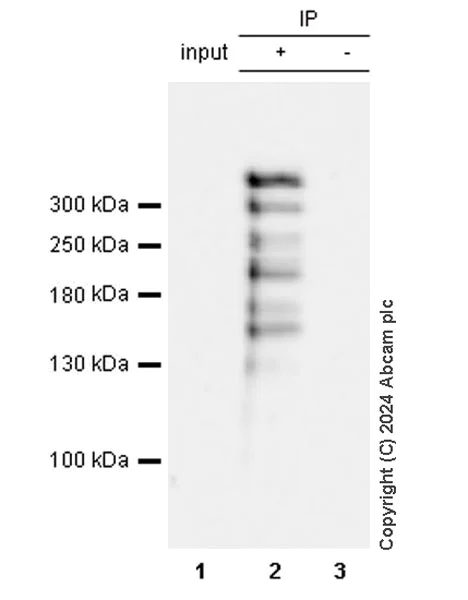
- IP
Supplier Data
Immunoprecipitation - Anti-RanBP2 antibody [RM2047] (AB316336)
RanBP2 was immunoprecipitated from 0.35 mg HeLa (human cervix adenocarcinoma epithelial cell) whole cell lysate with ab316336 at 1/30 dilution (2ug in 0.35mg lysates). Western blot was performed on the immunoprecipitate using ab316336 at 1/1000 dilution. VeriBlot for IP secondary antibody(HRP)(ab131366) was used at 1/5000 dilution.
Lane 1 : HeLa (human cervix adenocarcinoma epithelial cell) whole cell lysate
Lane 2 : ab316336 IP in HeLa (human cervix adenocarcinoma epithelial cell) whole cell lysate
Lane 3 : Rabbit monoclonal IgG (ab172730) instead of ab316336 in HeLa whole cell lysate
The bands beneath the target band (358 kDa) are likely to be degraded target fragments.
Lysates were freshly made and used for Western blotting immediately to minimize protein degradation.
All lanes:
Immunoprecipitation - Anti-RanBP2 antibody [RM2047] (ab316336) at 1/30 dilution
All lanes:
HeLa (human cervix adenocarcinoma epithelial cell) whole cell lysate
Secondary
All lanes:
Immunoprecipitation - VeriBlot for IP Detection Reagent (HRP) (<a href='/products/reagents/veriblot-for-ip-detection-reagent-hrp-ab131366'>ab131366</a>) at 1/5000 dilution
false
Exposure time: 10s
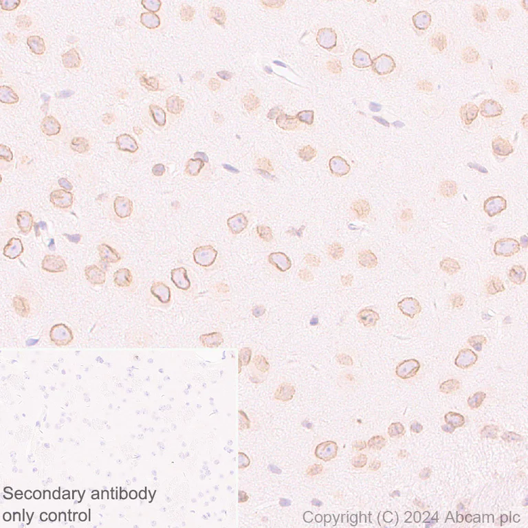
- IHC-P
Supplier Data
Immunohistochemistry (Formalin/PFA-fixed paraffin-embedded sections) - Anti-RanBP2 antibody [RM2047] (AB316336)
Immunohistochemical analysis of paraffin-embedded Mouse cerebrum tissue labeling RanBP2 with ab316336 at 1/1000 (0.501 ug/ml) dilution, followed by a ready to use Rabbit specific IHC polymer detection kit HRP/DAB (ab209101).
Positive staining on mouse cerebrum. The section was incubated with ab316336 for 30 mins at room temperature.
The immunostaining was performed on a Leica Biosystems BOND® RX instrument
Counterstained with Hematoxylin.
Secondary antibody only control : Secondary antibody is a ready to use Rabbit specific IHC polymer detection kit HRP/DAB (ab209101).
Heat mediated antigen retrieval was performed with Tris-EDTA buffer (pH 9.0, Epitope Retrieval Solution2) for 20 mins
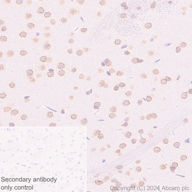
- IHC-P
Supplier Data
Immunohistochemistry (Formalin/PFA-fixed paraffin-embedded sections) - Anti-RanBP2 antibody [RM2047] (AB316336)
Immunohistochemical analysis of paraffin-embedded Rat cerebrum tissue labeling RanBP2 with ab316336 at 1/1000 (0.501 ug/ml) dilution, followed by a ready to use Rabbit specific IHC polymer detection kit HRP/DAB (ab209101).
Positive staining on rat cerebrum. The section was incubated with ab316336 for 30 mins at room temperature.
The immunostaining was performed on a Leica Biosystems BOND® RX instrument
Counterstained with Hematoxylin.
Secondary antibody only control : Secondary antibody is a ready to use Rabbit specific IHC polymer detection kit HRP/DAB (ab209101).
Heat mediated antigen retrieval was performed with Tris-EDTA buffer (pH 9.0, Epitope Retrieval Solution2) for 20 mins
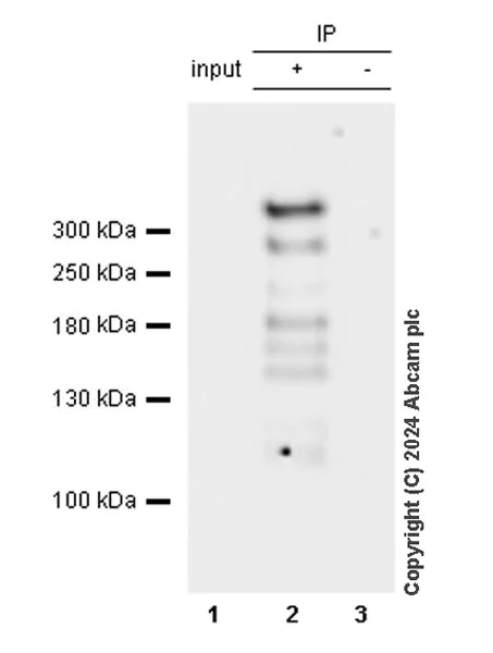
- IP
Supplier Data
Immunoprecipitation - Anti-RanBP2 antibody [RM2047] (AB316336)
RanBP2 was immunoprecipitated from 0.35 mg Neuro-2a (mouse neuroblastoma neuroblast) whole cell lysate with ab316336 at 1/30 dilution (2ug in 0.35mg lysates). Western blot was performed on the immunoprecipitate using ab316336 at 1/1000 dilution. VeriBlot for IP secondary antibody(HRP)(ab131366) was used at 1/5000 dilution.
Lane 1 : Neuro-2a (mouse neuroblastoma neuroblast) whole cell lysate
Lane 2 : ab316336 IP in Neuro-2a (mouse neuroblastoma neuroblast) whole cell lysate
Lane 3 : Rabbit monoclonal IgG (ab172730) instead of ab316336 in Neuro-2a whole cell lysate
The bands beneath the target band (358 kDa) are likely to be degraded target fragments.
Lysates were freshly made and used for Western blotting immediately to minimize protein degradation.
All lanes:
Immunoprecipitation - Anti-RanBP2 antibody [RM2047] (ab316336) at 1/30 dilution
All lanes:
Neuro-2a (mouse neuroblastoma neuroblast) whole cell lysate
Secondary
All lanes:
Immunoprecipitation - VeriBlot for IP Detection Reagent (HRP) (<a href='/products/reagents/veriblot-for-ip-detection-reagent-hrp-ab131366'>ab131366</a>) at 1/5000 dilution
false
Exposure time: 15s
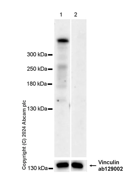
- WB
Supplier Data
Western blot - Anti-RanBP2 antibody [RM2047] (AB316336)
The bands beneath the target band (358 kDa) are likely to be degraded target fragments.
Lysates were freshly made and used for Western blotting immediately to minimize protein degradation.
In Western blot, Anti-Vinculin antibody [EPR8185] (ab129002) staining at 1/10000 dilution.
All lanes:
Western blot - Anti-RanBP2 antibody [RM2047] (ab316336) at 1/1000 dilution
Lane 1:
HeLa (human cervix adenocarcinoma epithelial cell) transfected with scrambled siRNA control whole cell lysate at 20 µg
Lane 2:
HeLa transfected with siRNA specifically targeting RanBP2 whole cell lysate at 20 µg
Secondary
All lanes:
Western blot - Goat Anti-Rabbit IgG H&L (HRP) (<a href='/products/secondary-antibodies/goat-rabbit-igg-h-l-hrp-ab97051'>ab97051</a>) at 1/200000 dilution
Observed band size: 358 kDa,124 kDa
false
Exposure time: 114s
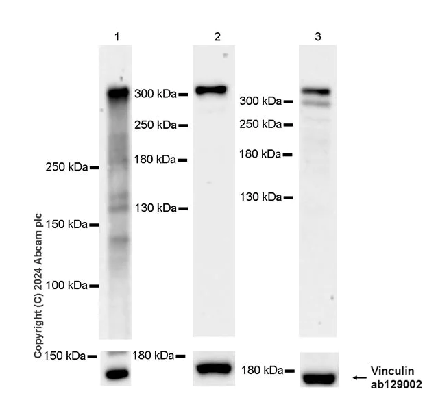
- WB
Supplier Data
Western blot - Anti-RanBP2 antibody [RM2047] (AB316336)
The bands beneath the target band (358 kDa) are likely to be degraded target fragments.
The lysates of lanes 2-3 were freshly made and used for Western blotting immediately to minimize protein degradation.
The lane 1 was developed using a high sensitivity ECL substrate.
Lanes 2-3 are applied with Goat Anti-Rabbit IgG, (H+L), Peroxidase conjugated (ab97051) at 1/20000 and lane 1 is applied with Goat Anti-Rabbit IgG (HRP) with minimal cross-reactivity with human IgG at 1/2000.
In Western blot, Anti-Vinculin antibody [EPR8185] (ab129002) staining at 1/10000 dilution.
Exposure time : Lane 1 : 180 seconds; lane 2 : 26 seconds; lane 3 : 180 seconds
All lanes:
Western blot - Anti-RanBP2 antibody [RM2047] (ab316336) at 1/1000 dilution
Lane 1:
Human testis tissue lysate at 20 µg
Lane 2:
Mouse testis tissue lysate at 20 µg
Lane 3:
Rat testis tissue lysate at 20 µg
Secondary
Lane 1:
Goat Anti-Rabbit IgG (HRP) with minimal cross-reactivity with human IgG at 1/2000 dilution
Lanes 2 - 3:
Western blot - Goat Anti-Rabbit IgG H&L (HRP) (<a href='/products/secondary-antibodies/goat-rabbit-igg-h-l-hrp-ab97051'>ab97051</a>) at 1/20000 dilution
Observed band size: 358 kDa,124 kDa
false
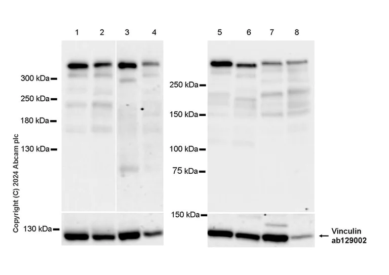
- WB
Supplier Data
Western blot - Anti-RanBP2 antibody [RM2047] (AB316336)
The bands beneath the target band (358 kDa) are likely to be degraded target fragments.
Lysates were freshly made and used for Western blotting immediately to minimize protein degradation.
In Western blot, Anti-Vinculin antibody [EPR8185] (ab129002) staining at 1/10000 dilution.
Exposure time : Lanes 1-2 : 10 seconds; lanes 3-4 : 180 seconds; lanes 5-8 : 26 seconds
All lanes:
Western blot - Anti-RanBP2 antibody [RM2047] (ab316336) at 1/1000 dilution
Lane 1:
HeLa (human cervix adenocarcinoma epithelial cell) whole cell lysate at 20 µg
Lane 2:
293T (human embryonic kidney epithelial cell) whole cell lysate at 20 µg
Lane 3:
NIH/3T3 (mouse embryonic fibroblast) whole cell lysate at 20 µg
Lane 4:
PC-12 (rat adrenal gland pheochromocytoma cell) whole cell lysate at 20 µg
Lane 5:
C6 (rat glial tumor glial cell) whole cell lysate at 20 µg
Lane 6:
Neuro-2a (mouse neuroblastoma neuroblast) whole cell lysate at 20 µg
Lane 7:
U-87 MG (human glioblastoma-astrocytoma epithelial cell) whole cell lysate at 20 µg
Lane 8:
SH-SY5Y (human neuroblastoma epithelial cell) whole cell lysate at 20 µg
Secondary
All lanes:
Western blot - Goat Anti-Rabbit IgG H&L (HRP) (<a href='/products/secondary-antibodies/goat-rabbit-igg-h-l-hrp-ab97051'>ab97051</a>) at 1/200000 dilution
Observed band size: 358 kDa,124 kDa
false
関連する標識済み抗体及び組成の異なる製品 (1)
-
Anti-RanBP2 antibody [RM2047] - BSA and Azide free
組合せ製品
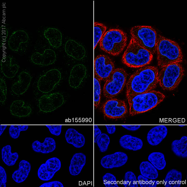
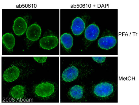
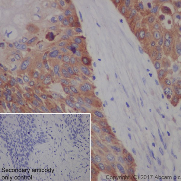
Primary Antibodies
AB214039
Anti-Transferrin Receptor antibody [EPR20584]
RabMAb|Recombinant|20ul selling size
- 5
- 1
Reactivity data
製品の詳細
What are recombinant multiclonals?
Recombinant multiclonals are a mixture of recombinant antibodies co-expressed from a library of heavy and light chains. They offer several advantages including:
- - The sensitivity of polyclonal antibodies by recognising multiple epitopes
- - High batch-to-batch consistency and reproducibility
- - Improved sensitivity and specificity
- - Long-term security of supply
- - Animal-free batch production
View our range of recombinant multiclonal antibodies.
Patented technology
Our RabMAb® technology is a patented hybridoma-based technology for making rabbit monoclonal antibodies. For details on our patents, please refer to RabMAb® patents.
出荷温度及び保存条件
製品の状態
精製方法
バッファー組成
出荷温度
短期保存期間
短期保存温度
長期保存温度
分注に関する情報
保管に関する情報
補足情報
This supplementary information is collated from multiple sources and compiled automatically.
Biological function summary
RanBP2 plays a significant role in multiple processes such as mitotic spindle assembly and chromatin condensation. It functions as part of the Ran GTPase system a critical complex for nucleocytoplasmic transport. In addition RanBP2 contributes to protein sumoylation where it acts as a scaffold for conjugating enzymes. It maintains nuclear envelope integrity and supports the positioning of centrosomes during cell division.
Pathways
RanBP2 is actively involved in the nucleocytoplasmic transport pathway ensuring the proper exchange of proteins and RNA between the nucleus and cytoplasm. It is critical to the Ran cycle where it works alongside proteins like RanGTP and RanGAP1 to regulate transport activities. The involvement of RanBP2 in the SUMOylation pathway also emphasizes its role in post-translational modification impacting various cellular events and signaling.
製品プロトコール
- Visit the General protocols
- Visit the Troubleshooting
ターゲットの情報
Abcam product promise
Please note: All products are 'FOR RESEARCH USE ONLY. NOT FOR USE IN DIAGNOSTIC OR THERAPEUTIC PROCEDURES'.
For licensing inquiries, please contact partnerships@abcam.com