Anti-PFKFB3 抗体 [EPR12594]
Anti-PFKFB3 antibody [EPR12594]
- RabMAb
- Recombinant
- KO Validated
- 20ul selling size
- 詳細を見る
5
(6 Reviews)
|
(105 Publications)
Anti-PFKFB3 antibody [EPR12594] (ab181861) is a rabbit monoclonal antibody detecting PFKFB3 in Western Blot, Flow Cytometry (Intra), IP, IHC-P, ICC/IF. Suitable for Human, Mouse, Rat.
- KO validated for confirmed specificity
- Biophysical QC for unrivalled batch-batch consistency
- Over 70 publications
別名を表示する
PFK/FBPase 3, Renal carcinoma antigen NY-REN-56, iPFK-2, PFKFB3

- Flow Cyt (Intra)
Unknown
Flow Cytometry (Intracellular) - Anti-PFKFB3 antibody [EPR12594] (AB181861)
Intracellular Flow Cytometry analysis of A431 (human epidermoid carcinoma) cells labeling PFKFB3 with purified ab181861 at 1/210 dilution (10ug/ml) (red). Cells were fixed with 4% paraformaldehyde and permeabilised with 90% methanol. A Goat anti rabbit IgG (Alexa Fluor® 488) (1/2000 dilution) was used as the secondary antibody. Rabbit monoclonal IgG (Black) was used as the isotype control, cells without incubation with primary antibody and secondary antibody (Blue) was used as the unlabeled control.

- ICC/IF
Supplier Data
Immunocytochemistry/ Immunofluorescence - Anti-PFKFB3 antibody [EPR12594] (AB181861)
Immunofluorescent analysis of 4% paraformaldehyde-fixed A431 cells labeling PFKFB3 with ab181861 at 1/100 dilution, followed by Goat anti rabbit IgG (Alexa Fluor®555) at 1/200 dilution. Counter stained with Dapi (blue).

- ICC/IF
Supplier Data
Immunocytochemistry/ Immunofluorescence - Anti-PFKFB3 antibody [EPR12594] (AB181861)
Immunofluorescent analysis of acetone-fixed HeLa cells labeling PFKFB3 with ab181861 at 1/100 dilution, followed by Goat anti rabbit IgG (Alexa Fluor®555) at 1/200 dilution. Counter stained with Dapi (blue).

- IHC-P
Supplier Data
Immunohistochemistry (Formalin/PFA-fixed paraffin-embedded sections) - Anti-PFKFB3 antibody [EPR12594] (AB181861)
Immunohistochemical analysis of paraffin-embedded human melanoma tissue labeling PFKFB3 with ab181861 at 1/50 dilution followed by prediluted HRP Polymer for Rabbit IgG. Counter stained with Hematoxylin.
Perform heat mediated antigen retrieval with EDTA buffer pH 9 before commencing with IHC staining protocol.

- IP
Supplier Data
Immunoprecipitation - Anti-PFKFB3 antibody [EPR12594] (AB181861)
Western blot analysis of PFKFB3 in HeLa cell lysate immunoprecipitated using ab181861 at 1/50 dilution (Lane 1). Lane 2 : Negative control.
Secondary antibody : Anti-Rabbit IgG (HRP), specific to the non-reduced form of IgG at 1/1500 dilution.
All lanes:
Immunoprecipitation - Anti-PFKFB3 antibody [EPR12594] (ab181861)
Predicted band size: 60 kDa
false

- IP
Lab
Immunoprecipitation - Anti-PFKFB3 antibody [EPR12594] (AB181861)
PFKFB3 was immunoprecipitated from 0.35 mg of AR42J (rat pancreatic tumor epithelial cell) whole cell lysate with ab181861 at 1/30 dilution. Western blot was performed from the immunoprecipitate using ab181861 at 1/1000 dilution. VeriBlot for IP Detection Reagent (HRP) (ab131366), was used for detection at 1/5000 dilution. Lane 1 : AR42J (rat pancreatic tumor epithelial cell) whole cell lysate 10 μg (Input). Lane 2 : ab181861 IP in AR42J whole cell lysate Lane 3 : Rabbit monoclonal IgG (ab172730) instead of ab181861 in AR42J whole cell lysate Blocking and dilution buffer and concentration : 5% NFDM/TBST. Exposure time : 180 seconds.
All lanes:
Immunoprecipitation - Anti-PFKFB3 antibody [EPR12594] (ab181861)
Lane 1:
AR42J (rat pancreatic tumor epithelial cell) whole cell lysate at 10 µg
Lane 2:
Anti-PFKFB3 antibody IP in AR42J whole cell lysate
Secondary
All lanes:
Immunoprecipitation - VeriBlot for IP Detection Reagent (HRP) (<a href='/products/reagents/veriblot-for-ip-detection-reagent-hrp-ab131366'>ab131366</a>) at 1/5000 dilution
Predicted band size: 60 kDa
Observed band size: 60 kDa
false

- IP
Lab
Immunoprecipitation - Anti-PFKFB3 antibody [EPR12594] (AB181861)
PFKFB3 was immunoprecipitated from 0.35 mg of Mouse skin tissue lysate with ab181861 at 1/30 dilution. Western blot was performed from the immunoprecipitate using ab181861 at 1/1000 dilution. VeriBlot for IP Detection Reagent (HRP) (ab131366), was used for detection at 1/5000 dilution.
Lane 1 : Mouse skin tissue lysate 10 μg (Input)
Lane 2 : ab181861 at 1/30 IP in Mouse skin tissue lysate
Lane 3 : Rabbit monoclonal IgG (ab172730) instead of ab181861 in Mouse skin tissue lysate
Blocking and dilution buffer and concentration : 5% NFDM/TBST
This blot was developed using a high sensitivity ECL substrate. The high-sensitivity ECL substrate used allows for the detection of proteins in the mid-femtogram range.
All lanes:
Immunoprecipitation - Anti-PFKFB3 antibody [EPR12594] (ab181861) at 1/1000 dilution
Lane 1:
Mouse skin tissue lysate at 10 µg
Lane 2:
ab181861 at 1/30 IP in Mouse skin tissue lysate at 1/30 dilution
Secondary
All lanes:
Immunoprecipitation - VeriBlot for IP Detection Reagent (HRP) (<a href='/products/reagents/veriblot-for-ip-detection-reagent-hrp-ab131366'>ab131366</a>) at 1/5000 dilution
Observed band size: 60 kDa
true
Exposure time: 180s

- WB
Lab
Western blot - Anti-PFKFB3 antibody [EPR12594] (AB181861)
Exposure time : Lane 1-2 : 26 seconds, Lane 3-7 : 48 seconds
Blocking buffer and concentration : 5% NFDM/TBST
Lane 3-7 were developed using a high sensitivity ECL substrate.
The expression level of PFKFB3 is upregulated during 3T3-L1 differentiation (PMID : 16306349).
The band at approximately 110 kDa is likely to be PFKFB3 dimer (PMID : 31889092).
All lanes:
Western blot - Anti-PFKFB3 antibody [EPR12594] (ab181861) at 1/1000 dilution
Lane 1:
A431 (human epidermoid carcinoma epithelial cell), whole cell lysate at 20 µg
Lane 2:
bEnd.3 (mouse brain endothelial cell), whole cell lysate at 20 µg
Lane 3:
NIH/3T3 (mouse embryonic fibroblast), whole cell lysate at 20 µg
Lane 4:
4T1 (mouse mammary gland carcinoma epithelial cell), whole cell lysate at 20 µg
Lane 5:
Undifferentiated 3T3-L1 (mouse embryonic fibroblast), whole cell lysate at 20 µg
Lane 6:
3T3-L1 (mouse embryonic fibroblast) differentiated into adipocyte-like cells, whole cell lysate at 20 µg
Lane 7:
C2C12 (mouse myoblast), whole cell lysate at 20 µg
Secondary
All lanes:
Western blot - Goat Anti-Rabbit IgG H&L (HRP) (<a href='/products/secondary-antibodies/goat-rabbit-igg-h-l-hrp-ab97051'>ab97051</a>) at 1/20000 dilution
Predicted band size: 60 kDa
Observed band size: 60 kDa
true

- WB
Lab
Western blot - Anti-PFKFB3 antibody [EPR12594] (AB181861)
Blocking and diluting buffer and concentration : 5% NFDM/TBST.
This blot was developed using a high sensitivity ECL substrate. The high-sensitivity ECL substrate used allows for the detection of proteins in the mid-femtogram range.
All lanes:
Western blot - Anti-PFKFB3 antibody [EPR12594] (ab181861) at 1/1000 dilution
Lane 1:
A431 (human epidermoid carcinoma epithelial cell) whole cell lysate at 20 µg
Lane 2:
Mouse skin tissue lysate at 20 µg
Lane 3:
Rat breast tissue lysate at 20 µg
Lane 4:
AR42J (rat pancreatic tumor epithelial cell) whole cell lysate at 20 µg
Lane 5:
L6 (rat skeletal muscle myoblast) whole cell lysate at 20 µg
Secondary
All lanes:
Western blot - Goat Anti-Rabbit IgG H&L (HRP) (<a href='/products/secondary-antibodies/goat-rabbit-igg-h-l-hrp-ab97051'>ab97051</a>) at 1/20000 dilution
Predicted band size: 60 kDa
Observed band size: 60 kDa
true
Exposure time: 15s

- WB
Lab
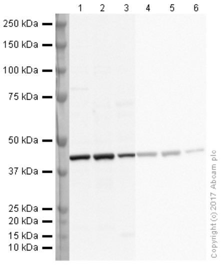
Western blot - Anti-PFKFB3 antibody [EPR12594] (AB181861)
Low expression : kidney tissue (PMID : 34660743).
In Western blot, Anti-GAPDH antibody [EPR16891] - Loading Control (ab181602) staining at 1/1000000 dilution.
All lanes:
Western blot - Anti-PFKFB3 antibody [EPR12594] (ab181861) at 1/1000 dilution
Lane 1:
Mouse lung tissue lysate at 20 µg
Lane 2:
Mouse liver tissue lysate at 20 µg
Lane 3:
Mouse heart tissue lysate at 20 µg
Lane 4:
Mouse kidney tissue lysate at 20 µg
Lane 5:
Rat lung tissue lysate at 20 µg
Lane 6:
Rat liver tissue lysate at 20 µg
Lane 7:
Rat heart tissue lysate at 20 µg
Lane 8:
Rat kidney tissue lysate at 20 µg
Secondary
All lanes:
Western blot - Goat Anti-Rabbit IgG H&L (HRP) (<a href='/products/secondary-antibodies/goat-rabbit-igg-h-l-hrp-ab97051'>ab97051</a>) at 1/20000 dilution
Observed band size: 60 kDa
true
Exposure time: 180s
関連する標識済み抗体及び組成の異なる製品 (3)
-
Anti-PFKFB3 antibody [EPR12594] - BSA and Azide free
-
519 Alexa Fluor® 488
Alexa Fluor® 488 Anti-PFKFB3 antibody [EPR12594]
-
665 Alexa Fluor® 647
Alexa Fluor® 647 Anti-PFKFB3 antibody [EPR12594]
代替製品

Primary Antibodies
AB232498
Anti-PFKFB3 (phospho S461) antibody [EPR19735] - BSA and Azide free
RabMAb|Recombinant

- 0
- 0

組合せ製品

Primary Antibodies
AB115730
Anti-Glucose Transporter GLUT1 antibody [EPR3915]
BOND RX™ Validated|20ul selling size|RabMAb|Recombinant|KO Validated

- 4
- 20
Primary Antibodies
AB209847
Anti-Hexokinase II antibody [EPR20839]
RabMAb|Recombinant|KO Validated|20ul selling size

- 5
- 2

Primary Antibodies
AB8227
Anti-beta Actin antibody - Loading Control
BOND RX™ Validated|Lab Essentials
- 5
- 104
Reactivity data
製品の詳細
What is this antibody validated in?
Anti-PFKFB3 antibody [EPR12594] (ab181861) is a rabbit recombinant monoclonal antibody and is validated for use in Western Blot (WB), Flow Cytometry (Intra), Flow Cytometry (Flow Cyt), Immunoprecipitation (IP), Immunohistochemistry (IHC-P), Immunocytochemistry/immunofluorescence (ICC/IF) in Human, Mouse, Rat samples.
What is the molecular weight of PFKFB3?
Anti-PFKFB3 [EPR12594] (ab181861) specifically detects a band for PFKFB3 (UniProt: Q16875) at a molecular weight of 60kDa.
Trusted by the scientific community
Anti-PFKFB3 [EPR12594] (ab181861) was first used in a scientific publication in 2014 and has been cited over 70 times in peer-reviewed journals.
Reviewed by scientists
Anti-PFKFB3 [EPR12594] (ab181861) has over 5 independent reviews from customers.
Specificity confirmed
The specificity of Anti-PFKFB3 antibody [EPR12594] (ab181861) has been confirmed by Western blot testing in PFKFB3 Knockout HAP1 cells.
Other related products
We have a range of other formats of antibody clone [EPR12594] also available for your convenience: ab181861, Alexa Fluor® 647 - ab203651, Alexa Fluor® 488 - ab203984, Carrier free - ab218121
Patented technology
Our RabMAb® technology is a patented hybridoma-based technology for making rabbit monoclonal antibodies. For details on our patents, please refer to RabMAb® patents.
What are the advantages of a recombinant monoclonal antibody?
This product is a recombinant monoclonal antibody, which offers several advantages including:
- - High batch-to-batch consistency and reproducibility
- - Improved sensitivity and specificity
- - Long-term security of supply
- - Animal-free batch production
For more information, read more on recombinant antibodies.
Trial sizes available!
Test your antibody or perform pre-screening before committing to a larger quantity. Sold in 20µl. Discover our selection of trial-size antibodies.
出荷温度及び保存条件
製品の状態
精製方法
バッファー組成
出荷温度
短期保存期間
短期保存温度
長期保存温度
分注に関する情報
保管に関する情報
補足情報
This supplementary information is collated from multiple sources and compiled automatically.
Biological function summary
PFKFB3 regulates cellular energy metabolism serving as a switch for converting carbohydrates into energy and building blocks for growth. This enzyme is part of a glycolytic complex coordinating energy needs during proliferation and survival. Its activity is important in processes demanding increased energy and anabolic precursors such as cell division. The enzyme's modulation affects fructose-26-bisphosphate levels influencing cellular responses to stress and nutrient availability directly impacting metabolic adaptation.
Pathways
PFKFB3 integrates into the glycolytic pathway and the pentose phosphate pathway. PFKFB3's control over fructose-26-bisphosphate levels influences phosphofructokinase-1 modulating the glycolytic pathway's rate. This modulation impacts energy production efficiency and the provision of metabolites required for nucleotide synthesis linking to the pentose phosphate pathway. Proline-rich AKT substrate of 40 kDa (PRAS40) and AKT may also interact indirectly to regulate PFKFB3 activity in these metabolic pathways.
製品プロトコール
- Visit the General protocols
- Visit the Troubleshooting
ターゲットの情報
文献 (105)
Recent publications for all applications. Explore the full list and refine your search
Pakistan journal of pharmaceutical sciences 38:1632-1646 PubMed40996179
2025
Applications
Unspecified application
Species
Unspecified reactive species
Clinical and translational medicine 15:e70441 PubMed40817604
2025
Applications
Unspecified application
Species
Unspecified reactive species
Cellular and molecular life sciences : CMLS 82:311 PubMed40801925
2025
Applications
Unspecified application
Species
Unspecified reactive species
Journal of cell communication and signaling 19:e70034 PubMed40692714
2025
Applications
Unspecified application
Species
Unspecified reactive species
Cancers 17: PubMed40427135
2025
Applications
Unspecified application
Species
Unspecified reactive species
Frontiers in immunology 16:1583587 PubMed40356921
2025
Applications
Unspecified application
Species
Unspecified reactive species
BMC cancer 25:374 PubMed40022029
2025
Applications
Unspecified application
Species
Unspecified reactive species
FASEB journal : official publication of the Federation of American Societies for Experimental Biology 39:e70332 PubMed39831513
2025
Applications
Unspecified application
Species
Unspecified reactive species
Cell & bioscience 14:144 PubMed39605072
2024
Applications
Unspecified application
Species
Unspecified reactive species
International journal of nanomedicine 19:9799-9819 PubMed39345912
2024
Applications
Unspecified application
Species
Unspecified reactive species
Abcam product promise
Please note: All products are 'FOR RESEARCH USE ONLY. NOT FOR USE IN DIAGNOSTIC OR THERAPEUTIC PROCEDURES'.
For licensing inquiries, please contact partnerships@abcam.com