Anti-Pepsin 抗体 [EPR28182-66] - BSA and Azide free
Anti-Pepsin antibody [EPR28182-66] - BSA and Azide free
- BOND RX™ Validated
- RabMAb
- Recombinant
- 詳細を見る
Be the first to review this product! Submit a review
|
(0 Publication)
Rabbit Recombinant Monoclonal PEPA5 antibody. Carrier free. Suitable for IHC-P, WB and reacts with Human, Transfected cell line - Human, Transfected cell lysate - Human, Recombinant fragment samples.
別名を表示する
Pepsin A-5, Pepsinogen-5, PGA5
- IHC-P
Supplier Data
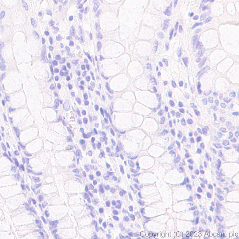
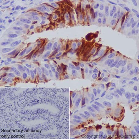
Immunohistochemistry (Formalin/PFA-fixed paraffin-embedded sections) - Anti-Pepsin antibody [EPR28182-66] - BSA and Azide free (AB312853)
This data was developed using ab312852, the same antibody clone in a different buffer formulation. Immunohistochemical analysis of paraffin-embedded Human colon tissue labelling Pepsin with ab312852 at 1/5000 (0.105 ug/ml) followed by a ready to use LeicaDS9800 (Bond™ Polymer Refine Detection). Negative control : no staining on human colon. The section was incubated with ab312852 for 10 mins at room temperature. The immunostaining was performed on a Leica Biosystems BOND® RX instrument Counterstained with Hematoxylin. Secondary antibody only control : Secondary antibody is a ready to use LeicaDS9800 (Bond™ Polymer Refine Detection). Heat mediated antigen retrieval was performed with Tris-EDTA buffer (pH 9.0, Epitope Retrieval Solution2) for 20 mins.
- IHC-P
Supplier Data
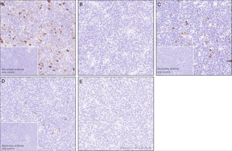
Immunohistochemistry (Formalin/PFA-fixed paraffin-embedded sections) - Anti-Pepsin antibody [EPR28182-66] - BSA and Azide free (AB312853)
This data was developed using ab312852, the same antibody clone in a different buffer formulation. Immunohistochemical analysis of paraffin-embedded (A) HEK-293T cells tissue labeling Pepsin with ab312852 at 1/1000 (0.528 ug/ml) followed by a ready to use LeicaDS9800 (Bond™ Polymer Refine Detection). Positive staining on (A) HEK-293T cells transfected with a human PGA3 expression vector containing a his tag, (C) HEK-293T cells transfected with a human PGA4 expression vector containing a his tag, and (D) HEK-293T cells transfected with a human PGA5 expression vector containing a his tag, no staining on (B) HEK-293T cells transfected with a human PGC expression vector containing a his tag and (E) HEK-293T cells transfected with empty vector containing a his tag. The section was incubated with ab312852 for 10 mins at room temperature. The immunostaining was performed on a Leica Biosystems BOND® RX instrument. Counterstained with Hematoxylin. Secondary antibody only control : Secondary antibody is a ready to use LeicaDS9800 (Bond™ Polymer Refine Detection). Heat mediated antigen retrieval was performed with Tris-EDTA buffer (pH 9.0, Epitope Retrieval Solution2) for 20 mins.
- IHC-P
Supplier Data
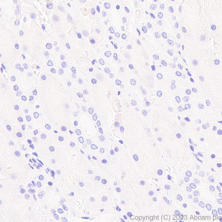
Immunohistochemistry (Formalin/PFA-fixed paraffin-embedded sections) - Anti-Pepsin antibody [EPR28182-66] - BSA and Azide free (AB312853)
This data was developed using ab312852, the same antibody clone in a different buffer formulation. Immunohistochemical analysis of paraffin-embedded Human kidney tissue labeling Pepsin with ab312852 at 1/5000 (0.105 ug/ml) followed by a ready to use LeicaDS9800 (Bond™ Polymer Refine Detection). Negative control : no staining on human kidney. The section was incubated with ab312852 for 10 mins at room temperature. The immunostaining was performed on a Leica Biosystems BOND® RX instrument. Counterstained with Hematoxylin. Secondary antibody only control : Secondary antibody is a ready to use LeicaDS9800 (Bond™ Polymer Refine Detection). Heat mediated antigen retrieval was performed with Tris-EDTA buffer (pH 9.0, Epitope Retrieval Solution2) for 20 mins.
- IHC-P
Supplier Data
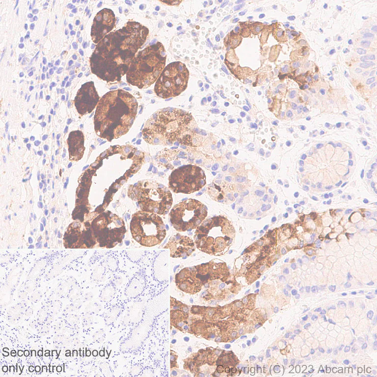
Immunohistochemistry (Formalin/PFA-fixed paraffin-embedded sections) - Anti-Pepsin antibody [EPR28182-66] - BSA and Azide free (AB312853)
This data was developed using ab312852, the same antibody clone in a different buffer formulation. Immunohistochemical analysis of paraffin-embedded Human stomach tissue labeling Pepsin with ab312852 at 1/5000 (0.105 ug/ml) followed by a ready to use LeicaDS9800 (Bond™ Polymer Refine Detection). Positive staining on human stomach. The section was incubated with ab312852 for 10 mins at room temperature. The immunostaining was performed on a Leica Biosystems BOND® RX instrument. Counterstained with Hematoxylin. Secondary antibody only control : Secondary antibody is a ready to use LeicaDS9800 (Bond™ Polymer Refine Detection). Heat mediated antigen retrieval was performed with Tris-EDTA buffer (pH 9.0, Epitope Retrieval Solution2) for 20 mins.
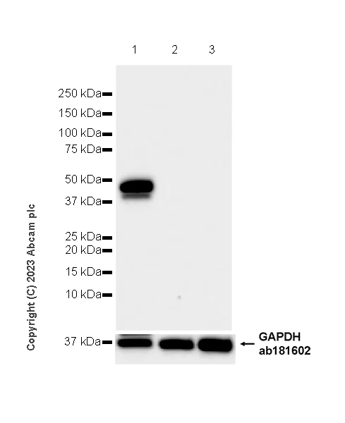
- WB
Supplier Data
Western blot - Anti-Pepsin antibody [EPR28182-66] - BSA and Azide free (AB312853)
This data was developed using ab312852, the same antibody clone in a different buffer formulation. Blocking and diluting buffer and concentration : 5% NFDM/TBST Negative control : kidney, skeletal muscle. The molecular weight observed is consistent with what has been described in the reference (PMID : 33620602). In Western blot, anti-GAPDH antibody (ab181602) loading control staining at 1/200000 dilution. Exposure time : 10 seconds
All lanes:
Western blot - Anti-Pepsin antibody [EPR28182-66] (<a href='/products/primary-antibodies/pepsin-antibody-epr28182-66-ab312852'>ab312852</a>) at 1/1000 dilution
Lane 1:
Human stomach tissue lysate at 20 µg
Lane 2:
Human kidney tissue lysate at 20 µg
Lane 3:
Human skeletal muscle tissue lysate at 20 µg
Secondary
All lanes:
Western blot - Goat Anti-Rabbit IgG H&L (HRP) (<a href='/products/secondary-antibodies/goat-rabbit-igg-h-l-hrp-ab97051'>ab97051</a>) at 1/100000 dilution
Observed band size: 42 kDa
false
Exposure time: 10s
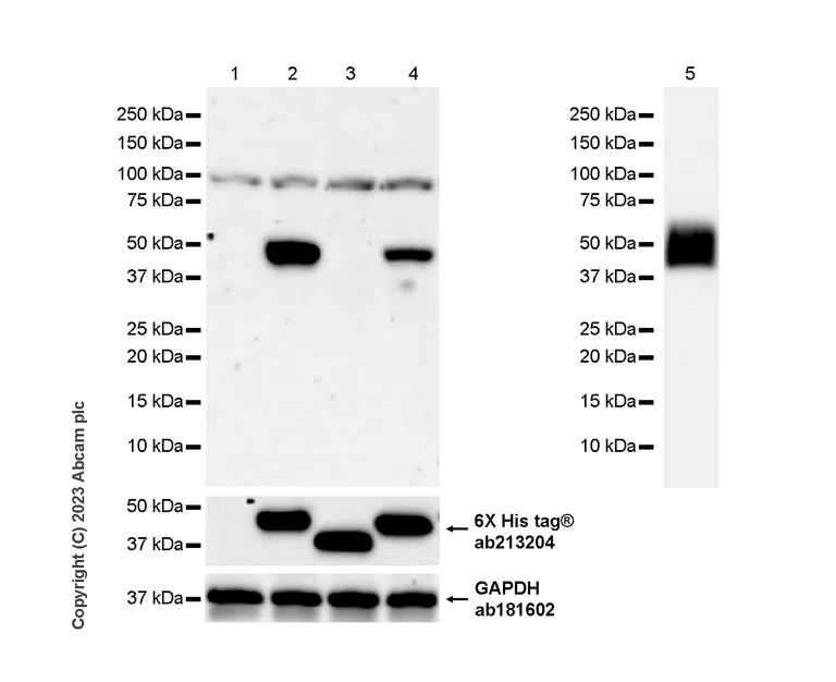
- WB
Supplier Data
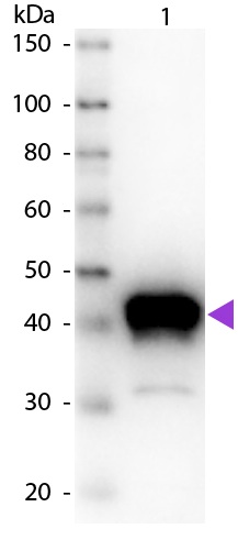
Western blot - Anti-Pepsin antibody [EPR28182-66] - BSA and Azide free (AB312853)
This data was developed using ab312852, the same antibody clone in a different buffer formulation. Blocking and diluting buffer and concentration : 5% NFDM/TBST This antibody demonstrates cross reactivity with human Pepsin A-3, Pepsin A-4 and Pepsin A-5. An additional band with unknown identity at 100 kDa was observed. In Western blot, anti-His antibody (ab213204) staining at 1/5000 dilution. Anti-GAPDH antibody (ab181602) loading control staining at 1/200000 dilution. Exposure time : Lane 1-4 : 180 seconds; Lane 5 : 10 seconds.
All lanes:
Western blot - Anti-Pepsin antibody [EPR28182-66] (<a href='/products/primary-antibodies/pepsin-antibody-epr28182-66-ab312852'>ab312852</a>) at 1/1000 dilution
Lane 1:
293T cells transfected with an empty vector containi a His-tag whole cell lysate at 20 µg
Lane 2:
293T cells transfected with a Human Pepsin A-3 expression vector containi a His-tag whole cell lysate at 2 µg
Lane 3:
293T cells transfected with a Human PGC expression vector containi a His-tag whole cell lysate at 2 µg
Lane 4:
293T cells transfected with a Human Pepsin A-4 expression vector containi a His-tag whole cell lysate at 10 µg
Lane 5:
Purified Pepsin A-5 recombinant protein (aa63-388) at 20 ng
Secondary
All lanes:
Western blot - Goat Anti-Rabbit IgG H&L (HRP) (<a href='/products/secondary-antibodies/goat-rabbit-igg-h-l-hrp-ab97051'>ab97051</a>) at 1/20000 dilution
Observed band size: 42 kDa
false
関連する標識済み抗体及び組成の異なる製品 (1)
-
Anti-Pepsin antibody [EPR28182-66]
組合せ製品
Primary Antibodies
AB207299
Anti-MMP7 antibody [EPR17888-71]
20ul selling size|RabMAb|Recombinant|KO Validated
- 0
- 0
Primary Antibodies
AB181602
Anti-GAPDH antibody [EPR16891] - Loading Control
KD Validated|RabMAb|Recombinant|Lab Essentials|20ul selling size

- 5
- 23
Reactivity data
製品の詳細
Patented technology
Our RabMAb® technology is a patented hybridoma-based technology for making rabbit monoclonal antibodies. For details on our patents, please refer to RabMAb® patents.
What are the advantages of a recombinant monoclonal antibody?
This product is a recombinant monoclonal antibody, which offers several advantages including:
- - High batch-to-batch consistency and reproducibility
- - Improved sensitivity and specificity
- - Long-term security of supply
- - Animal-free batch production
For more information, read more on recombinant antibodies.
Conjugation ready
Our carrier-free antibodies are typically supplied in a PBS-only formulation, purified and free of BSA, sodium azide and glycerol. This conjugation-ready format is designed for use with fluorochromes, metal isotopes, oligonucleotides, and enzymes, which makes them ideal for antibody labelling, functional and cell-based assays, flow-based assays (e.g. mass cytometry) and Multiplex Imaging applications.
Use our conjugation kits for antibody conjugates that are ready-to-use in as little as 20 minutes with 1 minute hands-on-time and 100% antibody recovery: available for fluorescent dyes, HRP, biotin and gold.
Compatibility
This product is compatible with the Maxpar® Antibody Labeling Kit from Fluidigm, without the need for antibody preparation. Maxpar® is a trademark of Fluidigm Canada Inc.
出荷温度及び保存条件
製品の状態
精製方法
バッファー組成
出荷温度
短期保存温度
長期保存温度
補足情報
This supplementary information is collated from multiple sources and compiled automatically.
Biological function summary
Pepsin fulfills an important role in the digestive process by breaking down larger protein molecules into smaller peptides facilitating nutrient absorption. It does not form part of a larger protein complex but operates independently within the gastric environment. Pepsin's enzymatic action targets specific peptide bonds ensuring efficient degradation of dietary proteins into absorbable forms. The specificity of its substrate binding ensures effective digestion and subsequent utilization of amino acids.
Pathways
Pepsin's activity integrates into proteolytic pathways particularly the catabolic processes in the gastrointestinal tract. It interacts with other digestive enzymes including trypsin and chymotrypsin which further hydrolyze peptides originated by pepsin. These interactions highlight its seamless fit in the digestive enzyme cascade optimized for effective protein turnover and nutrient release into the body. The pepsin pathway coordinates closely with the gastric and duodenal enzymatic processes.
製品プロトコール
- Visit the General protocols
- Visit the Troubleshooting
ターゲットの情報
Abcam product promise
Please note: All products are 'FOR RESEARCH USE ONLY. NOT FOR USE IN DIAGNOSTIC OR THERAPEUTIC PROCEDURES'.
For licensing inquiries, please contact partnerships@abcam.com