Anti-p21 抗体 [EPR3993] - BSA and Azide free
Anti-p21 antibody [EPR3993] - BSA and Azide free
- RabMAb
- Recombinant
- KO Validated
- 詳細を見る
Be the first to review this product! Submit a review
|
(3 Publications)
Rabbit Recombinant Monoclonal p21 antibody. Carrier free. Suitable for WB and reacts with Human, Mouse, Rat samples. Cited in 3 publications.
別名を表示する
CAP20, CDKN1, CIP1, MDA6, PIC1, SDI1, WAF1, CDKN1A, Cyclin-dependent kinase inhibitor 1, CDK-interacting protein 1, Melanoma differentiation-associated protein 6, p21, MDA-6
- WB
Lab
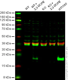
Western blot - Anti-p21 antibody [EPR3993] - BSA and Azide free (AB215971)
This data was developed using ab109199, the same antibody clone in a different buffer formulation.
Lane 1 Wild-type DLD-1 cell lysate (20 μg)
Lane 2 Wild-type DLD-1 20 μM 2,3-DCPE for 16hrs treated cell lysate (20 μg)
Lane 3 p21 knockout DLD-1 cell lysate (20 μg)
Lane 4 p21 knockout 20 μM 2,3-DCPE for 16hrs DLD-1 cell lysate (20 μg)
Lane 5 : HT1080 cell lysate (20 μg)
Lanes 1 - 5 Merged signal (red and green). Green - ab109199 observed at 20 kDa. Red - loading control, ab8245, observed at 37 kDa.
ab109199 was shown to recognize p21 in WT DLD-1 cells with 2,3-DCPE treatment along with additional cross-reactive bands. When p21 knockout DLD-1 cells +/- 2,3-DCPE treatment were used, no band was observed. Wild-type and p21 knockout samples were subjected to SDS-PAGE. ab109199 and ab8245 (loading control to GAPDH) were diluted 1/1000 and 1/10 000 respectively and incubated overnight at 4°C. Blots were developed with Goat anti-Rabbit IgG H&L (IRDye® 800CW) preadsorbed (ab216773) and Goat anti-Mouse IgG H&L (IRDye® 680RD) preadsorbed (ab216776) secondary antibodies at 1/10 000 dilution for 1 h at room temperature before imaging.
All lanes:
Western blot - Anti-p21 antibody [EPR3993] (<a href='/products/primary-antibodies/p21-antibody-epr3993-ab109199'>ab109199</a>)
Predicted band size: 18 kDa
Observed band size: 20 kDa
false
- WB
Lab
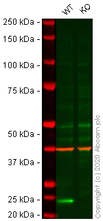
Western blot - Anti-p21 antibody [EPR3993] - BSA and Azide free (AB215971)
This data was developed using the same antibody clone in a different buffer formulation (ab109199).
Lanes 1- 2 : Merged signal (red and green). Green - ab109199 observed at 20 kDa. Red - Anti-GAPDH antibody [6C5] - Loading Control (ab8245) observed at 37 kDa.
ab109199 was shown to react with p21 in wild-type HCT116 cells in western blot. Loss of signal was observed when knockout cell line ab266860 (knockout cell lysate ab256870) was used. Wild-type HCT116 and CDKN1A knockout HCT116 cell lysates were subjected to SDS-PAGE. Membrane was blocked for 1 hour at room temperature in 0.1% TBST with 3% non-fat dried milk. ab109199 and Anti-GAPDH antibody [6C5] - Loading Control (ab8245) overnight at 4°C at a 1 in 1000 dilution and a 1 in 20000 dilution respectively. Blots were developed with Goat anti-Rabbit IgG H&L (IRDye®800CW) preadsorbed (ab216773) and Goat anti-Mouse IgG H&L (IRDye®680RD) preadsorbed (ab216776) secondary antibodies at 1 in 20000 dilution for 1 hour at room temperature before imaging.
All lanes:
Western blot - Anti-p21 antibody [EPR3993] (<a href='/products/primary-antibodies/p21-antibody-epr3993-ab109199'>ab109199</a>) at 1/1000 dilution
Lane 1:
Wild-type HCT116 cell lysate at 20 µg
Lane 2:
CDKN1A knockout HCT116 cell lysate at 20 µg
Lane 2:
Western blot - Human CDKN1A knockout HCT116 cell line (<a href='/products/cell-lines/human-cdkn1a-knockout-hct116-cell-line-ab266860'>ab266860</a>)
Predicted band size: 18 kDa,197 kDa,36 kDa,47 kDa,72 kDa,84 kDa
Observed band size: 150 kDa,20 kDa,40 kDa,45 kDa,47 kDa
false
- WB
Lab
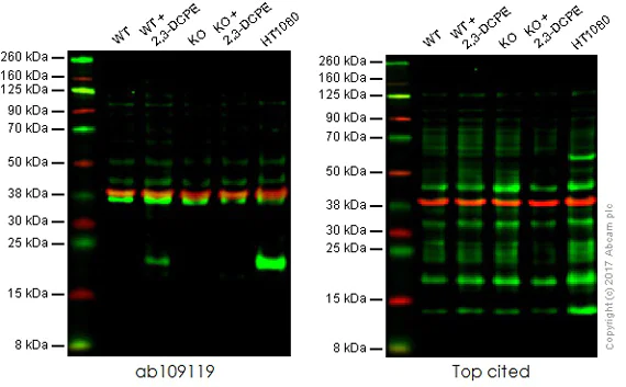
Western blot - Anti-p21 antibody [EPR3993] - BSA and Azide free (AB215971)
This data was developed using ab109199, the same antibody clone in a different buffer formulation.
Lane 1 Wild-type DLD-1 cell lysate (20 μg)
Lane 2 Wild-type DLD-1 20 μM 2,3-DCPE for 16hrs treated cell lysate (20 μg)
Lane 3 p21 knockout DLD-1 cell lysate (20 μg)
Lane 4 p21 knockout 20 μM 2,3-DCPE for 16hrs DLD-1 cell lysate (20 μg)
Lane 5 : HT1080 cell lysate (20 μg)
Lanes 1 - 5 Merged signal (red and green). Green - ab109199 observed at 20 kDa. Red - loading control, ab8245, observed at 37 kDa.
This western blot image is a comparison between ab109119 and a competitor's top cited rabbit polyclonal antibody.
All lanes:
Western blot - Anti-p21 antibody [EPR3993] (<a href='/products/primary-antibodies/p21-antibody-epr3993-ab109199'>ab109199</a>)
Predicted band size: 18 kDa
false
- WB
Unknown
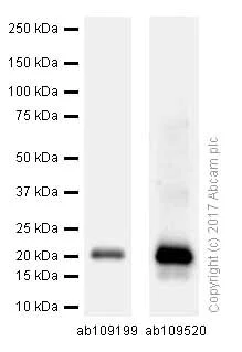
Western blot - Anti-p21 antibody [EPR3993] - BSA and Azide free (AB215971)
This data was developed using ab109199, the same antibody clone in a different buffer formulation.
Blocking and dilution buffer : 5% NFDM/TBST.
All lanes:
Western blot - Anti-p21 antibody [EPR3993] (<a href='/products/primary-antibodies/p21-antibody-epr3993-ab109199'>ab109199</a>)
All lanes:
MCF-7 (Human breast adenocarcinoma epithelial cell) whole cell lysates at 15 µg
Secondary
All lanes:
Western blot - Goat Anti-Rabbit IgG H&L (HRP) (<a href='/products/secondary-antibodies/goat-rabbit-igg-h-l-hrp-ab97051'>ab97051</a>) at 1/20000 dilution
Predicted band size: 18 kDa
false
Exposure time: 3min
- WB
Lab
Western blot - Anti-p21 antibody [EPR3993] - BSA and Azide free (AB215971)
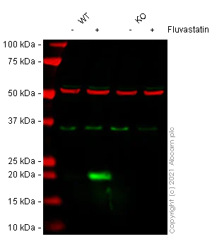
False colour image of Western blot : Anti-p21 antibody [EPR3993] staining at 1/1000 dilution, shown in green; Mouse anti-Alpha Tubulin [DM1A] (ab7291) loading control staining at 1/20000 dilution, shown in red. In Western blot, ab109199 was shown to bind specifically to p21. A band was observed at 21 kDa in wild-type y cell lysates with no signal observed at this size in CDKN2A knockout cell line ab255349 (knockout cell lysate ab263812). To generate this image, wild-type and CDKN2A knockout HeLa cell lysates were analysed. First, samples were run on an SDS-PAGE gel then transferred onto a nitrocellulose membrane. Membranes were blocked in 3 % milk in TBS-0.1 % Tween® 20 (TBS-T) before incubation with primary antibodies overnight at 4°C. Blots were washed four times in TBS-T, incubated with secondary antibodies for 1 h at room temperature, washed again four times then imaged.Secondary antibodies used were Goat anti-Rabbit IgG H&L (IRDye® 800CW) preabsorbed (ab216773) and Goat anti-Mouse IgG H&L (IRDye® 680RD) preabsorbed (ab216776) at 1/20000 dilution.
All lanes:
Western blot - Anti-p21 antibody [EPR3993] (<a href='/products/primary-antibodies/p21-antibody-epr3993-ab109199'>ab109199</a>) at 1/1000 dilution
Lane 1:
wild-type HeLa Vehicle Control Fluvastatin (0 uM, 24 h) cell lysate at 20 µg
Lane 2:
wild-type HeLa Treated Fluvastatin (50 uM, 24 h) cell lysate at 20 µg
Lane 2:
Western blot - Human CDKN1A knockout HeLa cell line (<a href='/products/cell-lines/human-cdkn1a-knockout-hela-cell-line-ab255349'>ab255349</a>)
Lane 3:
CDKN1A knockout HeLa Vehicle Control Fluvastatin (0 uM, 24 h) cell lysate at 20 µg
Lane 4:
CDKN1A knockout HeLa Treated Fluvastatin (50 uM, 24 h) cell lysate at 20 µg
Predicted band size: 18 kDa
Observed band size: 21 kDa
false
- WB
Lab
Western blot - Anti-p21 antibody [EPR3993] - BSA and Azide free (AB215971)
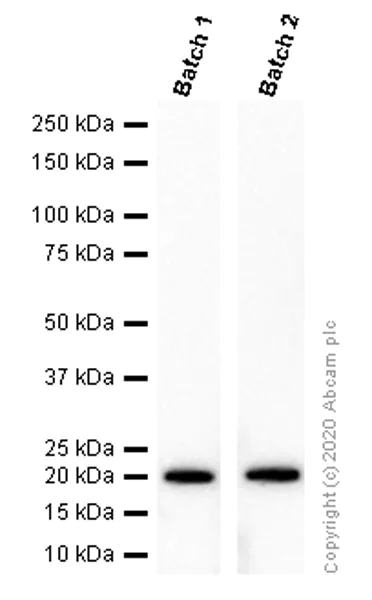
This data was developed using ab109199, the same antibody clone in a different buffer formulation. Different batches of ab109199 were tested on MCF7 (Human breast adenocarcinoma epithelial cell) lysate at 0.2 µg/ml. 15 µg of lysate was loaded in each lane. Bands observed at 21 kDa.
All lanes:
Western blot - Anti-p21 antibody [EPR3993] (<a href='/products/primary-antibodies/p21-antibody-epr3993-ab109199'>ab109199</a>)
Predicted band size: 18 kDa
false
- WB
Lab
Western blot - Anti-p21 antibody [EPR3993] - BSA and Azide free (AB215971)
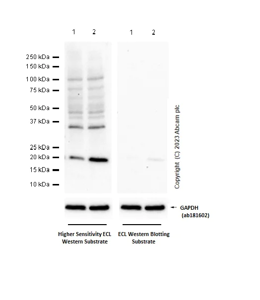
This data was developed using ab109199, the same antibody clone in a different buffer formulation. Blocking and diluting buffer and concentration : 5% NFDM/TBST. ab181602 was used as a GAPDH loading control. We recommend using higher or super higher sensitivity ECL substrate for detecting. Increase lysate amount can also help to get stronger signal.
All lanes:
Western blot - Anti-p21 antibody [EPR3993] (<a href='/products/primary-antibodies/p21-antibody-epr3993-ab109199'>ab109199</a>) at 1/1000 dilution
Lane 1:
PC-12(Rat adrenal gland pheochromocytoma) whole cell lysate at 20 µg
Lane 2:
PC-12(Rat adrenal gland pheochromocytoma) treated with 50ng/ml NFG for 48 hours whole cell lysate at 20 µg
Secondary
All lanes:
Western blot - Goat Anti-Rabbit IgG H&L (HRP) (<a href='/products/secondary-antibodies/goat-rabbit-igg-h-l-hrp-ab97051'>ab97051</a>) at 1/20000 dilution
Predicted band size: 18 kDa
Observed band size: 18 kDa
true
Exposure time: 180s
- WB
Lab
Western blot - Anti-p21 antibody [EPR3993] - BSA and Azide free (AB215971)
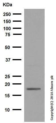
This data was developed using ab109199, the same antibody clone in a different buffer formulation.
Blocking and dilution buffer : 5% NFDM/TBST.
All lanes:
Western blot - Anti-p21 antibody [EPR3993] (<a href='/products/primary-antibodies/p21-antibody-epr3993-ab109199'>ab109199</a>) at 1/1000 dilution
All lanes:
PC-12 cell lysate at 10 µg
Secondary
All lanes:
Peroxidase-conjugated goat anti-rabbit IgG (H+L) at 1/1000 dilution
Predicted band size: 18 kDa
Observed band size: 21 kDa
false
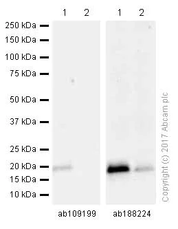
- WB
Lab
Western blot - Anti-p21 antibody [EPR3993] - BSA and Azide free (AB215971)
This data was developed using ab109199, the same antibody clone in a different buffer formulation.
All lanes:
Western blot - Anti-p21 antibody [EPR3993] (<a href='/products/primary-antibodies/p21-antibody-epr3993-ab109199'>ab109199</a>)
Lane 1:
RAW264.7 (Mouse Abelson murine leukemia virus-induced tumor macrophage) whole cell lysates at 15 µg
Lane 2:
Neuro-2a (Mouse neuroblastoma neuroblast) whole cell lysates at 15 µg
Secondary
All lanes:
Western blot - Goat Anti-Rabbit IgG H&L (HRP) (<a href='/products/secondary-antibodies/goat-rabbit-igg-h-l-hrp-ab97051'>ab97051</a>) at 1/20000 dilution
Predicted band size: 18 kDa
false
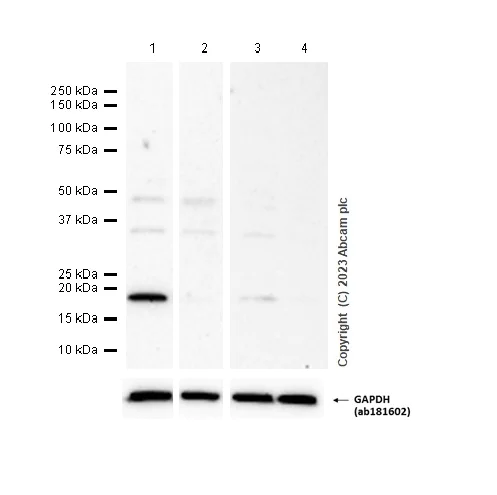
- WB
Lab
Western blot - Anti-p21 antibody [EPR3993] - BSA and Azide free (AB215971)
This data was developed using ab109199, the same antibody clone in a different buffer formulation. Blocking and diluting buffer and concentration : 5% NFDM/TBST. ab181602 was used as a GAPDH loading control.
All lanes:
Western blot - Anti-p21 antibody [EPR3993] (<a href='/products/primary-antibodies/p21-antibody-epr3993-ab109199'>ab109199</a>) at 1/1000 dilution
Lane 1:
Raw 264.7(Mouse Abelson murine leukemia virus-induced tumor macrophage) whole cell lysate at 20 µg
Lane 2:
NIH/3T3 (Mouse embryonic fibroblast) whole cell lysate at 20 µg
Lane 3:
C6 (Rat glial tumor glial cell) whole cell lysate at 20 µg
Lane 4:
PC-12(Rat adrenal gland pheochromocytoma) whole cell lysate at 20 µg
Secondary
All lanes:
Western blot - Goat Anti-Rabbit IgG H&L (HRP) (<a href='/products/secondary-antibodies/goat-rabbit-igg-h-l-hrp-ab97051'>ab97051</a>) at 1/20000 dilution
Predicted band size: 18 kDa
Observed band size: 18 kDa
false
Exposure time: 180s
関連する標識済み抗体及び組成の異なる製品 (1)
-
Anti-p21 antibody [EPR3993]
代替製品


Primary Antibodies
AB107099
BOND RX™ Validated|Lab Essentials|Recombinant

- 3
- 4
Primary Antibodies
AB188224
BOND RX™ Validated|RabMAb|Recombinant|Lab Essentials|20ul selling size

- 5
- 11
Primary Antibodies
AB109520
RabMAb|Recombinant|KO Validated|Lab Essentials|20ul selling size

- 5
- 17

Primary Antibodies
AB218311
Anti-p21 antibody [EPR362] - BSA and Azide free
RabMAb|Recombinant|KO Validated

- 0
- 0
Primary Antibodies
AB232512
Anti-p21 antibody [EPR18021] - BSA and Azide free
BOND RX™ Validated|RabMAb|Recombinant

- 4
- 2
組合せ製品


Primary Antibodies
AB211542
Anti-CDKN2A/p16INK4a antibody [EPR20418]
20ul selling size|RabMAb|Recombinant

- 5
- 8
Primary Antibodies
AB246550
Anti-p53 antibody [EPR20416-120]
RabMAb|Recombinant|Lab Essentials|20ul selling size|Advanced Validation

- 0
- 0
Primary Antibodies
AB181602
Anti-GAPDH antibody [EPR16891] - Loading Control
KD Validated|RabMAb|Recombinant|Lab Essentials|20ul selling size

- 5
- 23
Primary Antibodies
AB51243
Anti-ARPC5/p16 ARC antibody [EP1551Y]
RabMAb|Recombinant|KO Validated|20ul selling size

- 5
- 5
Reactivity data
製品の詳細
ab215971 is the carrier-free version of ab109199.
Patented technology
Our RabMAb® technology is a patented hybridoma-based technology for making rabbit monoclonal antibodies. For details on our patents, please refer to RabMAb® patents.
What are the advantages of a recombinant monoclonal antibody?
This product is a recombinant monoclonal antibody, which offers several advantages including:
- - High batch-to-batch consistency and reproducibility
- - Improved sensitivity and specificity
- - Long-term security of supply
- - Animal-free batch production
For more information, read more on recombinant antibodies.
Conjugation ready
Our carrier-free antibodies are typically supplied in a PBS-only formulation, purified and free of BSA, sodium azide and glycerol. This conjugation-ready format is designed for use with fluorochromes, metal isotopes, oligonucleotides, and enzymes, which makes them ideal for antibody labelling, functional and cell-based assays, flow-based assays (e.g. mass cytometry) and Multiplex Imaging applications.
Use our conjugation kits for antibody conjugates that are ready-to-use in as little as 20 minutes with 1 minute hands-on-time and 100% antibody recovery: available for fluorescent dyes, HRP, biotin and gold.
Compatibility
This product is compatible with the Maxpar® Antibody Labeling Kit from Fluidigm, without the need for antibody preparation. Maxpar® is a trademark of Fluidigm Canada Inc.
出荷温度及び保存条件
製品の状態
精製方法
バッファー組成
出荷温度
短期保存温度
長期保存温度
保管に関する情報
補足情報
This supplementary information is collated from multiple sources and compiled automatically.
Biological function summary
P21 plays a critical role in cell cycle regulation and DNA damage response. It associates with the p53 tumor suppressor protein forming an essential part of the p53 signaling pathway under stress conditions. p21 arrests cells in G1 phase allowing DNA repair or leading to cellular senescence. Through these mechanisms p21 integrates signals from stress and damage emphasizing its importance as a mediator in cellular checkpoint control.
Pathways
P21 is closely involved in the p53 and the Transforming Growth Factor-beta (TGF-β) signaling pathways. This integration helps in transmitting growth-inhibitory signals. p21 interacts with proteins such as p53 and cyclins ensuring precise regulation of the cell cycle and growth arrest when necessary. Triggered by p53 activation p21 contributes to preventing undisturbed cell proliferation acting as a safeguard against tumor formation.
製品プロトコール
- Visit the General protocols
- Visit the Troubleshooting
ターゲットの情報
文献 (3)
Recent publications for all applications. Explore the full list and refine your search
Scientific reports 14:30930 PubMed39730553
2024
Applications
Unspecified application
Species
Unspecified reactive species
Annals of translational medicine 11:72 PubMed36819526
2023
Applications
Unspecified application
Species
Unspecified reactive species
Aging 12:13115-13127 PubMed32614787
2020
Applications
Unspecified application
Species
Unspecified reactive species
Abcam product promise
Please note: All products are 'FOR RESEARCH USE ONLY. NOT FOR USE IN DIAGNOSTIC OR THERAPEUTIC PROCEDURES'.
For licensing inquiries, please contact partnerships@abcam.com