Anti-p21 抗体 [EPR362]
Anti-p21 antibody [EPR362]
- RabMAb
- Recombinant
- KO Validated
- Lab Essentials
- 20ul selling size
- 詳細を見る
5
(17 Reviews)
|
(555 Publications)
Anti-p21 antibody [EPR362] (ab109520) is a rabbit monoclonal antibody detecting p21 in Western Blot, Flow Cytometry (Intra), IP, IHC-P, ICC/IF. Suitable for Human.
- KO validated for confirmed specificity
- Biophysical QC for unrivalled batch-batch consistency
- Over 390 publications
別名を表示する
CAP20, CDKN1, CIP1, MDA6, PIC1, SDI1, WAF1, CDKN1A, Cyclin-dependent kinase inhibitor 1, CDK-interacting protein 1, Melanoma differentiation-associated protein 6, p21, MDA-6

- WB
Lab
Western blot - Anti-p21 antibody [EPR362] (AB109520)
Western blot : Anti-CDKN1A antibody [EPR362] (ab109520) staining at 1/1000 dilution, shown in green; Mouse anti-GAPDH antibody [6C5] (ab8245) loading control staining at 1/20000 dilution, shown in red. In Western blot, ab109520 was shown to bind specifically to CDKN1A. A band was observed at 21 kDa in wild-type HCT 116 cell lysates with no signal observed at this size in CDKN1A knockout cell line. To generate this image, wild-type and CDKN1A knockout HCT 116 cell lysates were analysed. First, samples were run on an SDS-PAGE gel then transferred onto a nitrocellulose membrane. Membranes were blocked in 3 % milk in TBS-0.1 % Tween® 20 (TBS-T) before incubation with primary antibodies overnight at 4 °C. Blots were washed four times in TBS-T, incubated with secondary antibodies for 1 h at room temperature, washed again four times then imaged. Secondary antibodies used were Goat anti-Rabbit IgG H&L 800CW and Goat anti-Mouse IgG H&L 680RD at 1/20000 dilution.
All lanes:
Western blot - Anti-p21 antibody [EPR362] (ab109520) at 1/1000 dilution
Lane 1:
Wild-type HCT 116 cell lysate at 20 µg
Lane 2:
CDKN1A knockout HCT 116 cell lysate at 20 µg
Lane 3:
Wild-type MCF7 cell lysate at 20 µg
Lane 4:
CDKN1A knockout MCF7 cell lysate at 20 µg
Lane 5:
Wild-type A549 cell lysate at 20 µg
Lane 6:
CDKN1A knockout A549 cell lysate at 20 µg
Secondary
All lanes:
Goat anti-Rabbit IgG H&L 800CW and Goat anti-Mouse IgG H&L 680RD at 1/20000 dilution
Predicted band size: 21 kDa
Observed band size: 21 kDa
false
- Flow Cyt (Intra)
Lab
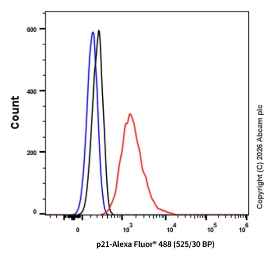
Flow Cytometry (Intracellular) - Anti-p21 antibody [EPR362] (AB109520)
Intracellular flow cytometric analysis of 90% methanol permeabilized HeLa cells labelling p21 with ab109520 at a dilution of 1/100 (red). Cells were fixed with 4% paraformaldehyde. Goat anti-Rabbit IgG H&L (Alexa Fluor® 488, ab150081) preadsorbed (1/5000) was used as the secondary antibody. Black - Isotype control, rabbit monoclonal IgG (ab172730). Blue - Unlabelled control, cells without incubation with primary and secondary antibodies.

- IHC-P
Lab
Immunohistochemistry (Formalin/PFA-fixed paraffin-embedded sections) - Anti-p21 antibody [EPR362] (AB109520)
Immunohistochemistry (Formalin/PFA-fixed paraffin-embedded sections) analysis of human thyroid carcinoma tissue labelling p21 with purified ab109520 at 1/100. Heat mediated antigen retrieval was performed using Tris/EDTA buffer pH 9. ab97051, a HRP-conjugated goat anti-rabbit IgG (H+L) was used as the secondary antibody (1/500). Negative control using PBS instead of primary antibody. Counterstained with hematoxylin.

- ICC/IF
Lab
Immunocytochemistry/ Immunofluorescence - Anti-p21 antibody [EPR362] (AB109520)
Immunocytochemistry/Immunofluorescence analysis of MCF7 cells labeling p21 with purified ab109520 at 1/1000. Cells were fixed with 4% paraformaldehyde and permeabilized with 0.1% Triton X-100. ab150077, an Alexa Fluor® 488-conjugated goat anti-rabbit IgG (1/500) was used as the secondary antibody. DAPI (blue) was used as the nuclear counterstain. ab7291, a mouse anti-tubulin (1/1000) and ab150120, an Alexa Fluor® 594-conjugated goat anti-mouse IgG (1/500) were also used.
Control 1 : primary antibody (1/1000) and secondary antibody, ab150120, an Alexa Fluor® 594-conjugated goat anti-mouse IgG (1/500).
Control 2 : ab7291 (1/1000) and secondary antibody, ab150077, an Alexa Fluor® 488-conjugated goat anti-rabbit IgG (1/500).

- ICC/IF
Lab
Immunocytochemistry/ Immunofluorescence - Anti-p21 antibody [EPR362] (AB109520)
Immunocytochemistry/ Immunofluorescence analysis of MCF7 (Human breast adenocarcinoma cell line) labeling p21 with ab109520 at 1/500 (2 μg/ml). ab150077, Alexa Fluor®488 Goat anti-Rabbit at 1/1000 (2 μg/ml) was used as secondary antibody. ab195889, Anti-alpha Tubulin antibody [DM1A] - Microtubule Marker (Alexa Fluor® 594) at 1/200 (2.5 μg/ml) was used as counterstain AbID. Cells were fixed with 4% Paraformaldehyde and permeabilised with 0.1% tritonX-100. Nuclei were counter stained blue with DAPI.
Confocal image showing nuclear staining on MCF7 cell line.

- IP
Lab
Immunoprecipitation - Anti-p21 antibody [EPR362] (AB109520)
ab109520 (purified) at 1/50 immunoprecipitating p21 in HEK293 whole cell lysate.
Lane 1 (input) : HEK293 whole cell lysate (10μg)
Lane 2 (+) : ab109520 + HEK293 whole cell lysate (10μg).
Lane 3 (-) : Rabbit monoclonal IgG (ab172730) instead of ab109520 in HEK293 whole cell lysate.
For western blotting, VeriBlot for IP Detection Reagent (HRP) (ab131366), was used for detection at 1/1500 dilution.
Blocking buffer and concentration : 5% NFDM/TBST.
Diluting buffer and concentration : 5% NFDM /TBST.
All lanes:
Immunoprecipitation - Anti-p21 antibody [EPR362] (ab109520)
Predicted band size: 18 kDa
Observed band size: 21 kDa
false

- WB
Lab
Western blot - Anti-p21 antibody [EPR362] (AB109520)
This blot was produced using 4-20% SDS-PAGE containing 15 μg of HEK-293 whole cell lysate per lane at 150V for 1hr before being transferred onto a 0.45 μm PVDF membrane at 75V for 1hr. The membrane was then blocked for 1hr using 5% NFDM/TBST, then incubated with ab109520 (1/2000) at room temperature for 1hr. After being washed three times in TBST, the membrane was incubated with Peroxidase conjugated goat anti-rabbit IgG (H+L) (ab97051) at 1/20,000 dilution for 1hr at room temperature. The membrane was washed three times again. Then the signal was developed using the ECL technique.
ab109520 was stored at a range of temperatures (+4°C, +22°C, +37°C) for 1 week before being tested in WB. The image shows the band intensity remains relatively constant across all storage temperatures, demonstrating that antibody activity is not affected under these conditions. This data was generated as part of a structured antibody stability study supporting Abcam’s transition from cold‑chain to ambient shipping.
All lanes:
Western blot - Anti-p21 antibody [EPR362] (ab109520) at 1/2000 dilution
All lanes:
HEK-293 whole cell lysate at 15 µg with NDFM/TBST
Secondary
All lanes:
Western blot - Goat Anti-Rabbit IgG H&L (HRP) (<a href='/products/secondary-antibodies/goat-rabbit-igg-h-l-hrp-ab97051'>ab97051</a>) at 1/20000 dilution
false
Exposure time: 40s

- WB
Lab
Western blot - Anti-p21 antibody [EPR362] (AB109520)
Different batches of ab109520 were tested on MCF7 (Human breast adenocarcinoma epithelial cell) lysate at 0.2 µg/ml. 15 µg of lysate was loaded in each lane. Bands observed at 21 kDa.
All lanes:
Western blot - Anti-p21 antibody [EPR362] (ab109520)
Predicted band size: 18 kDa
false

- WB
Lab
Western blot - Anti-p21 antibody [EPR362] (AB109520)
False colour image of Western blot : Anti-p21 antibody [EPR362] staining at 1/1000 dilution, shown in green; Mouse anti-Alpha Tubulin [DM1A] (ab7291) loading control staining at 1/20000 dilution, shown in red. In Western blot, ab109520 was shown to bind specifically to p21. A band was observed at 21 kDa in wild-type HeLa cell lysates with no signal observed at this size in CDKN1A knockout cell line ab255349 (knockout cell lysate ab263812). To generate this image, wild-type and CDKN1A knockout cell lysates were analysed. First, samples were run on an SDS-PAGE gel then transferred onto a nitrocellulose membrane. Membranes were blocked in 3 % milk in TBS-0.1 % Tween® 20 (TBS-T) before incubation with primary antibodies overnight at 4°C. Blots were washed four times in TBS-T, incubated with secondary antibodies for 1 h at room temperature, washed again four times then imaged.Secondary antibodies used were Goat anti-Rabbit IgG H&L (IRDye® 800CW) preabsorbed (ab216773) and Goat anti-Mouse IgG H&L (IRDye® 680RD) preabsorbed (ab216776) at 1/20000 dilution.
All lanes:
Western blot - Anti-p21 antibody [EPR362] (ab109520) at 1/1000 dilution
Lane 1:
wild-type HeLa Vehicle Control Fluvastatin (20 uM, 24 h) cell lysate at 20 µg
Lane 2:
wild-type HeLa Treated Fluvastatin (50 uM, 24 h) cell lysate at 20 µg
Lane 3:
CDKN1A knockout HeLa Vehicle Control Fluvastatin (20 uM, 24 h) cell lysate at 20 µg
Lanes 3 - 4:
Western blot - Human CDKN1A knockout HeLa cell line (<a href='/products/cell-lines/human-cdkn1a-knockout-hela-cell-line-ab255349'>ab255349</a>)
Lane 4:
CDKN1A knockout HeLa Treated Fluvastatin (50 uM, 24 h) cell lysate at 20 µg
Lane 5:
MCF7 cell lysate at 20 µg
Lane 6:
SH-SY5Y cell lysate at 20 µg
Predicted band size: 18 kDa
Observed band size: 21 kDa
false

- WB
Lab
Western blot - Anti-p21 antibody [EPR362] (AB109520)
Lane 1 : Wild-type DLD-1 cell lysate (20 μg)
Lane 2 : Wild-type DLD-1 20 μM 2,3-DCPE for 16hrs treated cell lysate (20 μg)
Lane 3 : p21 knockout DLD-1 cell lysate (20 μg)
Lane 4 : p21 knockout 20 μM 2,3-DCPE for 16hrs DLD-1 cell lysate (20 μg)
Lane 5 : HT1080 cell lysate (20 μg)
Lanes 1 - 5 : Merged signal (red and green). Green - ab109520 observed at 20 kDa. Red - loading control, ab8245, observed at 37 kDa.
ab109520 was shown to specifically recognize p21 in wild-type DLD-1 cells treated with 20 μM 2,3-DCPE. No band was observed when p21 knockout samples +/- 2,3-DCPE treatment were used. Wild-type and p21 knockout samples were subjected to SDS-PAGE. ab109520 and ab8245 (loading control to GAPDH) were diluted 1/1000 and 1/10 000 respectively and incubated overnight at 4°C. Blots were developed with Goat anti-Rabbit IgG H&L (IRDye® 800CW) preadsorbed (ab216773) and Goat anti-Mouse IgG H&L (IRDye® 680RD) preadsorbed (ab216776) secondary antibodies at 1/10 000 dilution for 1 h at room temperature before imaging.
All lanes:
Western blot - Anti-p21 antibody [EPR362] (ab109520)
Predicted band size: 18 kDa
Observed band size: 20 kDa
false

- WB
Lab
Western blot - Anti-p21 antibody [EPR362] (AB109520)
Lane 1 : Wild-type DLD-1 cell lysate (20 μg)
Lane 2 : Wild-type DLD-1 20 μM 2,3-DCPE for 16hrs treated cell lysate (20 μg)
Lane 3 : p21 knockout DLD-1 cell lysate (20 μg)
Lane 4 : p21 knockout 20 μM 2,3-DCPE for 16hrs DLD-1 cell lysate (20 μg)
Lane 5 : HT1080 cell lysate (20 μg)
Lanes 1 - 5 : Merged signal (red and green). Green - ab109520 observed at 20 kDa. Red - loading control, ab8245, observed at 37 kDa.
This western blot image is a comparison between ab109520 and a competitor's top cited rabbit polyclonal antibody.
All lanes:
Western blot - Anti-p21 antibody [EPR362] (ab109520)
Predicted band size: 18 kDa
false

- WB
Lab
Western blot - Anti-p21 antibody [EPR362] (AB109520)
Blocking and dilution buffer : 5% NFDM/TBST.
All lanes:
Western blot - Anti-p21 antibody [EPR362] (ab109520) at 1/2000 dilution
Lane 1:
MCF7 cell lysate at 20 µg
Lane 2:
HEK293 cell lysate at 20 µg
Lane 3:
U87-MG cell lysate at 20 µg
Secondary
All lanes:
Peroxidase-conjugated goat anti-rabbit IgG, (H+L) at 1/1000 dilution
Predicted band size: 18 kDa,24 kDa,57 kDa
Observed band size: 21 kDa
false

- WB
Lab
Western blot - Anti-p21 antibody [EPR362] (AB109520)
Blocking and dilution buffer : 5% NFDM/TBST.
All lanes:
Western blot - Anti-p21 antibody [EPR362] (ab109520) at 1/10000 dilution
All lanes:
LnCaP cell lysate at 20 µg
Secondary
All lanes:
Peroxidase-conjugated goat anti-rabbit IgG, (H+L) at 1/1000 dilution
Predicted band size: 18 kDa
Observed band size: 21 kDa
false
関連する標識済み抗体及び組成の異なる製品 (2)
-
Anti-p21 antibody [EPR362] - BSA and Azide free
-
519 Alexa Fluor® 488
Alexa Fluor® 488 Anti-p21 antibody [EPR362]
代替製品


Primary Antibodies
AB107099
BOND RX™ Validated|Lab Essentials|Recombinant

- 3
- 4
Primary Antibodies
AB109199
20ul selling size|KO Validated|RabMAb|Recombinant|Lab Essentials

- 4
- 12
Primary Antibodies
AB188224
BOND RX™ Validated|RabMAb|Recombinant|Lab Essentials|20ul selling size

- 5
- 11
Primary Antibodies
AB215971
Anti-p21 antibody [EPR3993] - BSA and Azide free
RabMAb|Recombinant|KO Validated

- 0
- 0

Primary Antibodies
AB232512
Anti-p21 antibody [EPR18021] - BSA and Azide free
BOND RX™ Validated|RabMAb|Recombinant

- 4
- 2
組合せ製品

Primary Antibodies
AB108349
Anti-CDKN2A/p16INK4a antibody [EPR1473] - C-terminal
BOND RX™ Validated|20ul selling size|KO Validated|RabMAb|Recombinant

- 4
- 7
Primary Antibodies
AB1101
BOND RX™ Validated|KO Validated|Lab Essentials

- 4
- 14
Primary Antibodies
AB184787
Anti-Caspase-3 antibody [EPR18297]
20ul selling size|RabMAb|Recombinant|KO Validated

- 3
- 3
Primary Antibodies
AB108357
Anti-Cdk4 antibody [EPR4513-32-7]
20ul selling size|RabMAb|Recombinant|KO Validated

- 5
- 1
Reactivity data
製品の詳細
Anti-p21 antibody [EPR362] (ab109520) was developed by Abcam using patented rabbit monoclonal antibody technology and is validated for use in Flow Cyt (Intra), ICC/IF, IHC-P, IP and WB.
Anti-p21 antibody [EPR362] (ab109520) was first used in a scientific publication in 2014 and has been cited over 391 times in peer reviewed journals. It's performance in Western Blot in human samples is trusted by the scientific community.
Abcam's high quality manufacturing and validation processes ensure Anti-p21 antibody [EPR362] (ab109520) has high sensitivity and specificity alongside high lot-to-lot consistency and reproducibility.
The specificity of Anti-p21 antibody [EPR362] (ab109520) has been confirmed by Western Blot testing in p21 knockout HeLa cells (ab255349).
Anti-p21 antibody [EPR362] (ab109520) has 13 independent reviews from customers.
Anti-p21 antibody [EPR362] (ab109520) specifically detects p21 (UniProt ID: P38936; Molecular weight: 18kDa) and is sold in a convenient trial size to enable initial testing (20 µL) and larger sizes for subsequent scaling up experiments (100 µL and 1 mL).
Conjugation-ready, carrier free format available for antibody clone EPR362 - ab218311.
Antibody clone EPR362 is also available pre-conjugated to a variety of labels for your convenience - Alexa Fluor® 488 (ab282187).
p21 is a cyclin-dependent kinase inhibitor that regulates cell cycle progression, promoting cell cycle arrest in response to DNA damage. It plays a dual role in oncology by both suppressing tumor growth through cell cycle inhibition and contributing to cancer progression when localized in the cytoplasm.
Patented technology
Our RabMAb® technology is a patented hybridoma-based technology for making rabbit monoclonal antibodies. For details on our patents, please refer to RabMAb® patents.
What are the advantages of a recombinant monoclonal antibody?
This product is a recombinant monoclonal antibody, which offers several advantages including:
- - High batch-to-batch consistency and reproducibility
- - Improved sensitivity and specificity
- - Long-term security of supply
- - Animal-free batch production
For more information, read more on recombinant antibodies.
出荷温度及び保存条件
製品の状態
精製方法
バッファー組成
出荷温度
短期保存期間
短期保存温度
長期保存温度
分注に関する情報
保管に関する情報
補足情報
This supplementary information is collated from multiple sources and compiled automatically.
Biological function summary
P21 plays a critical role in cell cycle regulation and DNA damage response. It associates with the p53 tumor suppressor protein forming an essential part of the p53 signaling pathway under stress conditions. p21 arrests cells in G1 phase allowing DNA repair or leading to cellular senescence. Through these mechanisms p21 integrates signals from stress and damage emphasizing its importance as a mediator in cellular checkpoint control.
Pathways
P21 is closely involved in the p53 and the Transforming Growth Factor-beta (TGF-β) signaling pathways. This integration helps in transmitting growth-inhibitory signals. p21 interacts with proteins such as p53 and cyclins ensuring precise regulation of the cell cycle and growth arrest when necessary. Triggered by p53 activation p21 contributes to preventing undisturbed cell proliferation acting as a safeguard against tumor formation.
製品プロトコール
- Visit the General protocols
- Visit the Troubleshooting
ターゲットの情報
文献 (555)
Recent publications for all applications. Explore the full list and refine your search
Cancer cell international 25:325 PubMed41044576
2025
Applications
Unspecified application
Species
Unspecified reactive species
Stem cell research & therapy 16:526 PubMed41024282
2025
Applications
Unspecified application
Species
Unspecified reactive species
Histology and histopathology 40:1985-2009 PubMed40959856
2025
Applications
Unspecified application
Species
Unspecified reactive species
Open life sciences 20:20251151 PubMed40822973
2025
Applications
Unspecified application
Species
Unspecified reactive species
International journal of molecular sciences 26: PubMed40806270
2025
Applications
Unspecified application
Species
Unspecified reactive species
Frontiers in pharmacology 16:1588722 PubMed40771929
2025
Applications
Unspecified application
Species
Unspecified reactive species
Science advances 11:eadv4238 PubMed40712009
2025
Applications
Unspecified application
Species
Unspecified reactive species
American journal of translational research 17:4187-4197 PubMed40672597
2025
Applications
Unspecified application
Species
Unspecified reactive species
Frontiers in bioengineering and biotechnology 13:1570270 PubMed40635690
2025
Applications
Unspecified application
Species
Unspecified reactive species
Oncology research 33:1695-1708 PubMed40612863
2025
Applications
Unspecified application
Species
Unspecified reactive species
Abcam product promise
Please note: All products are 'FOR RESEARCH USE ONLY. NOT FOR USE IN DIAGNOSTIC OR THERAPEUTIC PROCEDURES'.
For licensing inquiries, please contact partnerships@abcam.com