Anti-Nipah Virus Glycoprotein F antibody [CG11]
Anti-Nipah Virus Glycoprotein F antibody [CG11]
- Recombinant
- 詳細を見る
Be the first to review this product! Submit a review
|
(0 Publication)
Mouse Recombinant Monoclonal TLS/FUS antibody. Suitable for WB, I-ELISA and reacts with Transfected cell lysate - Henipavirus nipahense, Henipavirus nipahense samples.
別名を表示する
Fusion glycoprotein F0, Protein F, F
- I-ELISA
Supplier Data
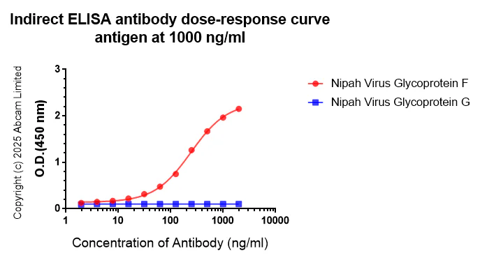
Indirect ELISA - Anti-Nipah Virus Glycoprotein F antibody [CG11] (AB325231)
Indirect ELISA analysis of ab325231 at 2000-0 ng/ml. The Secondary antibody used was Peroxidase AffiniPure Goat Anti-Mouse IgG, F(ab')₂ fragment specific at 1 : 20000 dilution dilution.
Antigen : Nipah Virus Glycoprotein F;Nipah Virus Glycoprotein G.
Antigen concentration : 1000 ng/ml
- WB
Supplier Data
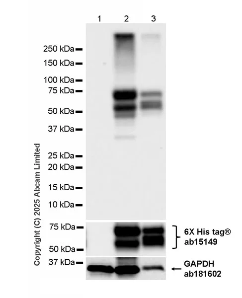
Western blot - Anti-Nipah Virus Glycoprotein F antibody [CG11] (AB325231)
Blocking and diluting buffer and concentration : 5% NFDM/TBST.
In Western blot, Anti-GAPDH antibody [EPR16891] - Loading Control (ab181602) staining at 1/200000 dilution.
In Western blot, Anti-6X His tag® antibody [AD1.1.10] (ab15149) staining at 1/1000 dilution.
All lanes:
Western blot - Anti-Nipah Virus Glycoprotein F antibody [CG11] (ab325231) at 1/1000 dilution
Lane 1:
293T (human embryonic kidney epithelial cell) transfected with an empty vector containing a myc-His-tag® whole cell lysate at 20 µg
Lane 2:
293T transfected with Nipah virus Fusion glycoprotein F0 expression vector containing a myc-His-tag® whole cell lysate at 20 µg
Lane 3:
293T transfected with Hendra virus (isolate Horse/Autralia/Hendra/1994) Fusion glycoprotein F0 expression vector containing a myc-His-tag® whole cell lysate at 2 µg
Secondary
All lanes:
Peroxidase-Conjugated Goat anti-Mouse IgG (H+L) at 1/10000 dilution
Observed band size: 60 kDa,36 kDa
false
Exposure time: 8s
組合せ製品
Primary Antibodies
AB183497
Anti-VSV-G tag antibody [EPR12997]
RabMAb|Recombinant|Lab Essentials

- 4
- 2
Primary Antibodies
AB271180
Rabbit monoclonal [EPR24334-118] to SARS-CoV-2 nucleocapsid protein
BOND RX™ Validated|20ul selling size|RabMAb|Recombinant

- 5
- 4
Primary Antibodies
AB183734
RabMAb|Recombinant|Lab Essentials|20ul selling size

- 5
- 24
Primary Antibodies
AB313343
Anti-Influenza A Virus Nucleoprotein antibody [EPR28253-17]
20ul selling size|RabMAb|Recombinant

- 5
- 1
Reactivity data
製品の詳細
What are the advantages of a recombinant monoclonal antibody?
This product is a recombinant monoclonal antibody, which offers several advantages including:
- - High batch-to-batch consistency and reproducibility
- - Improved sensitivity and specificity
- - Long-term security of supply
- - Animal-free batch production
For more information, read more on recombinant antibodies.
Want a custom formulation?
This antibody clone is manufactured by Abcam. If you require a custom buffer formulation or conjugation for your experiments, please contact orders@abcam.com
出荷温度及び保存条件
製品の状態
精製方法
バッファー組成
出荷温度
短期保存期間
短期保存温度
長期保存温度
分注に関する情報
保管に関する情報
製品プロトコール
- Visit the General protocols
- Visit the Troubleshooting
ターゲットの情報
Abcam product promise
Please note: All products are 'FOR RESEARCH USE ONLY. NOT FOR USE IN DIAGNOSTIC OR THERAPEUTIC PROCEDURES'.
For licensing inquiries, please contact partnerships@abcam.com