Anti-NCAM1 抗体 [CAL53]
Anti-NCAM1 antibody [CAL53]
- 20ul selling size
- RabMAb
- Recombinant
- 詳細を見る
5
(2 Reviews)
|
(4 Publications)
Anti-NCAM1 antibody [CAL53] (ab237708) is a rabbit monoclonal antibody detecting NCAM1 in Western Blot, IP, IHC-P, ICC/IF. Suitable for Human, Mouse, Rat.
- Biophysical QC for unrivalled batch-batch consistency
別名を表示する
CD56, NCAM, NCAM1, Neural cell adhesion molecule 1, N-CAM-1, NCAM-1
- IHC-P
Lab
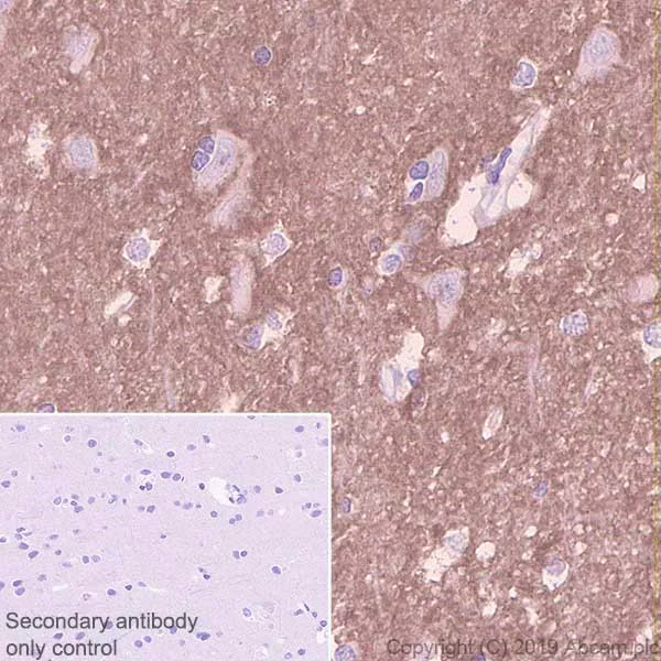
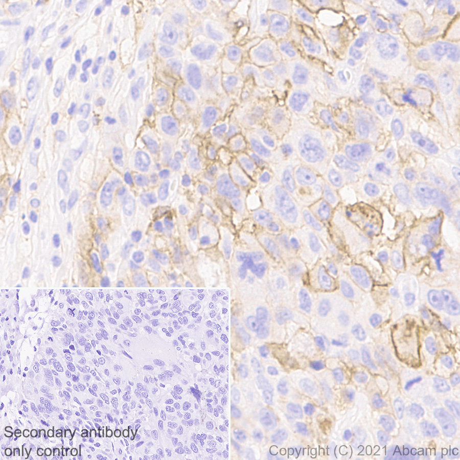
Immunohistochemistry (Formalin/PFA-fixed paraffin-embedded sections) - Anti-NCAM1 antibody [CAL53] (AB237708)
Immunohistochemical analysis of paraffin-embedded human cerebrum tissue labeling NCAM1 with ab237708 at 1/1000 dilution, followed by Goat Anti-Rabbit IgG H&L (HRP) ready to use. Positive staining on the human cerebrum is observed. Counter stained with hematoxylin.
Secondary antibody only control : Used PBS instead of primary antibody, secondary antibody is Goat Anti-Rabbit IgG H&L (HRP) ready to use.
Heat mediated antigen retrieval using ab93684 (Tris/EDTA buffer, pH 9.0).
- IP
Lab
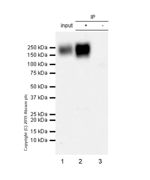
Immunoprecipitation - Anti-NCAM1 antibody [CAL53] (AB237708)
NCAM1 was immunoprecipitated from 0.35 mg of SH-SY5Y (Human neuroblastoma cell line from bone marrow) whole cell lysate with ab237708 at 1/20 dilution. Western blot was performed from the immunoprecipitate using ab237708 at 1/1000 dilution. VeriBlot for IP Detection Reagent (HRP) (ab131366), was used as secondary antibody at 1/5000 dilution.
Lane 1 : SH-SY5Y whole cell lysate 10 μg (Input).
Lane 2 : ab237708 IP in SH-SY5Y whole cell lysate.
Lane 3 : Rabbit monoclonal IgG (ab172730) instead of ab237708 in SH-SY5Y whole cell lysate.
Blocking and dilution buffer and concentration : 5% NFDM/TBST.
Exposure time : 5 seconds.
All lanes:
Immunoprecipitation - Anti-NCAM1 antibody [CAL53] (ab237708)
Predicted band size: 94 kDa
Observed band size: 140-180 kDa
false
- ICC/IF
Lab
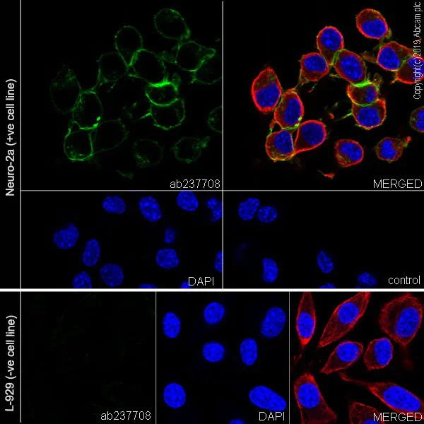
Immunocytochemistry/ Immunofluorescence - Anti-NCAM1 antibody [CAL53] (AB237708)
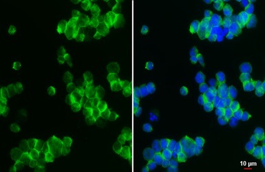
Immunofluorescent analysis of 4% paraformaldehyde-fixed, 0.1% Triton X-100 permeabilized Neuro-2a (mouse neuroblastoma cell line) cells labeling NCAM1 with ab237708 at 1/50 dilution, followed by Goat Anti-Rabbit IgG H&L (Alexa Fluor® 488) (ab150077) secondary antibody at 1/1000 dilution (green).Confocal image showing membranous staining in Neuro-2a cells. The nuclear counter stain is DAPI (blue). Tubulin is detected with Anti-alpha Tubulin antibody [DM1A] - Microtubule Marker (Alexa Fluor® 594) (ab195889) at 1/200 dilution (red).
PCS only control : Used PBS instead of primary antibody, followed by Goat Anti-Rabbit IgG H&L (Alexa Fluor® 488) (ab150077) secondary antibody at 1/1000 dilution.
Negative control : L-929 (PMID : 9696812).
- IHC-P
Lab
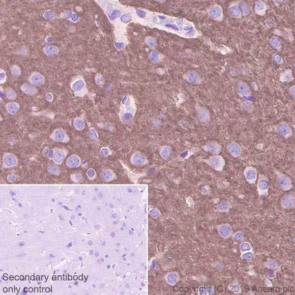
Immunohistochemistry (Formalin/PFA-fixed paraffin-embedded sections) - Anti-NCAM1 antibody [CAL53] (AB237708)
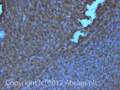
Immunohistochemical analysis of paraffin-embedded rat cerebrum tissue labeling NCAM1 with ab237708 at 1/1000 dilution, followed by Goat Anti-Rabbit IgG H&L (HRP) ready to use. Positive staining on islet in the rat cerebrum is observed. Counter stained with hematoxylin.
Secondary antibody only control : Used PBS instead of primary antibody, secondary antibody is Goat Anti-Rabbit IgG H&L (HRP) ready to use.
Heat mediated antigen retrieval using ab93684 (Tris/EDTA buffer, pH 9.0).
- IHC-P
Lab
Immunohistochemistry (Formalin/PFA-fixed paraffin-embedded sections) - Anti-NCAM1 antibody [CAL53] (AB237708)
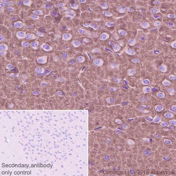
Immunohistochemical analysis of paraffin-embedded mouse cerebrum tissue labeling NCAM1 with ab237708 at 1/1000 dilution, followed by Goat Anti-Rabbit IgG H&L (HRP) ready to use. Positive staining on islet in the mouse cerebrum is observed. Counter stained with hematoxylin.
Secondary antibody only control : Used PBS instead of primary antibody, secondary antibody is Goat Anti-Rabbit IgG H&L (HRP) ready to use.
Heat mediated antigen retrieval using ab93684 (Tris/EDTA buffer, pH 9.0).
- WB
Lab
Western blot - Anti-NCAM1 antibody [CAL53] (AB237708)
Blocking and dilution buffer : 5% NFDM/TBST.
All lanes:
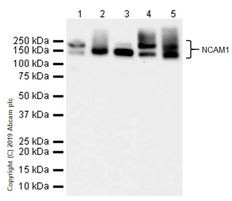
Western blot - Anti-NCAM1 antibody [CAL53] (ab237708) at 1/1000 dilution
Lane 1:
Human brain lysate at 20 µg
Lane 2:
Human cerebellum lysate at 20 µg
Lane 3:
Neuro-2a (mouse neuroblastoma cell line0 whole cell lysate at 20 µg
Lane 4:
Mouse cerebellum lysate at 20 µg
Lane 5:
Rat brain lysate at 20 µg
Secondary
All lanes:
Western blot - Goat Anti-Rabbit IgG H&L (HRP) (<a href='/products/secondary-antibodies/goat-rabbit-igg-h-l-hrp-ab97051'>ab97051</a>) at 1/100000 dilution
Predicted band size: 94 kDa
Observed band size: 140-180 kDa
false
Exposure time: 26s
関連する標識済み抗体及び組成の異なる製品 (1)
-
Anti-NCAM1 antibody [CAL53] - BSA and Azide free
代替製品

Primary Antibodies
AB137086
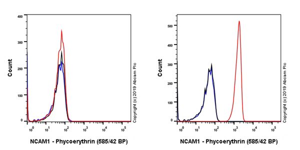
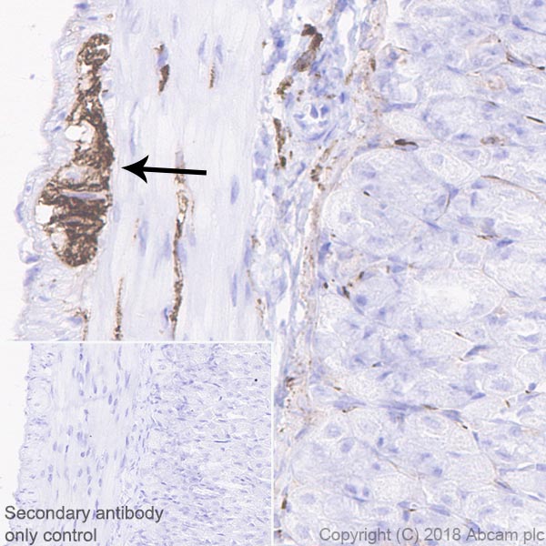
Anti-NCAM1 antibody [EP2566(2)]
20ul selling size|RabMAb|Recombinant

- 0
- 0
Primary Antibodies
AB75813
BOND RX™ Validated|20ul selling size|RabMAb|Recombinant

- 4
- 7
Primary Antibodies
AB215981
Anti-NCAM1 antibody [EP2567Y] - Low endotoxin, Azide free
BOND RX™ Validated|RabMAb|Recombinant

- 0
- 0
Primary Antibodies
AB214435
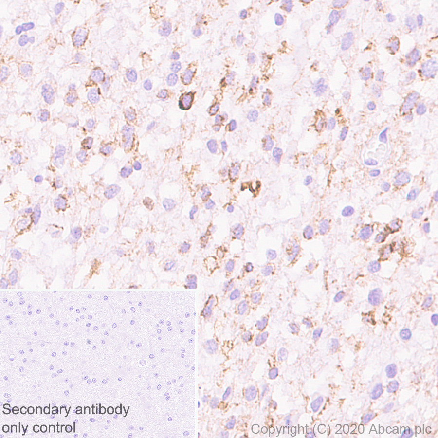
Anti-NCAM1 antibody [EP2567Y] - BSA and Azide free
BOND RX™ Validated|RabMAb|Recombinant
- 0
- 0
Primary Antibodies
AB214436
Anti-NCAM1 antibody [EPR2566] - BSA and Azide free
RabMAb|Recombinant

- 4
- 3

Primary Antibodies
AB248809
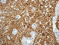
Anti-NCAM1 antibody [EP2566(2)] - BSA and Azide free
RabMAb|Recombinant
- 0
- 0

組合せ製品
Primary Antibodies
AB133616
BOND RX™ Validated|RabMAb|Recombinant|Lab Essentials|20ul selling size
- 5
- 20
Primary Antibodies
AB16669
Anti-CD3 epsilon antibody [SP7]
BOND RX™ Validated|RabMAb|Advanced Validation|Recombinant|Lab Essentials|20ul selling size

- 4
- 80
Primary Antibodies
AB182422
Anti-CD163 antibody [EPR19518]
BOND RX™ Validated|20ul selling size|RabMAb|Advanced Validation|Recombinant

- 5
- 17
Primary Antibodies
AB282458
BOND RX™ Validated|20ul selling size|RabMAb|Recombinant|KO Validated
- 0
- 0
Reactivity data
製品の詳細
What is this antibody validated in?
Anti-NCAM1 antibody [CAL53] (ab237708) is a rabbit recombinant monoclonal antibody and is validated for use in Western Blot (WB), Immunoprecipitation (IP), Immunohistochemistry (IHC-P), Immunocytochemistry/immunofluorescence (ICC/IF) in Human, Mouse, Rat samples.
What is the molecular weight of NCAM1?
Anti-NCAM1 [CAL53] (ab237708) specifically detects a band for NCAM1 (UniProt: P13591) at a molecular weight of 140kDa.
Other related products
We have a range of other formats of antibody clone [CAL53] also available for your convenience: ab237708, Carrier free - ab251595
What are the advantages of a recombinant monoclonal antibody?
This product is a recombinant monoclonal antibody, which offers several advantages including:
- - High batch-to-batch consistency and reproducibility
- - Improved sensitivity and specificity
- - Long-term security of supply
- - Animal-free batch production
For more information, read more on recombinant antibodies.
Trial sizes available!
Test your antibody or perform pre-screening before committing to a larger quantity. Sold in 20µl. Discover our selection of trial-size antibodies.
出荷温度及び保存条件
製品の状態
精製方法
精製に関する特記事項
バッファー組成
出荷温度
短期保存期間
短期保存温度
長期保存温度
分注に関する情報
保管に関する情報
補足情報
This supplementary information is collated from multiple sources and compiled automatically.
Biological function summary
NCAM1 influences neuronal growth survival and synaptic plasticity. This protein is part of the immunoglobulin superfamily and contributes to dynamic interactions during development. NCAM1 exists on cell membranes assisting in the formation and maintenance of tissue structures. In particular cell adhesion molecule CAL53 interacts with NCAM1 highlighting its involvement in synaptic connections. Additionally NCAM1 impacts cellular migration and differentiation particularly in myogenic processes involving MY31's pathway.
Pathways
The NCAM1 protein participates in the MAPK signaling pathway and the PI3K/AKT pathway both essential in cellular growth and survival. NCAM1 acts alongside ERIC1 aiding in the transduction of signals that regulate cytoskeletal rearrangement. These pathways influence cellular communication and responses to external stimuli further enhancing tissue development and regeneration mechanisms.
製品プロトコール
- Visit the General protocols
- Visit the Troubleshooting
ターゲットの情報
文献 (4)
Recent publications for all applications. Explore the full list and refine your search
Scientific reports 14:13432 PubMed38862586
2024
Applications
Unspecified application
Species
Unspecified reactive species
Signal transduction and targeted therapy 8:327 PubMed37661226
2023
Applications
Unspecified application
Species
Unspecified reactive species
Cancer science 114:2650-2663 PubMed36846943
2023
Applications
Unspecified application
Species
Unspecified reactive species
Cell reports. Medicine 3:100597 PubMed35492247
2022
Applications
Unspecified application
Species
Unspecified reactive species
Abcam product promise
Please note: All products are 'FOR RESEARCH USE ONLY. NOT FOR USE IN DIAGNOSTIC OR THERAPEUTIC PROCEDURES'.
For licensing inquiries, please contact partnerships@abcam.com