Anti-Myoferlin 抗体 [EPR18887] - BSA and Azide free
Anti-Myoferlin antibody [EPR18887] - BSA and Azide free
- RabMAb
- Recombinant
- KO Validated
- 詳細を見る
Be the first to review this product! Submit a review
|
(0 Publication)
Rabbit Recombinant Monoclonal Myoferlin antibody. Carrier free. Suitable for WB, ICC/IF, Flow Cyt (Intra) and reacts with Human, Mouse, Rat samples.
別名を表示する
FER1L3, KIAA1207, MYOF, Myoferlin, Fer-1-like protein 3
- Flow Cyt (Intra)
Supplier Data
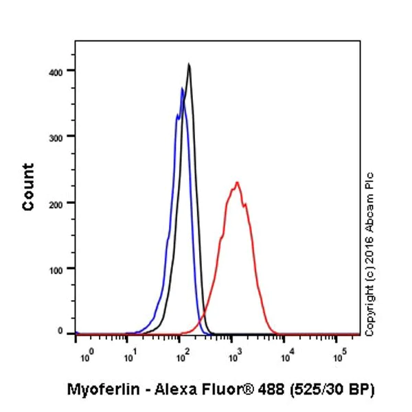
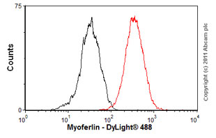
Flow Cytometry (Intracellular) - Anti-Myoferlin antibody [EPR18887] - BSA and Azide free (AB271928)
Intracellular flow cytometric analysis of 4% paraformaldehyde-fixed HeLa (human epithelial cell line from cervix adenocarcinoma) cells labeling Myoferlin with ab178386 at 1/400 dilution (red) compared with a rabbit monoclonal IgG isotype control (ab172730; black) and an unlabeled control (cells without incubation with primary antibody and secondary antibody; blue). Goat anti rabbit IgG (Alexa Fluor® 488) at 1/2000 dilution was used as the secondary antibody.
This data was developed using the same antibody clone in a different buffer formulation containing PBS, BSA, glycerol, and sodium azide (ab178386).
- ICC/IF
Supplier Data
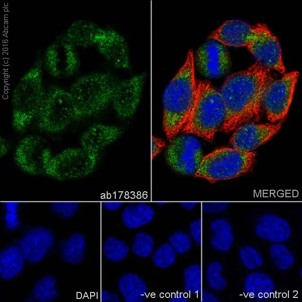
Immunocytochemistry/ Immunofluorescence - Anti-Myoferlin antibody [EPR18887] - BSA and Azide free (AB271928)
Immunofluorescent analysis of 100% methanol-fixed HeLa (human epithelial cell line from cervix adenocarcinoma) cells labeling Myoferlin with ab178386 at 1/100 dilution, followed by Goat anti-rabbit IgG (Alexa Fluor® 488) (ab150077) secondary antibody at 1/1000 dilution (green). Confocal image showing cytoplasmic staining on HeLa cells. The nuclear counter stain is DAPI (blue). Tubulin is detected with ab7291 (anti-Tubulin mouse mAb) at 1/1000 dilution and ab150120 (Alexa Fluor® 594 Goat anti-Mouse secondary) at 1/1000 dilution (red).
The negative controls are as follows :
-ve control 1 : ab178386 at 1/100 dilution followed by ab150120 (Alexa Fluor® 594 Goat anti-Mouse secondary) at 1/1000 dilution.
-ve control 2 : ab7291 (anti-Tubulin mouse mAb) at 1/1000 dilution followed by ab150077 (Alexa Fluor® 488 Goat Anti-Rabbit IgG H&L) at 1/1000 dilution.
This data was developed using the same antibody clone in a different buffer formulation containing PBS, BSA, glycerol, and sodium azide (ab178386).
- ICC/IF
Supplier Data
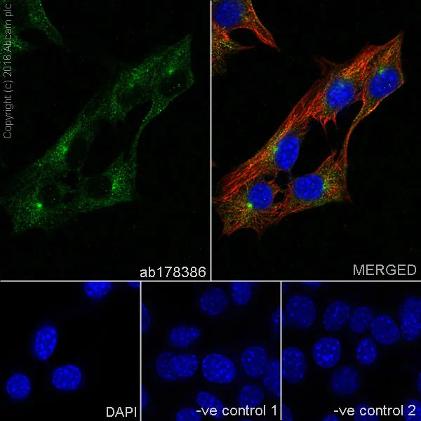
Immunocytochemistry/ Immunofluorescence - Anti-Myoferlin antibody [EPR18887] - BSA and Azide free (AB271928)
Immunofluorescent analysis of 100% methanol-fixed C2C12 (mouse myoblast cell line) cells labeling Myoferlin with ab178386 at 1/100 dilution, followed by Goat anti-rabbit IgG (Alexa Fluor® 488) (ab150077) secondary antibody at 1/1000 dilution (green). Confocal image showing cytoplasmic staining on C2C12 cells. The nuclear counter stain is DAPI (blue). Tubulin is detected with ab7291 (anti-Tubulin mouse mAb) at 1/1000 dilution and ab150120 (Alexa Fluor® 594 Goat anti-Mouse secondary) at 1/1000 dilution (red).
The negative controls are as follows :
-ve control 1 : ab178386 at 1/100 dilution followed by ab150120 (Alexa Fluor® 594 Goat anti-Mouse secondary) at 1/1000 dilution.
-ve control 2 : ab7291 (anti-Tubulin mouse mAb) at 1/1000 dilution followed by ab150077 (Alexa Fluor® 488 Goat Anti-Rabbit IgG H&L) at 1/1000 dilution.
This data was developed using the same antibody clone in a different buffer formulation containing PBS, BSA, glycerol, and sodium azide (ab178386).
- WB
Lab
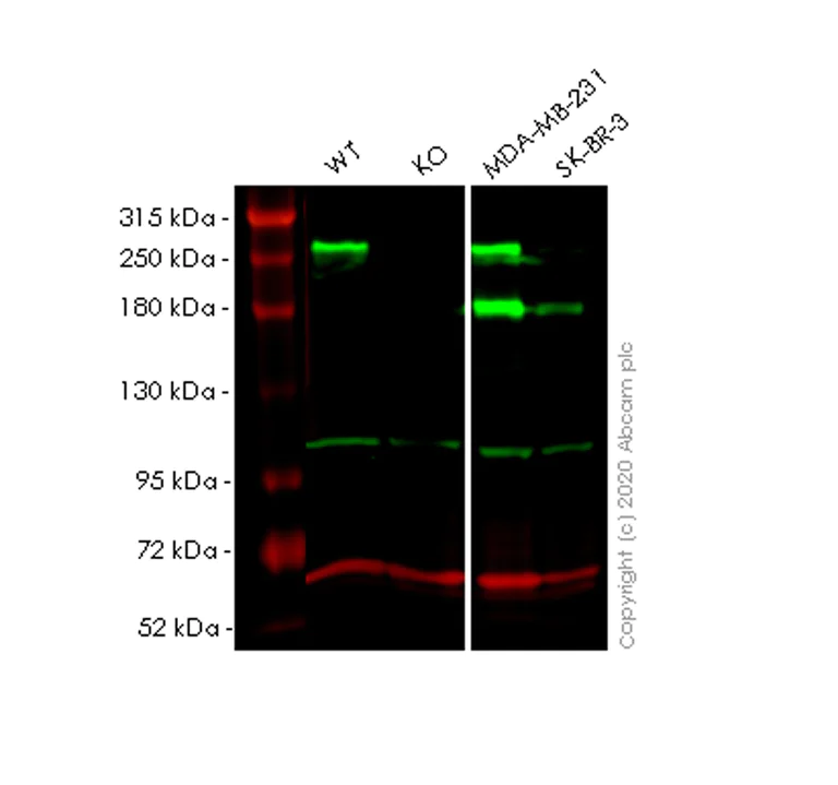
Western blot - Anti-Myoferlin antibody [EPR18887] - BSA and Azide free (AB271928)
This data was developed using the same antibody clone in a different buffer formulation (ab178386).
Lanes 1-4 : Merged signal (red and green). Green - ab178386 observed at 250,180 kDa. Red - loading control ab7291 observed at 50 kDa.
ab178386 Anti-Myoferlin antibody [EPR18887] was shown to specifically react with Myoferlin in wild-type HeLa cells. Loss of signal was observed when knockout cell line ab265782 (knockout cell lysate ab257547) was used. Wild-type and Myoferlin knockout samples were subjected to SDS-PAGE. ab178386 and Anti-alpha Tubulin antibody [DM1A] - Loading Control (ab7291) were incubated overnight at 4°C at 1 in 1000 dilution and 1 in 20000 dilution respectively. Blots were developed with Goat anti-Rabbit IgG H&L (IRDye® 800CW) preadsorbed (ab216773) and Goat anti-Mouse IgG H&L (IRDye® 680RD) preadsorbed (ab216776) secondary antibodies at 1 in 20000 dilution for 1 hour at room temperature before imaging.
All lanes:
Western blot - Anti-Myoferlin antibody [EPR18887] (<a href='/products/primary-antibodies/myoferlin-antibody-epr18887-ab178386'>ab178386</a>) at 1/1000 dilution
Lane 1:
Wild-type HeLa cell lysate at 20 µg
Lane 2:
MYOF knockout HeLa cell lysate at 20 µg
Lane 2:
Western blot - Human MYOF (Myoferlin) knockout HeLa cell line (<a href='/products/cell-lines/human-myof-myoferlin-knockout-hela-cell-line-ab265782'>ab265782</a>)
Lane 3:
MDA-MB-231 cell lysate at 20 µg
Lane 4:
SK-BR-3 cell lysate at 20 µg
Secondary
All lanes:
Western blot - Goat anti-Rabbit IgG H&L (IRDye® 800CW) preadsorbed (<a href='/products/secondary-antibodies/goat-rabbit-igg-h-l-irdye-800cw-preadsorbed-ab216773'>ab216773</a>) at 1/10000 dilution
Predicted band size: 235 kDa
Observed band size: 180 kDa,250 kDa
false
関連する標識済み抗体及び組成の異なる製品 (1)
-
Anti-Myoferlin antibody [EPR18887]
代替製品
組合せ製品
Primary Antibodies
AB124684
Anti-Dysferlin antibody [JAI-1-49-3]
20ul selling size|RabMAb|Recombinant
- 4
- 4
Primary Antibodies
AB183734
RabMAb|Recombinant|Lab Essentials|20ul selling size
- 5
- 24
Primary Antibodies
AB133741
Anti-Lamin B1 antibody [EPR8985(B)] - Nuclear Envelope Marker
RabMAb|Recombinant|KO Validated|Lab Essentials|20ul selling size
- 4
- 15
Primary Antibodies
AB182126
Anti-PKC delta antibody [EPR17075]
KO Validated|RabMAb|Recombinant|20ul selling size

- 4
- 7
Reactivity data
製品の詳細
ab271928 is the carrier-free version of ab178386.
Patented technology
Our RabMAb® technology is a patented hybridoma-based technology for making rabbit monoclonal antibodies. For details on our patents, please refer to RabMAb® patents.
What are the advantages of a recombinant monoclonal antibody?
This product is a recombinant monoclonal antibody, which offers several advantages including:
- - High batch-to-batch consistency and reproducibility
- - Improved sensitivity and specificity
- - Long-term security of supply
- - Animal-free batch production
For more information, read more on recombinant antibodies.
Conjugation ready
Our carrier-free antibodies are typically supplied in a PBS-only formulation, purified and free of BSA, sodium azide and glycerol. This conjugation-ready format is designed for use with fluorochromes, metal isotopes, oligonucleotides, and enzymes, which makes them ideal for antibody labelling, functional and cell-based assays, flow-based assays (e.g. mass cytometry) and Multiplex Imaging applications.
Use our conjugation kits for antibody conjugates that are ready-to-use in as little as 20 minutes with 1 minute hands-on-time and 100% antibody recovery: available for fluorescent dyes, HRP, biotin and gold.
Compatibility
This product is compatible with the Maxpar® Antibody Labeling Kit from Fluidigm, without the need for antibody preparation. Maxpar® is a trademark of Fluidigm Canada Inc.
出荷温度及び保存条件
製品の状態
精製方法
バッファー組成
出荷温度
短期保存温度
長期保存温度
保管に関する情報
補足情報
This supplementary information is collated from multiple sources and compiled automatically.
Biological function summary
The protein supports membrane dynamics and stabilization. Myoferlin is not part of a large complex but interacts with other proteins at the cellular membrane. It regulates cellular processes such as proliferation differentiation and endocytosis. In muscle cells it assists in the organization and function of the cytoskeleton maintaining proper cellular architecture and signaling.
Pathways
Myoferlin engages in critical roles in pathways related to cell membrane repair and trafficking. It associates with pathways like the myogenesis pathway and integrin signaling. In these pathways it interacts with other proteins such as dysferlin and caveolin-3 which facilitate membrane repair and receptor localization respectively. Myoferlin ensures efficient cellular response to membrane damage maintaining tissue integrity.
製品プロトコール
- Visit the General protocols
- Visit the Troubleshooting
ターゲットの情報
Abcam product promise
Please note: All products are 'FOR RESEARCH USE ONLY. NOT FOR USE IN DIAGNOSTIC OR THERAPEUTIC PROCEDURES'.
For licensing inquiries, please contact partnerships@abcam.com