Anti-MUC17 抗体 [EPR28482-85]
Anti-MUC17 antibody [EPR28482-85]
- BOND RX™ Validated
- 20ul selling size
- Recombinant
- RabMAb
- Advanced Validation
- 詳細を見る
Be the first to review this product! Submit a review
|
(0 Publication)
Rabbit Recombinant Monoclonal MUC17 antibody. Suitable for mIHC, WB, IHC-P, Flow Cyt (Intra), ICC/IF and reacts with Human samples.
別名を表示する
MUC3, MUC17, Mucin-17, MUC-17, Small intestinal mucin-3, MUC-3
- mIHC
Lab
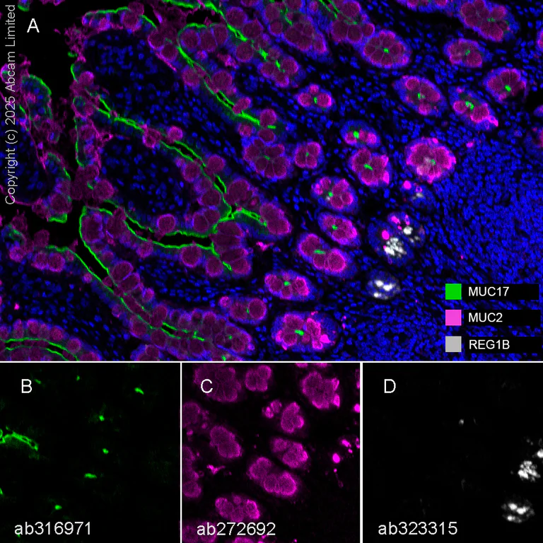
Multiplex immunohistochemistry - Anti-MUC17 antibody [EPR28482-85] (AB316971)
Multiplex immunohistochemistry analysis of formalin/PFA-fixed paraffin-embedded human small intestine tissue staining MUC17 with ab316971 at 1/500 dilution, ab272692 anti-MUC2 used at 1/2000 dilution and ab323315 anti-REG1B used at a 1/5000 dilution. Opal Polymer HRP Ms + Rb was used as a secondary antibody, and DAPI was used for a nuclear counter stain. Heat mediated antigen retrieval with Tris-EDTA buffer (pH 9.0, epitope retrieval solution2) for 20 mins.
Panel A : merged staining of anti-MUC17 (green; Opal™570), anti-MUC2 (magenta; Opal™690) and anti-REG1B (grey; Opal™520) on human small intestine.
Panel B : anti-MUC17 staining epithelium in human small intestine.
Panel C : anti-MUC2 staining goblet cells in human small intestine.
Panel D : anti-REG1B staining Paneth cells in human small intestine.
Nuclear DNA was labeled with DAPI (shown in blue).
The section was incubated in three rounds of staining : in the order of ab316971, ab272692 and ab323315 for 30 mins at room temperature. Each round was followed by a separate fluorescent tyramide signal amplification system.
The immunostaining was performed on a Leica Biosystems BOND® RX instrument with an Opal™ 4-color kit. Image acquisition was performed with Leica SP8 confocal microscope.
- ICC/IF
Supplier Data
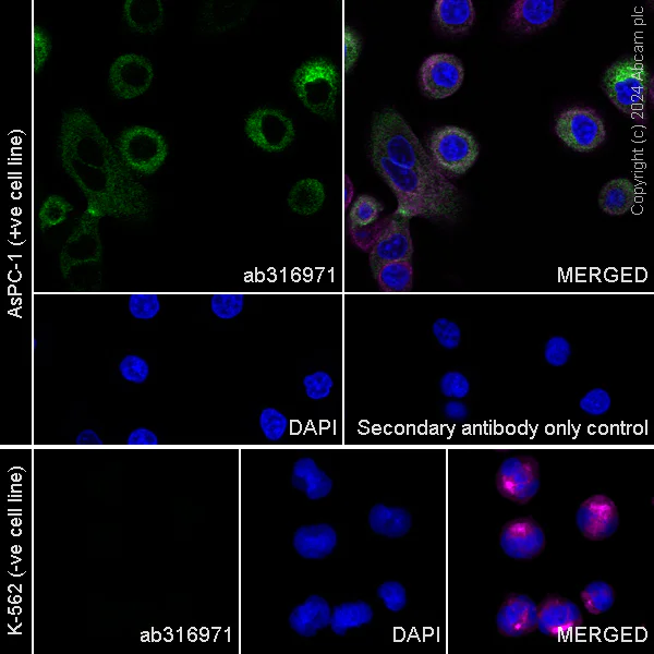
Immunocytochemistry/ Immunofluorescence - Anti-MUC17 antibody [EPR28482-85] (AB316971)
Immunofluorescent analysis of 4% Paraformaldehyde-fixed 0.1% TritonX-100 permeabilized AsPC-1 (human Pancreas epithelial cell) cells labelling MUC17 with ab316971 at 1/50 (10.2 ug/ml) dilution followed by ab150081 Goat Anti-Rabbit IgG H&L (Alexa Fluor® 488) preadsorbed antibody at 1/1000 (2 ug/mL) dilution (Green).
Confocal image showing cytoplasmic staining in AsPC-1 cell line (shown in green). The counterstain was observed in magenta. Nuclear DNA was labeled with DAPI (shown in blue).
Negative control : K-562 (PMID : 11855812)
Image was taken with a confocal microscope(Leica-Microsystems TCS SP8).
ab195889 Anti-alpha Tubulin mouse monoclonal antibody - Microtubule Marker (Alexa Fluor® 594) was used to counterstain tubulin at 1/200 (2.5 ug/mL) dilution (Magenta). The Nuclear counterstain was DAPI (Blue).
Secondary antibody only control : Secondary antibody is ab150081 Goat Anti-Rabbit IgG H&L (Alexa Fluor® 488) preadsorbed at 1/1000 (2 ug/mL) dilution.
- Flow Cyt (Intra)
Supplier Data
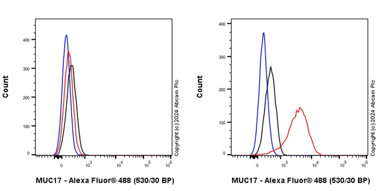
Flow Cytometry (Intracellular) - Anti-MUC17 antibody [EPR28482-85] (AB316971)
Flow cytometric analysis of 4% paraformaldehyde fixed 90% methanol permeabilized AsPC-1 (human Pancreas epithelial cell) / right and K-562 (human chronic myelogenous leukemia lymphoblast) / left cells labelling MUC17 with ab316971 at 1/500 dilution (0.1ug)/(Red) compared with a Rabbit monoclonal IgG (ab172730) (Black) isotype control and an unlabelled control (cells without incubation with primary antibody and secondary antibody) (Blue).
Goat Anti-Rabbit IgG (Alexa Fluor® 488, ab150081) at 1/5000 dilution was used as the secondary antibody.
Negative control : K-562
- IHC-P
Supplier Data
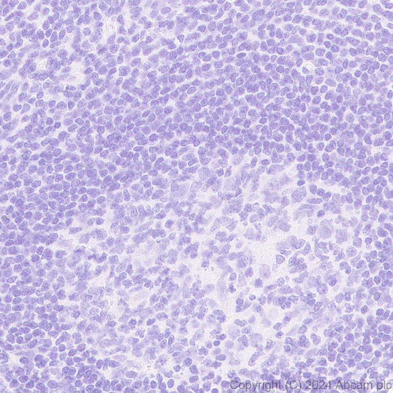
Immunohistochemistry (Formalin/PFA-fixed paraffin-embedded sections) - Anti-MUC17 antibody [EPR28482-85] (AB316971)
Immunohistochemical analysis of paraffin-embedded Human tonsil tissue labeling MUC17 with ab316971 at 1/500 (1.02 ug/ml) dilution, followed by a ready to use LeicaDS9800 (Bond™ Polymer Refine Detection).
Negative control : No staining on human tonsil.
The section was incubated with ab316971 for 30 mins at room temperature.
The immunostaining was performed on a Leica Biosystems BOND® RX instrument
Counterstained with Hematoxylin.
Secondary antibody only control : Secondary antibody is a ready to use LeicaDS9800 (Bond™ Polymer Refine Detection).
Heat mediated antigen retrieval was performed with Tris-EDTA buffer (pH 9.0, Epitope Retrieval Solution2) for 20 mins
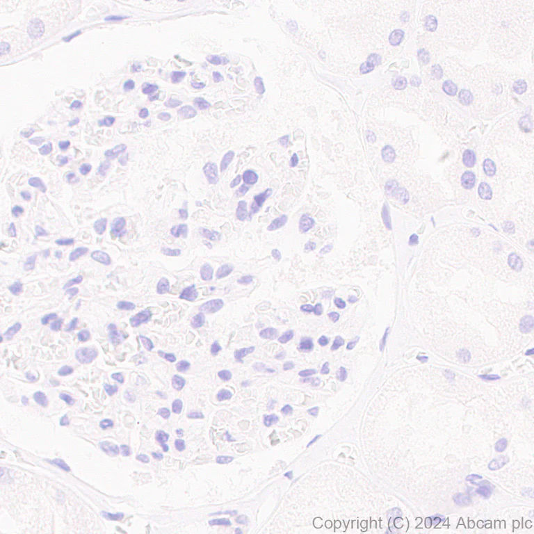
- IHC-P
Supplier Data
Immunohistochemistry (Formalin/PFA-fixed paraffin-embedded sections) - Anti-MUC17 antibody [EPR28482-85] (AB316971)
Immunohistochemical analysis of paraffin-embedded Human kidney tissue labeling MUC17 with ab316971 at 1/500 (1.02 ug/ml) dilution, followed by a ready to use LeicaDS9800 (Bond™ Polymer Refine Detection).
Negative control : No staining on human kidney.
The section was incubated with ab316971 for 30 mins at room temperature.
The immunostaining was performed on a Leica Biosystems BOND® RX instrument
Counterstained with Hematoxylin.
Secondary antibody only control : Secondary antibody is a ready to use LeicaDS9800 (Bond™ Polymer Refine Detection).
Heat mediated antigen retrieval was performed with Tris-EDTA buffer (pH 9.0, Epitope Retrieval Solution2) for 20 mins
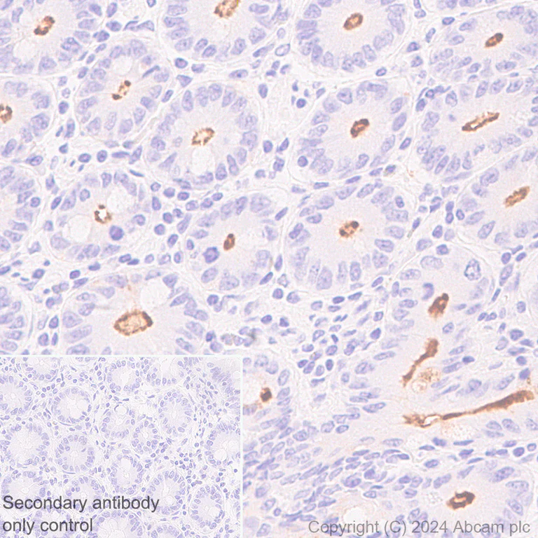
- IHC-P
Supplier Data
Immunohistochemistry (Formalin/PFA-fixed paraffin-embedded sections) - Anti-MUC17 antibody [EPR28482-85] (AB316971)
Immunohistochemical analysis of paraffin-embedded Human jejunum tissue labeling MUC17 with ab316971 at 1/500 (1.02 ug/ml) dilution, followed by a ready to use LeicaDS9800 (Bond™ Polymer Refine Detection).
Membranous staining on epithelium of human jejunum.
The section was incubated with ab316971 for 30 mins at room temperature.
The immunostaining was performed on a Leica Biosystems BOND® RX instrument
Counterstained with Hematoxylin.
Secondary antibody only control : Secondary antibody is a ready to use LeicaDS9800 (Bond™ Polymer Refine Detection).
Heat mediated antigen retrieval was performed with Tris-EDTA buffer (pH 9.0, Epitope Retrieval Solution2) for 20 mins
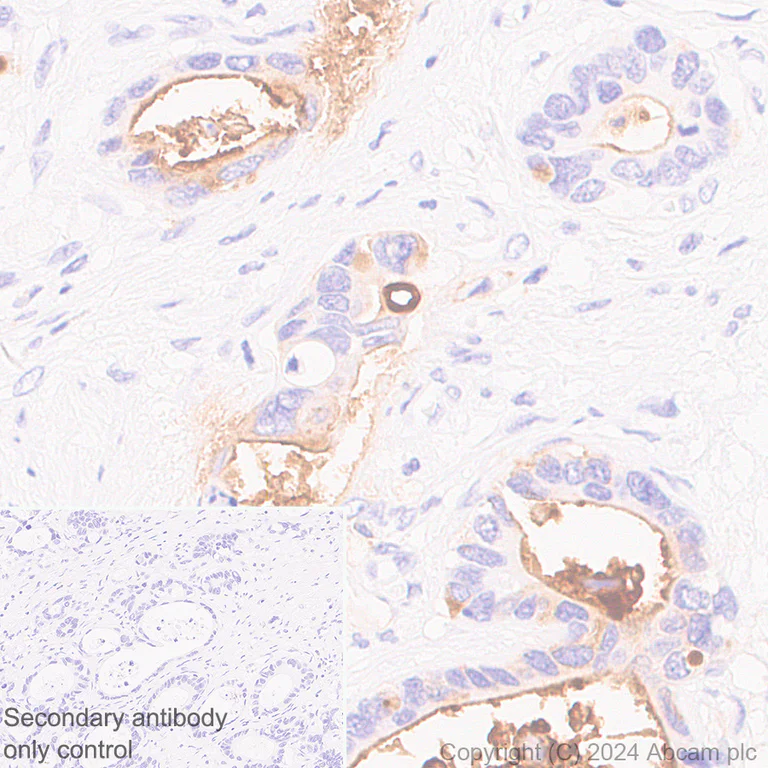
- IHC-P
Supplier Data
Immunohistochemistry (Formalin/PFA-fixed paraffin-embedded sections) - Anti-MUC17 antibody [EPR28482-85] (AB316971)
Immunohistochemical analysis of paraffin-embedded Human stomach cancer tissue labeling MUC17 with ab316971 at 1/500 (1.02 ug/ml) dilution, followed by a ready to use LeicaDS9800 (Bond™ Polymer Refine Detection).
Membranous staining on epithelium of human stomach cancer (PMID : 31262330).
The section was incubated with ab316971 for 30 mins at room temperature.
The immunostaining was performed on a Leica Biosystems BOND® RX instrument
Counterstained with Hematoxylin.
Secondary antibody only control : Secondary antibody is a ready to use LeicaDS9800 (Bond™ Polymer Refine Detection).
Heat mediated antigen retrieval was performed with Tris-EDTA buffer (pH 9.0, Epitope Retrieval Solution2) for 20 mins
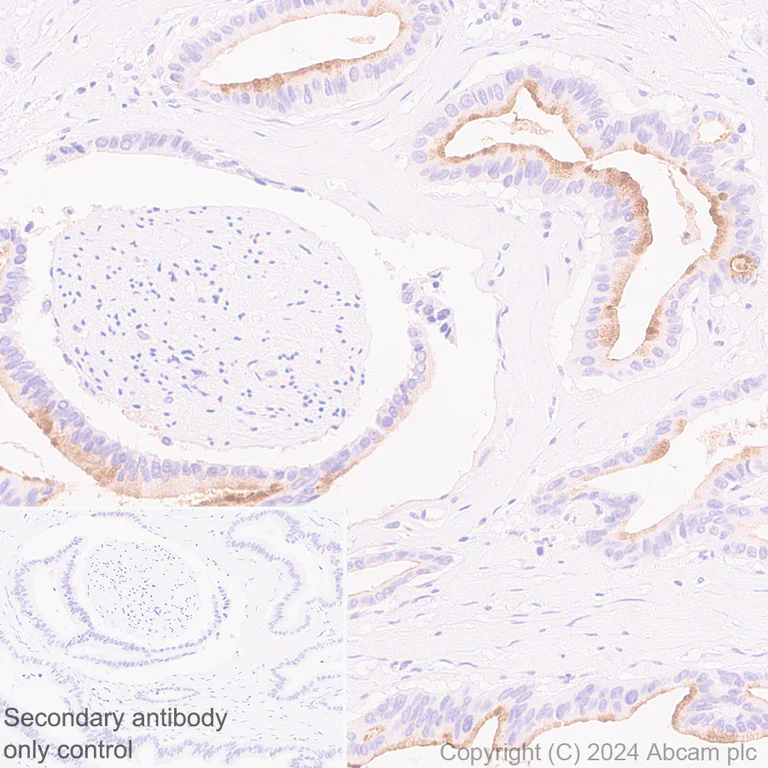
- IHC-P
Supplier Data
Immunohistochemistry (Formalin/PFA-fixed paraffin-embedded sections) - Anti-MUC17 antibody [EPR28482-85] (AB316971)
Immunohistochemical analysis of paraffin-embedded Human pancreas cancer tissue labeling MUC17 with ab316971 at 1/500 (1.02 ug/ml) dilution, followed by a ready to use LeicaDS9800 (Bond™ Polymer Refine Detection).
Membranous staining on epithelium of human pancreas cancer (PMID : 16737958).
The section was incubated with ab316971 for 30 mins at room temperature.
The immunostaining was performed on a Leica Biosystems BOND® RX instrument
Counterstained with Hematoxylin.
Secondary antibody only control : Secondary antibody is a ready to use LeicaDS9800 (Bond™ Polymer Refine Detection).
Heat mediated antigen retrieval was performed with Tris-EDTA buffer (pH 9.0, Epitope Retrieval Solution2) for 20 mins
- IHC-P
Supplier Data
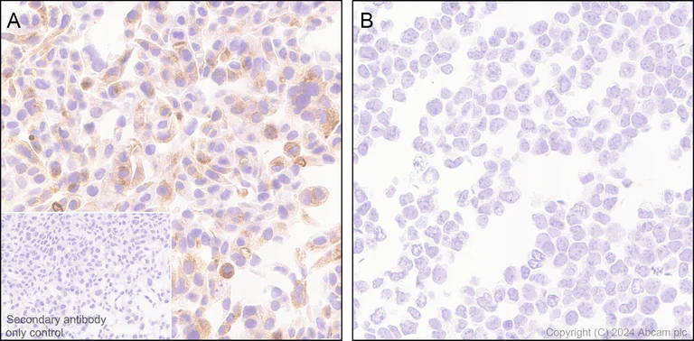
Immunohistochemistry (Formalin/PFA-fixed paraffin-embedded sections) - Anti-MUC17 antibody [EPR28482-85] (AB316971)
Immunohistochemical analysis of paraffin-embedded (A) AsPC-1 (human Pancreas epithelial cell), (B) K-562 (human chronic myelogenous leukemia lymphoblast) tissue labeling MUC17 with ab316971 at 1/500 (1.02 ug/ml) dilution, followed by a ready to use LeicaDS9800 (Bond™ Polymer Refine Detection).
Positive staining on panel (A) AsPC-1 cell pellet, no staining on panel (B) K-562 cell pellet.
The section was incubated with ab316971 for 30 mins at room temperature.
The immunostaining was performed on a Leica Biosystems BOND® RX instrument
Counterstained with Hematoxylin.
Secondary antibody only control : Secondary antibody is a ready to use LeicaDS9800 (Bond™ Polymer Refine Detection).
Heat mediated antigen retrieval was performed with Tris-EDTA buffer (pH 9.0, Epitope Retrieval Solution2) for 20 mins
- WB
Supplier Data
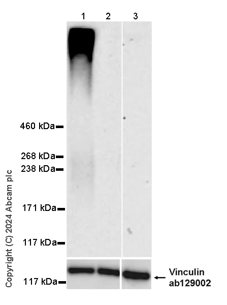
Western blot - Anti-MUC17 antibody [EPR28482-85] (AB316971)
Negative control : K-562, Hela (PMID : 11855812).
The expression profile/ molecular weight observed is consistent with what has been described in the literature (PMID : 11855812, 16737958).
In Western blot, Anti-Vinculin antibody [EPR8185] (ab129002) staining at 1/10000 dilution.
All lanes:
Western blot - Anti-MUC17 antibody [EPR28482-85] (ab316971) at 1/1000 dilution
Lane 1:
AsPC-1 (human Pancreas epithelial cell) whole cell lysate at 20 µg
Lane 2:
K-562 (human chronic myelogenous leukemia lymphoblast) whole cell lysate at 20 µg
Lane 3:
HeLa (human cervical adenocarcinoma epithelial cell) whole cell lysate at 20 µg
Secondary
All lanes:
Western blot - Goat Anti-Rabbit IgG H&L (HRP) (<a href='/products/secondary-antibodies/goat-rabbit-igg-h-l-hrp-ab97051'>ab97051</a>) at 1/20000 dilution
Observed band size: 500 kDa,124 kDa
false
Exposure time: 180s
- WB
Supplier Data
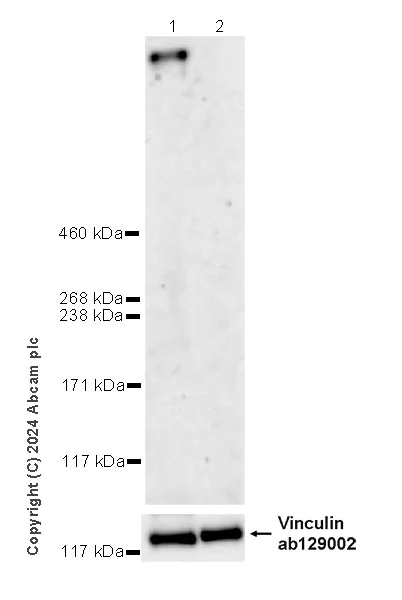
Western blot - Anti-MUC17 antibody [EPR28482-85] (AB316971)
Negative control : kidney (PMID : 11855812).
In Western blot, Anti-Vinculin antibody [EPR8185] (ab129002) staining at 1/10000 dilution.
All lanes:
Western blot - Anti-MUC17 antibody [EPR28482-85] (ab316971) at 1/1000 dilution
Lane 1:
Human small intestine tissue lysate at 20 µg
Lane 2:
Human kidney tissue lysate at 20 µg
Secondary
All lanes:
Western blot - Goat Anti-Rabbit IgG H&L (HRP) (<a href='/products/secondary-antibodies/goat-rabbit-igg-h-l-hrp-ab97051'>ab97051</a>) at 1/2000 dilution
Observed band size: 500 kDa,124 kDa
true
Exposure time: 180s
関連する標識済み抗体及び組成の異なる製品 (1)
-
Anti-MUC17 antibody [EPR28482-85] - BSA and Azide free
組合せ製品
Primary Antibodies
AB137132
Anti-PD1 antibody [EPR4877(2)]
BOND RX™ Validated|20ul selling size|RabMAb|Recombinant

- 5
- 5
Primary Antibodies
AB134162
RabMAb|Recombinant|KO Validated|20ul selling size
- 0
- 0
Primary Antibodies
AB108403
20ul selling size|RabMAb|KO Validated|Recombinant

- 0
- 1
Primary Antibodies
AB109190
Anti-LI Cadherin antibody [EPR3996]
BOND RX™ Validated|RabMAb|Recombinant|20ul selling size
- 0
- 0
Reactivity data
製品の詳細
Patented technology
Our RabMAb® technology is a patented hybridoma-based technology for making rabbit monoclonal antibodies. For details on our patents, please refer to RabMAb® patents.
What are the advantages of a recombinant monoclonal antibody?
This product is a recombinant monoclonal antibody, which offers several advantages including:
- - High batch-to-batch consistency and reproducibility
- - Improved sensitivity and specificity
- - Long-term security of supply
- - Animal-free batch production
For more information, read more on recombinant antibodies.
出荷温度及び保存条件
製品の状態
精製方法
バッファー組成
出荷温度
短期保存期間
短期保存温度
長期保存温度
分注に関する情報
保管に関する情報
製品プロトコール
- Visit the General protocols
- Visit the Troubleshooting
ターゲットの情報
Abcam product promise
Please note: All products are 'FOR RESEARCH USE ONLY. NOT FOR USE IN DIAGNOSTIC OR THERAPEUTIC PROCEDURES'.
For licensing inquiries, please contact partnerships@abcam.com