Anti-MSH6 抗体 [SP93]
Anti-MSH6 antibody [SP93]
- BOND RX™ Validated
- RabMAb
- Recombinant
- KO Validated
- 詳細を見る
Be the first to review this product! Submit a review
|
(1 Publication)
Anti-MSH6 antibody [SP93] (ab99889) is a rabbit monoclonal antibody detecting MSH6 in Western Blot, Flow Cytometry (Intra), IHC-P, ICC/IF. Suitable for Human, Mouse.
- KO validated for confirmed specificity
- Biophysical QC for unrivalled batch-batch consistency
- Trusted since 2010
別名を表示する
GTBP, MSH6, DNA mismatch repair protein Msh6, hMSH6, G/T mismatch-binding protein, MutS protein homolog 6, MutS-alpha 160 kDa subunit, GTMBP, p160
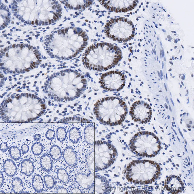
- IHC-P
Lab
Immunohistochemistry (Formalin/PFA-fixed paraffin-embedded sections) - Anti-MSH6 antibody [SP93] (AB99889)
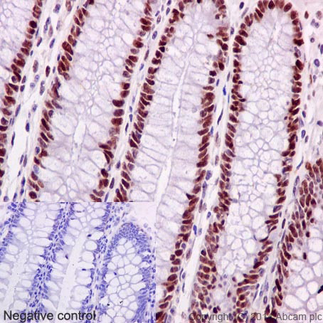
Immunohistochemical analysis of formalin fixed paraffin embedded human colon labelling MSH6 with ab99889 at a dilution of 1/400. The immunostaining was performed on a Ventana DISCOVERY ULTRA (Roche Tissue Diagnostics) instrument with a OptiView DAB IHC Detection Kit. Heat mediated antigen retrieval was performed with DISCOVERY cell conditioning solution (CC1) 100°C, pH8.5 for 32mins. ab99889 anti-MSH6 antibody [SP93] was incubated for 16mins at 37°C. Sections were counterstained with Hematoxylin II. Image inset shows absence of staining in secondary antibody only control.
Customers are encouraged to optimise antigen retrieval conditions, antibody concentration, incubation times and temperature for best results in their own IHC assay workflow (automated and manual).
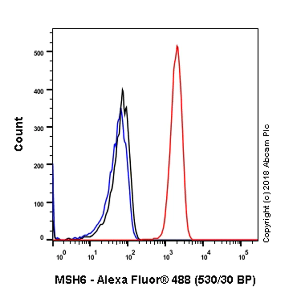
- Flow Cyt (Intra)
Unknown
Flow Cytometry (Intracellular) - Anti-MSH6 antibody [SP93] (AB99889)
Flow cytometry analysis of Hela (human cervix adenocarcinoma) labeling MSH6 with purified ab99889 at 1/20 dilution (5.05 μg/ml) (red). Cells were fixed with 4% paraformaldehyde and permeabilised with 90% methanol. Goat anti rabbit IgG (Alexa Fluor® 488, ab150077) at 1/2000 dilution was used as a secondary antibody. Isotype control - Rabbit monoclonal IgG (ab172730) (Black). Unlableled control - Unlabelled cells (blue).
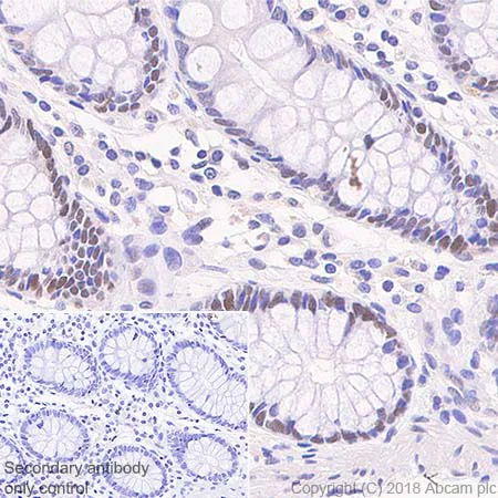
- IHC-P
Unknown
Immunohistochemistry (Formalin/PFA-fixed paraffin-embedded sections) - Anti-MSH6 antibody [SP93] (AB99889)
Immunohistochemistry (Formalin/PFA-fixed paraffin-embedded sections) analysis of Human colon tissue sections labeling MSH6 with ab99889 at 1/100 dilution (1.0 μg/ml). Heat mediated antigen retrieval with Tris-EDTA buffer (pH 9.0, epitope retrieval solution 2) for 10mins. Rabbit specific IHC polymer detection kit HRP/DAB (ab209101) was used as the secondary antibody. Hematoxylin was used as a counterstain. Sporadically nuclear staining on human colon, performed on a Leica Biosystems BOND™ RX instrument.
The section was incubated with ab99889 for 30 mins at room temperature.
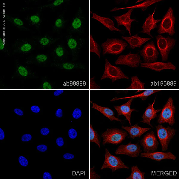
- ICC/IF
Unknown
Immunocytochemistry/ Immunofluorescence - Anti-MSH6 antibody [SP93] (AB99889)
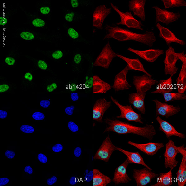
ab99889 staining MSH6 in HeLa cells. The cells were fixed with 100% methanol (5min), permeabilized with 0.1% Triton X-100 for 5 minutes and then blocked with 1% BSA/10% normal goat serum/0.3M glycine in 0.1% PBS-Tween for 1h. The cells were then incubated with ab99889 at 1/250 dilution and ab195889 at 1/250 dilution (shown in pseudocolour red) overnight at +4°C, followed by a further incubation at room temperature for 1h with a goat secondary antibody to Rabbit IgG (Alexa Fluor® 488) (ab150081) at 2 μg/ml (shown in green). Nuclear DNA was labelled in blue with DAPI.
Image was taken with a confocal microscope (Leica-Microsystems, TCS SP8).
- IHC-P
Unknown
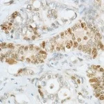
Immunohistochemistry (Formalin/PFA-fixed paraffin-embedded sections) - Anti-MSH6 antibody [SP93] (AB99889)
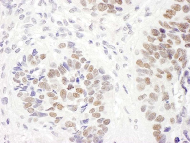
Staining of MSH6 in a formalin fixed, paraffin embedded Human rectal carcinoma tissue using ab99889 at a dilution of 1/100.
- ICC/IF
Unknown
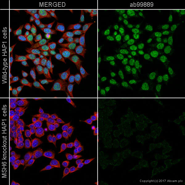
Immunocytochemistry/ Immunofluorescence - Anti-MSH6 antibody [SP93] (AB99889)
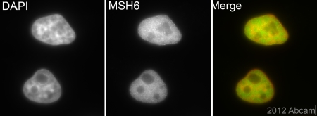
ab99889 staining MSH6 in wild-type HAP1 cells (top panel) and MSH6 knockout HAP1 cells (bottom panel). The cells were fixed with 100% methanol (5min), permeabilized with 0.1% Triton X-100 for 5 minutes and then blocked with 1% BSA/10% normal goat serum/0.3M glycine in 0.1% PBS-Tween for 1h. The cells were then incubated with ab99889 at 1/250 dilution and ab195889 at 1/250 dilution (shown in pseudocolour red) overnight at +4°C, followed by a further incubation at room temperature for 1h with a goat secondary antibody to Rabbit IgG (Alexa Fluor® 488) (ab150081) at 2 μg/ml (shown in green). Nuclear DNA was labelled in blue with DAPI.
Image was taken with a confocal microscope (Leica-Microsystems, TCS SP8).
- IHC-P
Unknown
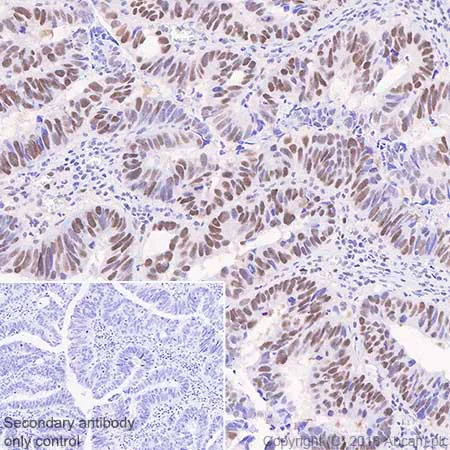
Immunohistochemistry (Formalin/PFA-fixed paraffin-embedded sections) - Anti-MSH6 antibody [SP93] (AB99889)
Immunohistochemistry (Formalin/PFA-fixed paraffin-embedded sections) analysis of Human colon carcinoma tissue sections labeling MSH6 with ab99889 at 1/100 dilution (1.0 μg/ml). Heat mediated antigen retrieval with Tris-EDTA buffer (pH 9.0, epitope retrieval solution 2) for 10mins. Rabbit specific IHC polymer detection kit HRP/DAB (ab209101) was used as the secondary antibody. Hematoxylin was used as a counterstain. Nuclear staining on human colon carcinoma, performed on a Leica Biosystems BOND™ RX instrument.
The section was incubated with ab99889 for 30 mins at room temperature.
- IHC-P
Unknown
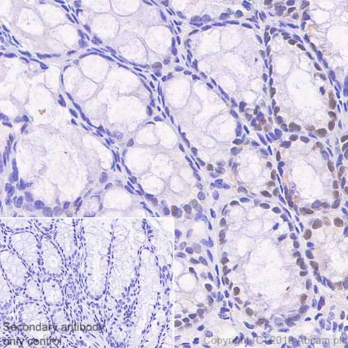
Immunohistochemistry (Formalin/PFA-fixed paraffin-embedded sections) - Anti-MSH6 antibody [SP93] (AB99889)
Immunohistochemistry (Formalin/PFA-fixed paraffin-embedded sections) analysis of Mouse colon
tissue sections labeling MSH6 with ab99889 at 1/100 dilution (1.0 μg/ml). Heat mediated antigen retrieval with Tris-EDTA buffer (pH 9.0, epitope retrieval solution 2) for 10mins. Rabbit specific IHC polymer detection kit HRP/DAB (ab209101) was used as the secondary antibody. Hematoxylin was used as a counterstain. Sporadically nuclear staining on mouse colon, performed on a Leica Biosystems BOND™ RX instrument.
The section was incubated with ab99889 for 30 mins at room temperature.
- WB
Unknown
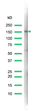
Western blot - Anti-MSH6 antibody [SP93] (AB99889)
ab99889 used in the image is the undiluted version of ab99890.
All lanes:
Western blot - Anti-MSH6 antibody [SP93] (ab99889) at 1/100 dilution
All lanes:
Lysate prepared from A431 cells
Predicted band size: 153 kDa
Observed band size: 170 kDa
false
- WB
Supplier Data
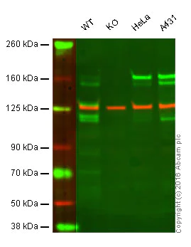
Western blot - Anti-MSH6 antibody [SP93] (AB99889)
Lane 1 : Wild-type HAP1 cell lysate (20 μg)
Lane 2 : MSH6 knockout HAP1 cell lysate (20 μg)
Lane 3 : HeLa cell lysate (20 μg)
Lane 4 : A431 cell lysate (20 μg)
Lanes 1 - 4 : Merged signal (red and green). Green - ab99889 observed at 160 kDa. Red - loading control, ab18058, observed at 124 kDa.
ab99889 was shown to specifically react with MSH6 in wild-type HAP1 cells along with additional cross reactive bands. No band was observed in MSH6 knockout samples. Wild-type and MSH6 knockout samples were subjected to SDS-PAGE. ab99889 and ab18058 (loading control to Vinculin) were diluted at 1 μg/ml and 1/10 000 respectively and incubated overnight at 4°C. Blots were developed with Goat anti-Rabbit IgG H&L (IRDye® 800CW) preadsorbed ab216773 and Goat anti-Mouse IgG H&L (IRDye® 680RD) preadsorbed ab216776 secondary antibodies at 1/10 000 dilution for 1 h at room temperature before imaging.
All lanes:
Western blot - Anti-MSH6 antibody [SP93] (ab99889)
Predicted band size: 153 kDa
false
関連する標識済み抗体及び組成の異なる製品 (1)
-
Anti-MSH6 antibody [SP93] - BSA and Azide free
代替製品
Primary Antibodies
AB198334
Alexa Fluor® 647 Anti-MSH6 antibody [EPR3945]
RabMAb|Recombinant|KO Validated
- 3
- 2
Primary Antibodies
AB208940
RabMAb|Recombinant|KO Validated|Trial Size

- 0
- 0
Primary Antibodies
AB214454
Anti-MSH6 antibody [EPR3945] - BSA and Azide free
BOND RX™ Validated|RabMAb|Recombinant|KO Validated
- 0
- 0
Primary Antibodies
AB92471
20ul selling size|BOND RX™ Validated|RabMAb|Recombinant|KO Validated|Trial Size
- 5
- 5
組合せ製品
Primary Antibodies
AB52635
Anti-Cytokeratin 5 antibody [EP1601Y] - Cytoskeleton Marker
BOND RX™ Validated|RabMAb|Recombinant|Lab Essentials
- 5
- 11
Primary Antibodies
AB133616
BOND RX™ Validated|RabMAb|Recombinant|Lab Essentials|20ul selling size
- 5
- 20
Primary Antibodies
AB16663
20ul selling size|BOND RX™ Validated|RabMAb|Recombinant|KO Validated|Lab Essentials

- 4
- 23
Reactivity data
製品の詳細
What is this antibody validated in?
Anti-MSH6 antibody [SP93] (ab99889) is a rabbit recombinant monoclonal antibody and is validated for use in Western Blot (WB), Flow Cytometry (Intra), Immunohistochemistry (IHC-P), Immunocytochemistry/immunofluorescence (ICC/IF) in Human, Mouse samples.
Collaboration
Anti-MSH6 [SP93] (ab99889) is a clone from the portfolio of Spring Bioscience (Roche) SP clones which have been optimised for immunohistochemistry (IHC).
Specificity confirmed
The specificity of Anti-MSH6 antibody [SP93] (ab99889) has been confirmed by Western blot testing in MSH6 Knockout HAP1 cells.
Other related products
We have a range of other formats of antibody clone [SP93] also available for your convenience: ab99889, Carrier free - ab238805, ab279563
What are the advantages of a recombinant monoclonal antibody?
This product is a recombinant monoclonal antibody, which offers several advantages including:
- - High batch-to-batch consistency and reproducibility
- - Improved sensitivity and specificity
- - Long-term security of supply
- - Animal-free batch production
For more information, read more on recombinant antibodies.
出荷温度及び保存条件
製品の状態
精製方法
精製に関する特記事項
バッファー組成
出荷温度
短期保存温度
長期保存温度
補足情報
This supplementary information is collated from multiple sources and compiled automatically.
Biological function summary
MSH6 functions as part of the MMR complex which is essential for maintaining genomic stability. The MutSα complex where MSH6 pairs with MSH2 operates along with other proteins in the MMR pathway to correct DNA replication errors. MSH6 is also known to interact with PCNA a DNA polymerase processivity factor which facilitates its role in the repair process.
Pathways
MSH6 participates prominently in the DNA mismatch repair pathway. This pathway is critical for correcting DNA errors and preventing mutations during replication. In association with MLH1-PMS2 (MutLα complex) MSH6 ensures that DNA integrity is preserved. Additionally MSH6 is involved in the base excision repair (BER) pathway where it collaborates with other repair proteins to fix small base lesions.
製品プロトコール
- Visit the General protocols
- Visit the Troubleshooting
ターゲットの情報
文献 (1)
Recent publications for all applications. Explore the full list and refine your search
Modern pathology : an official journal of the Unit 30:1144-1151 PubMed28548127
2017
Applications
Unspecified application
Species
Unspecified reactive species
Abcam product promise
Please note: All products are 'FOR RESEARCH USE ONLY. NOT FOR USE IN DIAGNOSTIC OR THERAPEUTIC PROCEDURES'.
For licensing inquiries, please contact partnerships@abcam.com