Anti-MelanA 抗体 [EPR20380]
Anti-MelanA antibody [EPR20380]
- RabMAb
- Recombinant
- 詳細を見る
5
(15 Reviews)
|
(34 Publications)
Anti-MelanA antibody [EPR20380] (ab210546) is a rabbit monoclonal antibody detecting MelanA in Western Blot, Flow Cytometry (Intra), IHC-P, ICC/IF. Suitable for Human, Mouse.
- Biophysical QC for unrivalled batch-batch consistency
- Over 20 publications
別名を表示する
MART1, MLANA, Melanoma antigen recognized by T-cells 1, MART-1, Antigen LB39-AA, Antigen SK29-AA, Protein Melan-A

- IHC-P
Supplier Data
Immunohistochemistry (Formalin/PFA-fixed paraffin-embedded sections) - Anti-MelanA antibody [EPR20380] (AB210546)
Immunohistochemical analysis of paraffin-embedded human malignant melanoma tissue labeling MelanA with ab210546 at 1/1000 dilution, followed by Goat Anti-Rabbit IgG H&L (HRP) (ab97051) at 1/500 dilution.
Cytoplasmic staining on human malignant melanoma is observed [PMID : 17445277] [PMID : 21840568].
Counter stained with Hematoxylin.
Secondary antibody only control : Used PBS instead of primary antibody, secondary antibody is Goat Anti-Rabbit IgG H&L (HRP) (ab97051) at 1/500 dilution.
Perform heat mediated antigen retrieval with Tris/EDTA buffer pH 9.0 before commencing with IHC staining protocol.

- ICC/IF
Supplier Data
Immunocytochemistry/ Immunofluorescence - Anti-MelanA antibody [EPR20380] (AB210546)
Immunofluorescent analysis of 4% paraformaldehyde-fixed, 0.1% Triton X-100 permeabilized MeWo (Human malignant melanoma cell line) cells labeling MelanA with ab210546 at 1/1000 dilution, followed by Goat anti-rabbit IgG (Alexa Fluor® 488) (ab150077) secondary antibody at 1/1000 dilution (green).
Confocal image showing cytoplasmic staining on MeWo cell line.
The nuclear counterstain is DAPI (blue).
Tubulin is detected with ab195889 (Anti-alpha Tubulin antibody [DM1A] - Microtubule Marker (Alexa Fluor® 594)) at 1/200 dilution (red).
Secondary antibody only control : Used PBS instead of primary antibody, secondary antibody is Goat anti-rabbit IgG (Alexa Fluor® 488) (ab150077) at 1/1000 dilution.

- IHC-P
Supplier Data
Immunohistochemistry (Formalin/PFA-fixed paraffin-embedded sections) - Anti-MelanA antibody [EPR20380] (AB210546)
Immunohistochemical analysis of paraffin-embedded human metastatic malignant melanoma tissue labeling MelanA with ab210546 at 1/1000 dilution, followed by Goat Anti-Rabbit IgG H&L (HRP) (ab97051) at 1/500 dilution.
Cytoplasmic staining on human metastatic malignant melanoma is observed [PMID : 17445277] [PMID : 21840568].
Counter stained with Hematoxylin.
Secondary antibody only control : Used PBS instead of primary antibody, secondary antibody is Goat Anti-Rabbit IgG H&L (HRP) (ab97051) at 1/500 dilution.
Perform heat mediated antigen retrieval with Tris/EDTA buffer pH 9.0 before commencing with IHC staining protocol.

- IHC-P
Supplier Data
Immunohistochemistry (Formalin/PFA-fixed paraffin-embedded sections) - Anti-MelanA antibody [EPR20380] (AB210546)
Immunohistochemical analysis of paraffin-embedded human liver cancer tissue labeling MelanA with ab210546 at 1/1000 dilution, followed by Goat Anti-Rabbit IgG H&L (HRP) (ab97051) at 1/500 dilution.
Negative control : no staining on human liver cancer [PMID : 10096358].
Counter stained with Hematoxylin.
Perform heat mediated antigen retrieval with Tris/EDTA buffer pH 9.0 before commencing with IHC staining protocol.

- Flow Cyt (Intra)
Lab
Flow Cytometry (Intracellular) - Anti-MelanA antibody [EPR20380] (AB210546)
Flow cytometry overlay histogram showing left Malme-3M positive cells and right negative HEK293 stained with ab210546 (red line). The cells were fixed with 80% methanol (5 min) and then permeabilised with 0.1 % PBS-Triton X-100 for 15 min. The cells were then incubated in 1x PBS containing 10% normal goat serum to block non-specific protein-protein interaction followed by the antibody (ab210546) (1x 10⁶ in 100μl at 0.2μg/ml (1/10500)) for 30min at 22°C.
The secondary antibody Goat Anti-Rabbit IgG H&L (Alexa Fluor® 488) preadsorbed was incubated at 1/4000 for 30min at 22°C
Isotype control antibody (black line) was Recombinant Rabbit IgG, monoclonal [EPR25A] - Isotype Control used at the same concentration and conditions as the primary antibody. Unlabelled sample (blue line) was also used as a control.
Acquisition of >5000 events were collected using a 50 mW Blue laser (488nm) and 525/40 bandpass filter.
This antibody gave a positive signal in Malme-3M Fixed with 4% formaldehyde (10 min) / permeabilised with 0.1 % PBS-Triton X-100 for 15 min under the same conditions.

- IHC-P
Supplier Data
Immunohistochemistry (Formalin/PFA-fixed paraffin-embedded sections) - Anti-MelanA antibody [EPR20380] (AB210546)
Immunohistochemical analysis of paraffin-embedded mouse hair bulb tissue labeling MelanA with ab210546 at 1/1000 dilution, followed by Goat Anti-Rabbit IgG H&L (HRP) (ab97051) at 1/500 dilution.
Cytoplasmic staining on melanocytes of mouse hair bulb is observed.
Counter stained with Hematoxylin.
Secondary antibody only control : Used PBS instead of primary antibody, secondary antibody is Goat Anti-Rabbit IgG H&L (HRP) (ab97051) at 1/500 dilution.
Perform heat mediated antigen retrieval with Tris/EDTA buffer pH 9.0 before commencing with IHC staining protocol.

- ICC/IF
Supplier Data
Immunocytochemistry/ Immunofluorescence - Anti-MelanA antibody [EPR20380] (AB210546)
Immunofluorescent analysis of 4% paraformaldehyde-fixed, 0.1% Triton X-100 permeabilized B16F0 (Mouse melanoma cell line) cells labeling MelanA with ab210546 at 1/1000 dilution, followed by Goat anti-rabbit IgG (Alexa Fluor® 488) (ab150077) secondary antibody at 1/1000 dilution (green).
Confocal image showing cytoplasmic staining on B16F0 cell line.
The nuclear counterstain is DAPI (blue).
Tubulin is detected with ab195889 (Anti-alpha Tubulin antibody [DM1A] - Microtubule Marker (Alexa Fluor® 594)) at 1/200 dilution (red).
Secondary antibody only control : Used PBS instead of primary antibody, secondary antibody is Goat anti-rabbit IgG (Alexa Fluor® 488) (ab150077) at 1/1000 dilution.

- Flow Cyt (Intra)
Supplier Data
Flow Cytometry (Intracellular) - Anti-MelanA antibody [EPR20380] (AB210546)
Intracellular flow cytometric analysis of 4% paraformaldehyde-fixed B16F0 (Mouse melanoma cell line) cells labeling MelanA with ab210546 at 1/500 dilution (red) compared with a rabbit monoclonal IgG isotype control (ab172730; black) and an unlabeled control (cells without incubation with primary antibody and secondary antibody; blue). Goat anti rabbit IgG (Alexa Fluor® 488) at 1/2000 dilution was used as the secondary antibody.

- WB
Supplier Data
Western blot - Anti-MelanA antibody [EPR20380] (AB210546)
Blocking/Dilution buffer : 5% NFDM/TBST.
Exposure time : Lane 1 and 2 : 10 seconds; Lane 3 and 4 : 3 seconds.
A-375 doesn't express MelanA. The expression profile observed is consistent with what has been described in the literature (PMID : 12670905).
All lanes:
Western blot - Anti-MelanA antibody [EPR20380] (ab210546) at 1/2000 dilution
Lane 1:
Human melanoma lysate at 20 µg
Lane 2:
A-375 (Human malignant melanoma cell line) whole cell lysate at 20 µg
Lane 3:
B16F0 (Mouse melanoma cell line) whole cell lysate at 20 µg
Lane 4:
MeWo (Human malignant melanoma cell line) whole cell lysate at 20 µg
Secondary
All lanes:
Western blot - Goat Anti-Rabbit IgG H&L (HRP) (<a href='/products/secondary-antibodies/goat-rabbit-igg-h-l-hrp-ab97051'>ab97051</a>) at 1/100000 dilution
Predicted band size: 13 kDa
Observed band size: 18 kDa
false
- WB
CiteAb
Western blot - Anti-MelanA antibody [EPR20380] (AB210546)
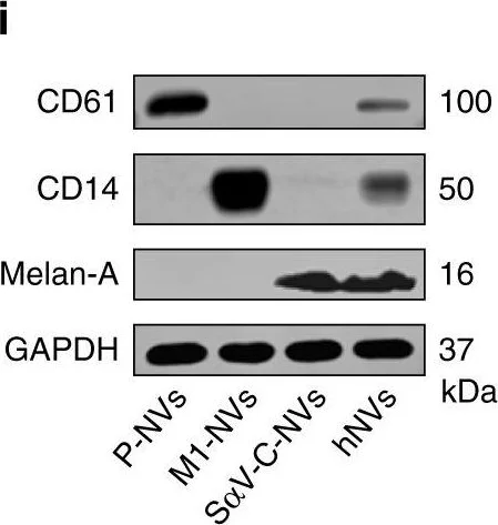
Western Blotting using Anti-MelanA antibody [EPR20380], ab210546. Publication image from Sun, W. et al., 2020, Nat Commun, 32999291. Legend direct from paper.
Schematic and characterization of hNVs.a Schematic showing the hNVs consist of engineered SαV-C-NVs, M1-NVs, and P-NVs. b Schematic showing the hNVs efficiently interact with CTCs in the blood, accumulate in the post-surgical tumor bed, repolarize TAMs towards M1 phenotype, and block the CD47-SIRPα ‘don’t eat me’ pathway, thus promoting macrophage phagocytosis of cancer cells, as well as boosting antitumor Tcell immunity. c, d Immunofluorescence imaging c and flow cytometry analysis d of SαV expression on original and engineered B16F10 cells. Scale bar, 10 µm. e Relative mRNA expression of Cd86, Tnf, Il6, and Inos in M0 cells, M1 cells, M0-NVs and M1-NVs. f Hydrodynamic size and zeta potential of P-NVs, M1-NVs, SαV-C-NVs, and hNVs measured by DLS. g TEM images of hNVs. Scale bar, 100 nm and 50 nm in the left and right panel, respectively. The samples were negatively stained with uranyl acetate. h Immunofluorescence images of hNVs. Scale bar, 10 µm. i Western blot analysis of specific protein CD61, CD14, and Melan-A in the samples of P-NVs, M1-NVs, SαV-C-NVs, and hNVs. All data are presented as mean ± S.D. (n = 3). Statistical significance was calculated via unpaired two-tailed t test. **P < 0.01; ***P < 0.001.
false
関連する標識済み抗体及び組成の異なる製品 (4)
-
Anti-MelanA antibody [EPR20380] - BSA and Azide free
-
660 APC
APC Anti-MelanA antibody [EPR20380]
-
665 Alexa Fluor® 647
Alexa Fluor® 647 Anti-MelanA antibody [EPR20380]
-
578 PE
PE Anti-MelanA antibody [EPR20380]
代替製品


Primary Antibodies
AB51061
Anti-MelanA antibody [EP1422Y]
20ul selling size|BOND RX™ Validated|RabMAb|Recombinant

- 5
- 1


Primary Antibodies
AB170090
Anti-MelanA antibody [EP1422Y] - Low endotoxin, Azide free
BOND RX™ Validated|RabMAb|Recombinant

- 0
- 0
Primary Antibodies
AB271839
Anti-MelanA antibody [EP1422Y] - BSA and Azide free
BOND RX™ Validated|RabMAb|Recombinant

- 5
- 2
組合せ製品

Primary Antibodies
AB137078
Anti-Melanoma gp100 antibody [EP4863(2)]
20ul selling size|BOND RX™ Validated|RabMAb|Recombinant

- 5
- 14

Primary Antibodies
AB303530
Anti-MiTF antibody [EPR26363-10]
BOND RX™ Validated|20ul selling size|RabMAb|Recombinant

- 4
- 1
Primary Antibodies
AB124964
Anti-alpha smooth muscle Actin antibody [EPR5368]
BOND RX™ Validated|RabMAb|Recombinant|KO Validated|20ul selling size

- 5
- 19
Reactivity data
製品の詳細
What is this antibody validated in?
Anti-MelanA antibody [EPR20380] (ab210546) is a rabbit recombinant monoclonal antibody and is validated for use in Western Blot (WB), Flow Cytometry (Intra), Immunohistochemistry (IHC-P), Immunocytochemistry/immunofluorescence (ICC/IF) in Human, Mouse samples.
What is the molecular weight of MelanA?
Anti-MelanA [EPR20380] (ab210546) specifically detects a band for MelanA (UniProt: Q16655) at a molecular weight of 13kDa.
Trusted by the scientific community
Anti-MelanA [EPR20380] (ab210546) was first used in a scientific publication in 2016 and has been cited over 20 times in peer-reviewed journals.
Reviewed by scientists
Anti-MelanA [EPR20380] (ab210546) has over 15 independent reviews from customers.
Other related products
We have a range of other formats of antibody clone [EPR20380] also available for your convenience: ab210546, Carrier free - ab222483, APC - ab225498, PE - ab225499, Alexa Fluor® 647 - ab225500
Patented technology
Our RabMAb® technology is a patented hybridoma-based technology for making rabbit monoclonal antibodies. For details on our patents, please refer to RabMAb® patents.
What are the advantages of a recombinant monoclonal antibody?
This product is a recombinant monoclonal antibody, which offers several advantages including:
- - High batch-to-batch consistency and reproducibility
- - Improved sensitivity and specificity
- - Long-term security of supply
- - Animal-free batch production
For more information, read more on recombinant antibodies.
出荷温度及び保存条件
製品の状態
精製方法
バッファー組成
出荷温度
短期保存期間
短期保存温度
長期保存温度
分注に関する情報
保管に関する情報
製品プロトコール
- Visit the General protocols
- Visit the Troubleshooting
ターゲットの情報
文献 (34)
Recent publications for all applications. Explore the full list and refine your search
Nature communications 16:8822 PubMed41044182
2025
Applications
Unspecified application
Species
Unspecified reactive species
NAR cancer 7:zcaf026 PubMed40918647
2025
Applications
Unspecified application
Species
Unspecified reactive species
Journal of cosmetic dermatology 24:e70155 PubMed40162532
2025
Applications
Unspecified application
Species
Unspecified reactive species
Frontiers in bioengineering and biotechnology 12:1437817 PubMed39493304
2024
Applications
Unspecified application
Species
Unspecified reactive species
Investigative ophthalmology & visual science 65:7 PubMed38833258
2024
Applications
Unspecified application
Species
Unspecified reactive species
Oncology reports 51: PubMed38551165
2024
Applications
Unspecified application
Species
Unspecified reactive species
Scientific reports 13:15508 PubMed37726363
2023
Applications
Unspecified application
Species
Unspecified reactive species
Nature medicine 29:1123-1134 PubMed37106167
2023
Applications
Unspecified application
Species
Unspecified reactive species
International journal of molecular sciences 24: PubMed37175529
2023
Applications
Unspecified application
Species
Unspecified reactive species
The Journal of pathology 260:84-96 PubMed36852981
2023
Applications
Unspecified application
Species
Unspecified reactive species
Abcam product promise
Please note: All products are 'FOR RESEARCH USE ONLY. NOT FOR USE IN DIAGNOSTIC OR THERAPEUTIC PROCEDURES'.
For licensing inquiries, please contact partnerships@abcam.com