Anti-MAT1A + MAT2A 抗体 [EPR19048] - BSA and Azide free
Anti-MAT1A + MAT2A antibody [EPR19048] - BSA and Azide free
- RabMAb
- Recombinant
- 詳細を見る
Be the first to review this product! Submit a review
|
(0 Publication)
Rabbit Recombinant Monoclonal MAT2A antibody. Carrier free. Suitable for IP, WB and reacts with Human, Mouse, Rat, Recombinant full length protein - Human samples.
別名を表示する
AMS2, MATA2, MAT2A, S-adenosylmethionine synthase isoform type-2, AdoMet synthase 2, Methionine adenosyltransferase 2, Methionine adenosyltransferase II, MAT 2, MAT-II, AMS1, MATA1, MAT1A, S-adenosylmethionine synthase isoform type-1, AdoMet synthase 1, Methionine adenosyltransferase 1, Methionine adenosyltransferase I/III, MAT 1, MAT-I/III
- IP
Supplier Data
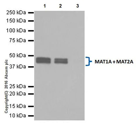
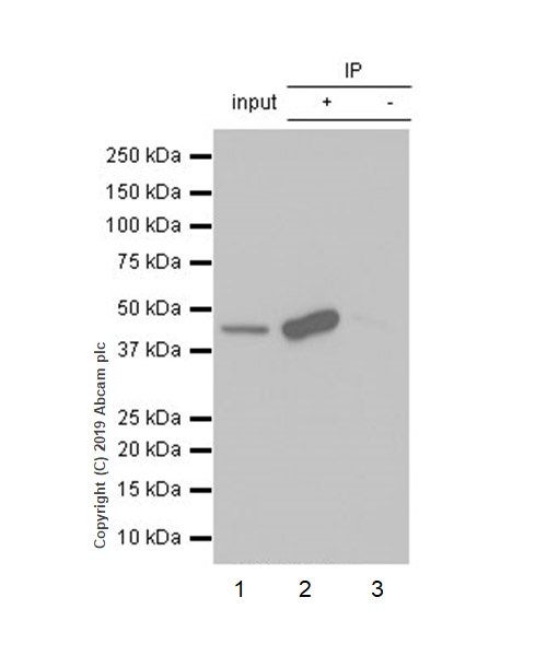
Immunoprecipitation - Anti-MAT1A + MAT2A antibody [EPR19048] - BSA and Azide free (AB251153)
This data was developed using ab192846, the same antibody clone in a different buffer formulation.
MAT2A was immunoprecipitated from 0.35 mg of Ramos (human Burkitt's lymphoma cell line) whole cell lysate with ab192846 at 1/30 dilution. Western blot was performed from the immunoprecipitate using ab192846 at 1/1000 dilution. VeriBlot for IP Detection Reagent (HRP) (ab131366), was used for detection at 1/10000 dilution.
Lane 1 : Ramos whole cell lysate 10 μg (Input).
Lane 2 : ab192846 IP in Ramos whole cell lysate.
Lane 3 : Rabbit monoclonal IgG (ab172730) instead of ab192846 in Ramos whole cell lysate.
Blocking and dilution buffer : 5% NFDM/TBST.
Exposure time : 1 second.
All lanes:
Immunoprecipitation - Anti-MAT1A + MAT2A antibody [EPR19048] (<a href='/products/primary-antibodies/mat1a-mat2a-antibody-epr19048-ab192846'>ab192846</a>)
Observed band size: 44 kDa
false
- WB
Supplier Data
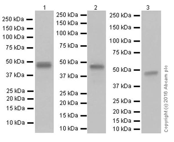
Western blot - Anti-MAT1A + MAT2A antibody [EPR19048] - BSA and Azide free (AB251153)
This data was developed using ab192846, the same antibody clone in a different buffer formulation.
Blocking and dilution buffer : 5% NFDM/TBST.
Lane 1:
Western blot - Anti-MAT1A + MAT2A antibody [EPR19048] (<a href='/products/primary-antibodies/mat1a-mat2a-antibody-epr19048-ab192846'>ab192846</a>) at 1/1000 dilution
Lanes 2 - 3:
Western blot - Anti-MAT1A + MAT2A antibody [EPR19048] (<a href='/products/primary-antibodies/mat1a-mat2a-antibody-epr19048-ab192846'>ab192846</a>) at 1/5000 dilution
Lane 1:
Human fetal liver tissue lysate at 10 µg
Lane 2:
Mouse liver tissue lysate at 10 µg
Lane 3:
Rat liver tissue lysate at 10 µg
Secondary
Lane 1:
Goat Anti-Rabbit IgG Peroxidase Conjugate, specific to the non-reduced form of IgG at 1/10000 dilution
Lanes 2 - 3:
Western blot - Goat Anti-Rabbit IgG H&L (HRP) (<a href='/products/secondary-antibodies/goat-rabbit-igg-h-l-hrp-ab97051'>ab97051</a>) at 1/100000 dilution
Predicted band size: 44 kDa
Observed band size: 44 kDa
true
Exposure time: 1s
- WB
Supplier Data
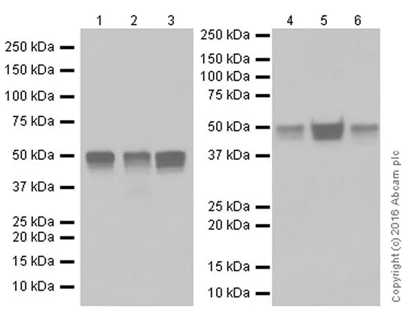
Western blot - Anti-MAT1A + MAT2A antibody [EPR19048] - BSA and Azide free (AB251153)
This data was developed using ab192846, the same antibody clone in a different buffer formulation.
Blocking and dilution buffer : 5% NFDM/TBST.
Lanes 1 - 3:
Western blot - Anti-MAT1A + MAT2A antibody [EPR19048] (<a href='/products/primary-antibodies/mat1a-mat2a-antibody-epr19048-ab192846'>ab192846</a>) at 1/10000 dilution
Lanes 4 - 6:
Western blot - Anti-MAT1A + MAT2A antibody [EPR19048] (<a href='/products/primary-antibodies/mat1a-mat2a-antibody-epr19048-ab192846'>ab192846</a>) at 1/1000 dilution
Lane 1:
Ramos (human Burkitt's lymphoma cell line) whole cell lysate at 10 µg
Lane 2:
U937 (human histiocytic lymphoma cell line) whole cell lysate at 10 µg
Lane 3:
HEK-293T (human epithelial cell line from embryonic kidney transformed with large T antigen) whole cell lysate at 10 µg
Lane 4:
Human fetal heart tissue lysate at 10 µg
Lane 5:
Human fetal kidney tissue lysate at 10 µg
Lane 6:
Human fetal spleen tissue lysate at 10 µg
Secondary
Lanes 1 - 3:
Western blot - Goat Anti-Rabbit IgG H&L (HRP) (<a href='/products/secondary-antibodies/goat-rabbit-igg-h-l-hrp-ab97051'>ab97051</a>) at 1/100000 dilution
Lanes 4 - 6:
Goat Anti-Rabbit IgG Peroxidase Conjugate, specific to the non-reduced form of IgG at 1/10000 dilution
Predicted band size: 44 kDa
Observed band size: 44 kDa
true
Exposure time: 3s
- WB
Supplier Data
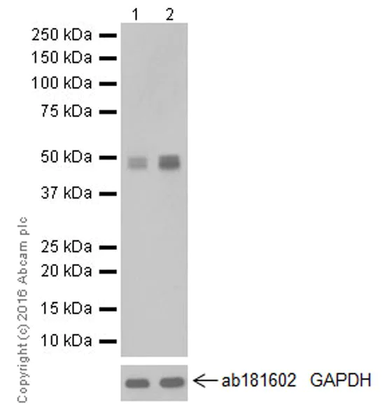
Western blot - Anti-MAT1A + MAT2A antibody [EPR19048] - BSA and Azide free (AB251153)
This data was developed using ab192846, the same antibody clone in a different buffer formulation.
Blocking and dilution buffer : 5% NFDM/TBST.
The expression profile observed is consistent with what has been described in the literature (PMID : 18695670 and 22879628).
All lanes:
Western blot - Anti-MAT1A + MAT2A antibody [EPR19048] (<a href='/products/primary-antibodies/mat1a-mat2a-antibody-epr19048-ab192846'>ab192846</a>) at 1/20000 dilution
Lane 1:
Untreated HepG2 (human liver hepatocellular carcinoma cell line) whole cell lysate at 10 µg
Lane 2:
HepG2 (human liver hepatocellular carcinoma cell line) treated with 500 ng/ml Lipopolysaccharides for 4 hours, whole cell lysate at 10 µg
Secondary
All lanes:
Western blot - Goat Anti-Rabbit IgG H&L (HRP) (<a href='/products/secondary-antibodies/goat-rabbit-igg-h-l-hrp-ab97051'>ab97051</a>) at 1/100000 dilution
Predicted band size: 44 kDa
Observed band size: 44 kDa
true
Exposure time: 5s
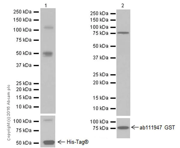
- WB
Supplier Data
Western blot - Anti-MAT1A + MAT2A antibody [EPR19048] - BSA and Azide free (AB251153)
This data was developed using ab192846, the same antibody clone in a different buffer formulation.
Blocking and dilution buffer : 5% NFDM/TBST.
Exposure times : Lane 1 : 1 second; Lane 2 : 2 seconds.
Human MAT1A recombinant protein contains aa1-395 with a His-Tag®. This recombinant protein was made in-house.
All lanes:
Western blot - Anti-MAT1A + MAT2A antibody [EPR19048] (<a href='/products/primary-antibodies/mat1a-mat2a-antibody-epr19048-ab192846'>ab192846</a>) at 1/20000 dilution
Lane 1:
Human MAT1A recombinant protein at 0.01 µg
Lane 2:
Human MAT2A recombinant protein at 0.01 µg
Secondary
All lanes:
Western blot - Goat Anti-Rabbit IgG H&L (HRP) (<a href='/products/secondary-antibodies/goat-rabbit-igg-h-l-hrp-ab97051'>ab97051</a>) at 1/100000 dilution
Predicted band size: 44 kDa
Observed band size: 44 kDa,75 kDa
true
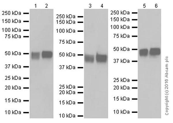
- WB
Supplier Data
Western blot - Anti-MAT1A + MAT2A antibody [EPR19048] - BSA and Azide free (AB251153)
This data was developed using ab192846, the same antibody clone in a different buffer formulation.
Blocking and dilution buffer : 5% NFDM/TBST.
Exposure times : Lanes 1-2 : 5 seconds; Lanes 3-6 : 3 seconds.
All lanes:
Western blot - Anti-MAT1A + MAT2A antibody [EPR19048] (<a href='/products/primary-antibodies/mat1a-mat2a-antibody-epr19048-ab192846'>ab192846</a>) at 1/1000 dilution
Lane 1:
Mouse kidney tissue lysate at 10 µg
Lane 2:
Mouse spleen tissue lysate at 10 µg
Lane 3:
Rat brain tissue lysate at 10 µg
Lane 4:
Rat kidney tissue lysate at 10 µg
Lane 5:
C6 (rat glial tumor cell line) whole cell lysate at 10 µg
Lane 6:
NIH/3T3 (Mouse embryonic fibroblast cell line) whole cell lysate at 10 µg
Secondary
All lanes:
Western blot - Goat Anti-Rabbit IgG H&L (HRP) (<a href='/products/secondary-antibodies/goat-rabbit-igg-h-l-hrp-ab97051'>ab97051</a>) at 1/100000 dilution
Predicted band size: 44 kDa
Observed band size: 44 kDa
true
関連する標識済み抗体及び組成の異なる製品 (1)
-
Anti-MAT1A + MAT2A antibody [EPR19048]
組合せ製品
Primary Antibodies
AB129176
Anti-MAT1A + MAT2A antibody [EPR7938]
20ul selling size|RabMAb|Recombinant
- 0
- 0
Primary Antibodies
AB215036
20ul selling size|RabMAb|Recombinant|Advanced Validation
- 5
- 5
Primary Antibodies
AB179434
Anti-Ubiquitin (linkage-specific K63) antibody [EPR8590-448]
20ul selling size|RabMAb|Recombinant

- 5
- 6
Reactivity data
製品の詳細
ab251153 is the carrier-free version of ab192846.
Patented technology
Our RabMAb® technology is a patented hybridoma-based technology for making rabbit monoclonal antibodies. For details on our patents, please refer to RabMAb® patents.
What are the advantages of a recombinant monoclonal antibody?
This product is a recombinant monoclonal antibody, which offers several advantages including:
- - High batch-to-batch consistency and reproducibility
- - Improved sensitivity and specificity
- - Long-term security of supply
- - Animal-free batch production
For more information, read more on recombinant antibodies.
Conjugation ready
Our carrier-free antibodies are typically supplied in a PBS-only formulation, purified and free of BSA, sodium azide and glycerol. This conjugation-ready format is designed for use with fluorochromes, metal isotopes, oligonucleotides, and enzymes, which makes them ideal for antibody labelling, functional and cell-based assays, flow-based assays (e.g. mass cytometry) and Multiplex Imaging applications.
Use our conjugation kits for antibody conjugates that are ready-to-use in as little as 20 minutes with 1 minute hands-on-time and 100% antibody recovery: available for fluorescent dyes, HRP, biotin and gold.
Compatibility
This product is compatible with the Maxpar® Antibody Labeling Kit from Fluidigm, without the need for antibody preparation. Maxpar® is a trademark of Fluidigm Canada Inc.
出荷温度及び保存条件
製品の状態
バッファー組成
出荷温度
短期保存温度
長期保存温度
保管に関する情報
補足情報
This supplementary information is collated from multiple sources and compiled automatically.
Biological function summary
Methionine adenosyltransferase 1A and 2A function to catalyze the transfer of an adenosyl group from ATP to methionine forming SAMe. This reaction is part of a critical metabolic process acting as a source of methyl groups for methylation reactions. MAT1A commonly works as part of a homotetrameric complex while MAT2A can form both dimers and tetramers. This flexibility in complex formation helps regulate the production of SAMe based on cellular demands linking it to vital biomolecules synthesis.
Pathways
Methionine salvage pathways and polyamine biosynthesis include the significant involvement of MAT1A and MAT2A. These enzymes provide the necessary SAMe for the transmethylation and transsulfuration pathways connecting to other proteins such as GNMT and SAH hydrolase. The enzymes' activity influences the levels of important intermediates impacting these pathways and resulting metabolic outputs.
製品プロトコール
- Visit the General protocols
- Visit the Troubleshooting
ターゲットの情報
追加のターゲット
Abcam product promise
Please note: All products are 'FOR RESEARCH USE ONLY. NOT FOR USE IN DIAGNOSTIC OR THERAPEUTIC PROCEDURES'.
For licensing inquiries, please contact partnerships@abcam.com