Anti-LAMP1 抗体 [EPR24395-31]
Anti-LAMP1 antibody [EPR24395-31] - Lysosome Marker
- BOND RX™ Validated
- RabMAb
- KO Validated
- Recombinant
- 20ul selling size
- 詳細を見る
Be the first to review this product! Submit a review
|
(7 Publications)
Anti-LAMP1 antibody [EPR24395-31] (ab278043) is a rabbit monoclonal antibody detecting LAMP1 in Western Blot, Flow Cytometry (Intra), IHC-P, ICC/IF. Suitable for Human.
- KO validated for confirmed specificity
- Biophysical QC for unrivalled batch-batch consistency
別名を表示する
CD107a, Lysosome-associated membrane glycoprotein 1, LAMP-1, Lysosome-associated membrane protein 1, CD107 antigen-like family member A, LAMP1

- ICC/IF
Lab
Immunocytochemistry/ Immunofluorescence - Anti-LAMP1 antibody [EPR24395-31] - Lysosome Marker (AB278043)
Immunofluorescent analysis of 4% Paraformaldehyde-fixed, 0.1% TritonX-100 permeabilized HeLa cells labelling LAMP1 with ab278043 at 1/100 (5.45 ug/ml) dilution, followed by ab150077 Goat Anti-Rabbit IgG H&L (Alexa Fluor® 488) antibody at 1/1000 (green). Confocal image showing cytoplasmic staining in HeLa. ab25630 Anti-LAMP1 mouse monoclonal antibody was used to counterstain at 1/500 (red). The Nuclear counterstain was DAPI (blue).
Secondary antibody only control : Secondary antibody is ab150077 Goat Anti-Rabbit IgG H&L (Alexa Fluor® 488) at 1/1000 dilution

- IHC-P
Lab
Immunohistochemistry (Formalin/PFA-fixed paraffin-embedded sections) - Anti-LAMP1 antibody [EPR24395-31] - Lysosome Marker (AB278043)
Immunohistochemical analysis of paraffin-embedded Human lung cancer tissue labelling LAMP1 with ab278043 at 1/5000 (0.109 ug/ml) dilution followed by a ready to use LeicaDS9800 (Bond® Polymer Refine Detection) was used. Cytoplasmic staining in human lung cancer. The section was incubated with ab278043 for 30 mins at room temperature. The immunostaining was performed on a Leica Biosystems BOND® RX instrument with Hematoxylin. Secondary antibody only control : Secondary antibody is a ready to use LeicaDS9800 (Bond® Polymer Refine Detection) was used.
Heat mediated antigen retrieval with Tris-EDTA buffer (pH 9.0, epitope retrieval solution2) for 20 mins

- IHC-P
Lab
Immunohistochemistry (Formalin/PFA-fixed paraffin-embedded sections) - Anti-LAMP1 antibody [EPR24395-31] - Lysosome Marker (AB278043)
Immunohistochemical analysis of paraffin-embedded Human liver tissue labeling LAMP1 with ab278043 at 1/5000 (0.109 ug/ml) dilution followed by a ready to use LeicaDS9800 (Bond® Polymer Refine Detection) was used. Cytoplasmic staining in human liver. The section was incubated with ab278043 for 30 mins at room temperature. The immunostaining was performed on a Leica Biosystems BOND® RX instrument with Hematoxylin. Secondary antibody only control : Secondary antibody is a ready to use LeicaDS9800 (Bond® Polymer Refine Detection) was used.
Heat mediated antigen retrieval with Tris-EDTA buffer (pH 9.0, epitope retrieval solution2) for 20 mins

- IHC-P
Lab
Immunohistochemistry (Formalin/PFA-fixed paraffin-embedded sections) - Anti-LAMP1 antibody [EPR24395-31] - Lysosome Marker (AB278043)
Immunohistochemical analysis of paraffin-embedded Human pancreas tissue labelling LAMP1 with ab278043 at 1/5000 (0.109 ug/ml) dilution followed by a ready to use LeicaDS9800 (Bond® Polymer Refine Detection) was used. Cytoplasmic staining in human pancreas. The section was incubated with ab278043 for 30 mins at room temperature. The immunostaining was performed on a Leica Biosystems BOND® RX instrument with Hematoxylin. Secondary antibody only control : Secondary antibody is a ready to use LeicaDS9800 (Bond® Polymer Refine Detection) was used.
Heat mediated antigen retrieval with Tris-EDTA buffer (pH 9.0, epitope retrieval solution2) for 20 mins

- IHC-P
Lab
Immunohistochemistry (Formalin/PFA-fixed paraffin-embedded sections) - Anti-LAMP1 antibody [EPR24395-31] - Lysosome Marker (AB278043)
Immunohistochemical analysis of paraffin-embedded Human tonsil labelling LAMP1 with ab278043 at 1/5000 (0.109 ug/ml) dilution followed by a ready to use LeicaDS9800 (Bond® Polymer Refine Detection) was used. Cytoplasmic staining in human tonsil. The section was incubated with ab278043 for 30 mins at room temperature. The immunostaining was performed on a Leica Biosystems BOND® RX instrument with Hematoxylin. Secondary antibody only control : Secondary antibody is a ready to use LeicaDS9800 (Bond® Polymer Refine Detection) was used.
Heat mediated antigen retrieval with Tris-EDTA buffer (pH 9.0, epitope retrieval solution2) for 20 mins.

- Flow Cyt (Intra)
Lab
Flow Cytometry (Intracellular) - Anti-LAMP1 antibody [EPR24395-31] - Lysosome Marker (AB278043)
Intracellular flow cytometric analysis of HeLa (human cervix adenocarcinoma epithelial cell) cells labelling LAMP1 with ab278043 at 1/500 dilution (red) compared with a Rabbit monoclonal IgG (ab172730) (Black) isotype control and an unlabelled control (cells without incubation with primary antibody and secondary antibody) (Blue). Goat anti rabbit IgG (Alexa Fluor® 488, ab150077) at 1/2000 dilution was used as the secondary antibody.

- mIHC
Supplier Data
Multiplex immunohistochemistry - Anti-LAMP1 antibody [EPR24395-31] - Lysosome Marker (AB278043)
Multiplex immunohistochemistry analysis of formalin/PFA-fixed paraffin-embedded human thyroid tissue staining FUCA1 with ab323794 at a 1 : 1000 (0.511 ug/ml) dilution, ab278043 anti-LAMP1 used at 1 : 5000 (0.103 ug/ml) dilution.
Panel A : merged staining of anti-FUCA1 (green; Opal™520), anti-LAMP1 (magenta; Opal™570) on human thyroid.
Panel B : anti-FUCA1 showed granular staining in human thyroid.
Panel C : ant-LAMP1 staining lysosome in human thyroid.
Nuclear DNA was labeled with DAPI (shown in blue).
The section was incubated in three rounds of staining : in the order of ab323794 and ab278043 for 30 mins at room temperature. Each round was followed by a separate fluorescent tyramide signal amplification system.
The immunostaining was performed on a Leica Biosystems BOND® RX instrument with an Opal™ 4-color kit. Image acquisition was performed with Leica SP8 confocal microscope.
Nuclear counter stain with DAPI.
Heat mediated antigen retrieval with Tris-EDTA buffer (pH 9.0, epitope retrieval solution2) for 20 mins.
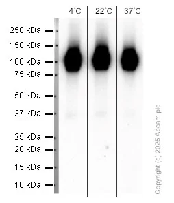
- WB
Lab
Western blot - Anti-LAMP1 antibody [EPR24395-31] - Lysosome Marker (AB278043)
This blot was produced using 4-20% SDS-PAGE containing 15 μg of HeLa whole cell lysate per lane at 150V for 1hr before being transferred onto a 0.45 μm PVDF membrane at 75V for 1hr. The membrane was then blocked for 1hr using 5% NFDM/TBST, then incubated with ab278043 (1/2000) at room temperature for 1hr. After being washed three times in TBST, the membrane was incubated with Peroxidase conjugated goat anti-rabbit IgG (H+L) (ab97051) at 1/50,000 dilution for 1hr at room temperature. The membrane was washed three times again. Then the signal was developed using the ECL technique.
ab278043 was stored at a range of temperatures (+4°C, +22°C, +37°C) for 1 week before being tested in WB. The image shows the band intensity remains relatively constant across all storage temperatures, demonstrating that antibody activity is not affected under these conditions. This data was generated as part of a structured antibody stability study supporting Abcam’s transition from cold‑chain to ambient shipping.
All lanes:
Western blot - Anti-LAMP1 antibody [EPR24395-31] - Lysosome Marker (ab278043) at 1/2000 dilution
All lanes:
HeLa whole cell lysate at 15 µg with NDFM/TBST
Secondary
All lanes:
Western blot - Goat Anti-Rabbit IgG H&L (HRP) (<a href='/products/secondary-antibodies/goat-rabbit-igg-h-l-hrp-ab97051'>ab97051</a>) at 1/50000 dilution
false
Exposure time: 40s

- WB
Lab
Western blot - Anti-LAMP1 antibody [EPR24395-31] - Lysosome Marker (AB278043)
Blocking and Diluting buffer : 5% NFDM/TBST
LAMP1 is a glycoprotein. The molecular weight observed is consistent with what has been described in the literature (PMID : 27061067, 15111122).
Exposure time : 3 minutes
All lanes:
Western blot - Anti-LAMP1 antibody [EPR24395-31] - Lysosome Marker (ab278043) at 1/1000 dilution
Lane 1:
HeLa (human cervix adenocarcinoma epithelial cell) whole cell lysate at 20 µg
Lane 2:
HepG2 (human hepatocellular carcinoma epithelial cell) whole cell lysate at 20 µg
Lane 3:
MCF7 (human breast adenocarcinoma epithelial cell) whole cell lysate at 20 µg
Secondary
All lanes:
Western blot - Goat Anti-Rabbit IgG H&L (HRP) (<a href='/products/secondary-antibodies/goat-rabbit-igg-h-l-hrp-ab97051'>ab97051</a>) at 1/50000 dilution
Predicted band size: 45 kDa
Observed band size: 110 kDa
false
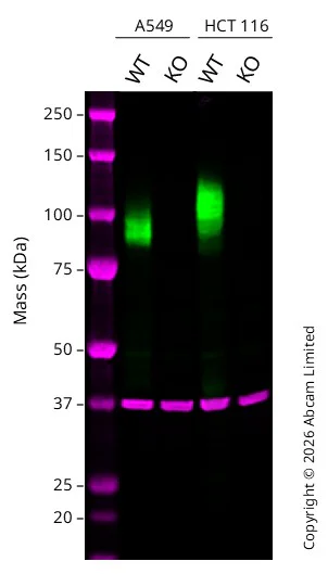
- WB
Lab
Western blot - Anti-LAMP1 antibody [EPR24395-31] - Lysosome Marker (AB278043)
Western blot : Rabbit monoclonal [EPR24395-31] to LAMP1 ab278043 staining at 1/1000 dilution, shown in green; Mouse anti GAPDH ab8245 loading control staining at 1/20000 dilution, shown in magenta. A band was observed at 80-120 kDa in Wild-type A549 and HCT 116 cell lysates with no signal observed at this size in LAMP1 knockout A549 cell line and LAMP1 knockout HCT 116 cell line. To generate this image, samples were run on an SDS-PAGE gel then transferred onto a nitrocellulose membrane. Membranes were blocked in 3 % Milk in TBS-0.1 % Tween® 20 (TBS-T) before incubation with primary antibodies overnight at 4 °C. Blots were washed four times in TBS-T, incubated with secondary antibodies for 1 h at room temperature, washed again four times then imaged. Secondary antibodies used were Goat anti-Rabbit 800CW and Goat anti-Mouse 680RD at 1/20,000 dilution.
Lanes 1 - 4:
Western blot - Anti-LAMP1 antibody [EPR24395-31] - BSA and Azide free (<a href='/products/primary-antibodies/lamp1-antibody-epr24395-31-bsa-and-azide-free-ab278046'>ab278046</a>) at 1/1000 dilution
Lanes 1 - 4:
Western blot - Anti-LAMP1 antibody [EPR24395-31] - Lysosome Marker (ab278043) at 1/1000 dilution
Lane 1:
Wild-type A549 cell lysates at 20 µg
Lane 2:
Western blot - Human LAMP1 knockout A549 cell line (<a href='/products/cell-lines/human-lamp1-knockout-a549-cell-line-ab288919'>ab288919</a>) at 20 µg
Lane 3:
Wild-type HCT 116 ab277289 cell lysates at 20 µg
Lane 4:
Western blot - Human LAMP1 knockout HCT116 cell line (<a href='/products/cell-lines/human-lamp1-knockout-hct116-cell-line-ab289151'>ab289151</a>) at 20 µg
Secondary
All lanes:
Goat anti-Rabbit 800CW & Goat anti-Mouse 680RD at 1/20000 dilution
Predicted band size: 45 kDa
Observed band size: 80-120 kDa
false

- WB
Lab
Western blot - Anti-LAMP1 antibody [EPR24395-31] - Lysosome Marker (AB278043)
Blocking and Diluting buffer : 5% NFDM/TBST
LAMP1 is a glycoprotein. The molecular weight observed is consistent with what has been described in the literature (PMID : 27061067, 15111122).
Exposure time : 3 minutes
All lanes:
Western blot - Anti-LAMP1 antibody [EPR24395-31] - Lysosome Marker (ab278043) at 1/1000 dilution
Lane 1:
Human kidney tissue lysate at 20 µg
Lane 2:
Human spleen tissue lysate at 20 µg
Lane 3:
Human colon cancer tissue lysate at 20 µg
Secondary
All lanes:
Western blot - VeriBlot for IP Detection Reagent (HRP) (<a href='/products/reagents/veriblot-for-ip-detection-reagent-hrp-ab131366'>ab131366</a>) at 1/1000 dilution
Predicted band size: 45 kDa
Observed band size: 110 kDa
false

- WB
Lab
Western blot - Anti-LAMP1 antibody [EPR24395-31] - Lysosome Marker (AB278043)
Western blot : Anti-LAMP1 antibody [EPR24395-31] staining at 1/1000 dilution, shown in green; Mouse anti-GAPDH antibody [6C5] (ab8245) loading control staining at 1/20000 dilution, shown in red. In Western blot, ab278043 was shown to bind specifically to LAMP1. A band was observed at 100-120 kDa in wild-type HCT 116 cell lysates with no signal observed at this size in LAMP1 knockout cell line. To generate this image, wild-type and LAMP1 knockout HCT 116 cell lysates were analysed. First, samples were run on an SDS-PAGE gel then transferred onto a nitrocellulose membrane. Membranes were blocked in 3% milk in TBS-0.1% Tween® 20 (TBS-T) before incubation with primary antibodies overnight at 4°C. Blots were washed four times in TBS-T, incubated with secondary antibodies for 1 h at room temperature, washed again four times then imaged. Secondary antibodies used were Goat anti-Rabbit IgG H&L 800CW and Goat anti-Mouse IgG H&L 680RD at 1/20000 dilution.
All lanes:
Western blot - Anti-LAMP1 antibody [EPR24395-31] - Lysosome Marker (ab278043) at 1/1000 dilution
Lane 1:
Wild-type HCT 116 cell lysate at 20 µg
Lane 2:
LAMP1 knockout HCT 116 cell lysate at 20 µg
Lane 3:
HeLa cell lysate at 20 µg
Lane 4:
MCF7 cell lysate at 20 µg
Observed band size: 100-120 kDa
false
関連する標識済み抗体及び組成の異なる製品 (7)
-
Anti-LAMP1 antibody [EPR24395-31] - BSA and Azide free
-
660 APC
APC Anti-LAMP1 antibody [EPR24395-31] - Lysosome Marker
-
421 Alexa Fluor® 405
Donkey Anti-Rabbit IgG H&L (Alexa Fluor® 405)
-
421 Alexa Fluor® 405
Donkey Anti-Rabbit IgG H&L (Alexa Fluor® 405) preadsorbed
-
519 Alexa Fluor® 488
Alexa Fluor® 488 Anti-LAMP1 antibody [EPR24395-31] - Lysosome Marker
-
665 Alexa Fluor® 647
Alexa Fluor® 647 Anti-LAMP1 antibody [EPR24395-31] - Lysosome Marker
-
578 PE
PE Anti-LAMP1 antibody [EPR24395-31] - Lysosome Marker
組合せ製品

Primary Antibodies
AB192890
Anti-LC3B antibody [EPR18709] - Autophagosome Marker
BOND RX™ Validated|RabMAb|Recombinant|KO Validated|Lab Essentials|20ul selling size

- 5
- 19

Primary Antibodies
AB183734
RabMAb|Recombinant|Lab Essentials|20ul selling size

- 5
- 24
Reactivity data
製品の詳細
What is this antibody validated in?
Anti-LAMP1 antibody [EPR24395-31] (ab278043) is a rabbit recombinant monoclonal antibody and is validated for use in Western Blot (WB), Flow Cytometry (Intra), Immunohistochemistry (IHC-P), Immunocytochemistry/immunofluorescence (ICC/IF) in Human samples.
What is the molecular weight of LAMP1?
Anti-LAMP1 [EPR24395-31] (ab278043) specifically detects a band for LAMP1 (UniProt: P11279) at a molecular weight of 45kDa.
Specificity confirmed
The specificity of Anti-LAMP1 antibody [EPR24395-31] (ab278043) has been confirmed by Flow Cytometry (Intracellular) testing in LAMP1 Knockout HAP1 cells.
Patented technology
Our RabMAb® technology is a patented hybridoma-based technology for making rabbit monoclonal antibodies. For details on our patents, please refer to RabMAb® patents.
What are the advantages of a recombinant monoclonal antibody?
This product is a recombinant monoclonal antibody, which offers several advantages including:
- - High batch-to-batch consistency and reproducibility
- - Improved sensitivity and specificity
- - Long-term security of supply
- - Animal-free batch production
For more information, read more on recombinant antibodies.
Trial sizes available!
Test your antibody or perform pre-screening before committing to a larger quantity. Sold in 20µl. Discover our selection of trial-size antibodies.
出荷温度及び保存条件
製品の状態
精製方法
バッファー組成
出荷温度
短期保存期間
短期保存温度
長期保存温度
分注に関する情報
保管に関する情報
補足情報
This supplementary information is collated from multiple sources and compiled automatically.
Biological function summary
LAMP1 functions to protect lysosomal membranes from the harsh environment inside the lysosome. It forms part of a protective glycocalyx composed of highly glycosylated proteins supporting lysosomal stability. This protein interacts closely with other lysosome markers and assists in the fusion of vesicles with lysosomes making it fundamental to lysosomal processes. As a lysosome marker LAMP1 helps identify lysosomal compartments during studies involving microscopy and LAMP1 staining.
Pathways
The protein LAMP1 participates in the autophagy and endocytic pathways. It acts collaboratively within the lysosomal degradation route involving proteins like LAMP2 which also supports lysosomal function. LAMP1 influences these pathways by mediating the fusion of autophagosomes or endosomes with lysosomes thereby ensuring efficient breakdown of cellular debris and macromolecules.
製品プロトコール
- Visit the General protocols
- Visit the Troubleshooting
ターゲットの情報
文献 (7)
Recent publications for all applications. Explore the full list and refine your search
Acta biochimica et biophysica Sinica : PubMed40726277
2025
Applications
Unspecified application
Species
Unspecified reactive species
Journal of translational medicine 23:631 PubMed40474178
2025
Applications
Unspecified application
Species
Unspecified reactive species
Cell death discovery 11:243 PubMed40393958
2025
Applications
Unspecified application
Species
Unspecified reactive species
PLoS pathogens 21:e1013184 PubMed40367285
2025
Applications
Unspecified application
Species
Unspecified reactive species
NPJ Parkinson's disease 11:61 PubMed40155632
2025
Applications
Unspecified application
Species
Unspecified reactive species
The EMBO journal 44:1829-1865 PubMed39930267
2025
Applications
Unspecified application
Species
Unspecified reactive species
Cellular and molecular life sciences : CMLS 79:488 PubMed35984564
2022
Applications
Unspecified application
Species
Unspecified reactive species
Abcam product promise
Please note: All products are 'FOR RESEARCH USE ONLY. NOT FOR USE IN DIAGNOSTIC OR THERAPEUTIC PROCEDURES'.
For licensing inquiries, please contact partnerships@abcam.com