Anti-KIAA0319L 抗体 [EPR28329-62]
Anti-KIAA0319L antibody [EPR28329-62]
- KD Validated
- BOND RX™ Validated
- 20ul selling size
- RabMAb
- Recombinant
- KO Validated
- 詳細を見る
Be the first to review this product! Submit a review
|
(1 Publication)
Rabbit Recombinant Monoclonal KIAA0319L antibody. Suitable for WB, IHC-P and reacts with Human, Mouse, Rat samples. Cited in 1 publication.
別名を表示する
AAVR, KIAA1837, PP791, KIAA0319L, Dyslexia-associated protein KIAA0319-like protein, Adeno-associated virus receptor

- IHC-P
Supplier Data
Immunohistochemistry (Formalin/PFA-fixed paraffin-embedded sections) - Anti-KIAA0319L antibody [EPR28329-62] (AB315027)
Immunohistochemical analysis of paraffin-embedded Human cerebrum tissue labeling KIAA0319L with ab315027 at 1/500 (1.038 ug/ml) followed by a ready to use LeicaDS9800 (Bond™ Polymer Refine Detection). Positive staining on human cerebrum. The section was incubated with ab315027 for 30 mins at room temperature. The immunostaining was performed on a Leica Biosystems BOND® RX instrument. Counterstained with Hematoxylin.
Secondary antibody only control : Secondary antibody is a ready to use LeicaDS9800 (Bond™ Polymer Refine Detection).
Heat mediated antigen retrieval was performed with Tris-EDTA buffer (pH 9.0, Epitope Retrieval Solution2) for 20 mins.

- IHC-P
Supplier Data
Immunohistochemistry (Formalin/PFA-fixed paraffin-embedded sections) - Anti-KIAA0319L antibody [EPR28329-62] (AB315027)
Immunohistochemical analysis of paraffin-embedded Mouse cerebrum tissue labeling KIAA0319L with ab315027 at 1/2000 (0.26 ug/ml) followed by a ready to use LeicaDS9800 (Bond™ Polymer Refine Detection). Positive staining on mouse cerebrum. The section was incubated with ab315027 for 30 mins at room temperature. The immunostaining was performed on a Leica Biosystems BOND® RX instrument. Counterstained with Hematoxylin.
Secondary antibody only control : Secondary antibody is a ready to use LeicaDS9800 (Bond™ Polymer Refine Detection).
Heat mediated antigen retrieval was performed with Tris-EDTA buffer (pH 9.0, Epitope Retrieval Solution2) for 20 mins.

- IHC-P
Supplier Data
Immunohistochemistry (Formalin/PFA-fixed paraffin-embedded sections) - Anti-KIAA0319L antibody [EPR28329-62] (AB315027)
Immunohistochemical analysis of paraffin-embedded Rat cerebrum tissue labeling KIAA0319L with ab315027 at 1/2000 (0.26 ug/ml) followed by a ready to use LeicaDS9800 (Bond™ Polymer Refine Detection). Positive staining on rat cerebrum. The section was incubated with ab315027 for 30 mins at room temperature. The immunostaining was performed on a Leica Biosystems BOND® RX instrument. Counterstained with Hematoxylin.
Secondary antibody only control : Secondary antibody is a ready to use LeicaDS9800 (Bond™ Polymer Refine Detection).
Heat mediated antigen retrieval was performed with Tris-EDTA buffer (pH 9.0, Epitope Retrieval Solution2) for 20 mins.
- WB
Lab
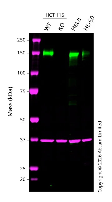
Western blot - Anti-KIAA0319L antibody [EPR28329-62] (AB315027)
Western blot : Anti-KIAA0319L antibody [EPR28329-62] ab315027 staining at 1/1000 dilution, shown in green; Mouse anti GAPDH ab8245 loading control staining at 1/20000 dilution, shown in magenta. A band was observed at 150 kDa in Wild-type HCT 116 cell lysates with no signal observed at this size in KIAA0319L knockout HCT 116 cell line. To generate this image, samples were run on an SDS-PAGE gel then transferred onto a nitrocellulose membrane. Membranes were blocked in 5% Milk in TBS-0.1 % Tween® 20 (TBS-T) before incubation with primary antibodies overnight at 4 °C. Blots were washed four times in TBS-T, incubated with secondary antibodies for 1 h at room temperature, washed again four times then imaged. Secondary antibodies used were Goat anti-Rabbit 800CW and Goat anti-Mouse 680RD at 1/20,000 dilution.
All lanes:
Western blot - Anti-KIAA0319L antibody [EPR28329-62] (ab315027) at 1/1000 dilution
Lane 1:
Wild-type HCT 116 cell lysates at 20 µg
Lane 2:
KIAA0319L knockout HCT 116 cell lysates at 20 µg
Lane 3:
HeLa cell lysates at 20 µg
Lane 4:
HL-60 cell lysates at 20 µg
Secondary
All lanes:
Goat anti-Rabbit 800CW & Goat anti-Mouse 680RD at 1/20000 dilution
Predicted band size: 150 kDa
Observed band size: 150 kDa
false

- WB
Supplier Data
Western blot - Anti-KIAA0319L antibody [EPR28329-62] (AB315027)
Blocking and diluting buffer and concentration : 5% NFDM/TBST.
Negative control : HL-60 (PMID : 28679762).
The observed Mw of ~150KDa is higher than the predicted Mw due to glycosylation (PMID : 28679762).
In Western blot, Anti-GAPDH antibody [EPR16891] - Loading Control (ab181602) staining at 1/200000 dilution.
All lanes:
Western blot - Anti-KIAA0319L antibody [EPR28329-62] (ab315027) at 1/1000 dilution
Lane 1:
HeLa (human cervical adenocarcinoma epithelial cell) transfected with scrambled siRNA control whole cell lysate at 20 µg
Lane 2:
HeLa transfected with siRNA specifically targeting KIAA0319L whole cell lysate at 20 µg
Lane 3:
HeLa (human cervical adenocarcinoma epithelial cell) whole cell lysate at 20 µg
Lane 4:
Huh7 (human hepatocellular carcinoma epithelial cell) whole cell lysate at 20 µg
Lane 5:
HL-60 (human acute promyelocytic leukemia promyeloblast) whole cell lysate at 20 µg
Lane 6:
Neuro-2a (mouse neuroblastoma neuroblast) whole cell lysate at 20 µg
Lane 7:
NIH/3T3 (mouse embryonic fibroblast) whole cell lysate at 20 µg
Lane 8:
C6 (rat glial tumor glial cell) whole cell lysate at 20 µg
Lane 9:
L6 (rat skeletal muscle myoblast) whole cell lysate at 20 µg
Secondary
All lanes:
Western blot - Goat Anti-Rabbit IgG H&L (HRP) (<a href='/products/secondary-antibodies/goat-rabbit-igg-h-l-hrp-ab97051'>ab97051</a>) at 1/100000 dilution
Observed band size: 150 kDa,36 kDa
false
Exposure time: 48s

- WB
Supplier Data
Western blot - Anti-KIAA0319L antibody [EPR28329-62] (AB315027)
Blocking and diluting buffer and concentration : 5% NFDM/TBST.
Deglycosylation induced a shift in the observed molecular weight from ~150KDa to ~100KDa (PMID : 28679762).
In Western blot, Anti-GAPDH antibody [EPR16891] - Loading Control (ab181602) staining at 1/200000 dilution.
All lanes:
Western blot - Anti-KIAA0319L antibody [EPR28329-62] (ab315027) at 1/1000 dilution
Lane 1:
Untreated HeLa (human cervical adenocarcinoma epithelial cell) whole cell lysate at 20 µg
Lane 2:
HeLa whole cell lysate treated with Protein Deglycosylation MIX II at 20 µg
Secondary
All lanes:
Western blot - Goat Anti-Rabbit IgG H&L (HRP) (<a href='/products/secondary-antibodies/goat-rabbit-igg-h-l-hrp-ab97051'>ab97051</a>) at 1/100000 dilution
Observed band size: 150 kDa,100 kDa,36 kDa
false
Exposure time: 48s

- WB
Supplier Data
Western blot - Anti-KIAA0319L antibody [EPR28329-62] (AB315027)
Blocking and diluting buffer and concentration : 5% NFDM/TBST.
The observed Mw of ~150KDa is higher than the predicted Mw due to glycosylation (PMID : 28679762).
In Western blot, Anti-GAPDH antibody [EPR16891] - Loading Control (ab181602) staining at 1/200000 dilution.
All lanes:
Western blot - Anti-KIAA0319L antibody [EPR28329-62] (ab315027) at 1/1000 dilution
Lane 1:
Human hippocampus tissue lysate at 20 µg
Lane 2:
Mouse brain tissue lysate at 20 µg
Lane 3:
Rat brain tissue lysate at 20 µg
Secondary
All lanes:
Western blot - Goat Anti-Rabbit IgG H&L (HRP) (<a href='/products/secondary-antibodies/goat-rabbit-igg-h-l-hrp-ab97051'>ab97051</a>) at 1/100000 dilution
Observed band size: 150 kDa,36 kDa
false
Exposure time: 180s
関連する標識済み抗体及び組成の異なる製品 (1)
-
Anti-KIAA0319L antibody [EPR28329-62] - BSA and Azide free
組合せ製品
Primary Antibodies
AB172730
Rabbit IgG, monoclonal [EPR25A] - Isotype Control
KD Validated|RabMAb|Advanced Validation|Recombinant

- 0
- 0
Primary Antibodies
AB7291
Anti-alpha Tubulin antibody [DM1A] - Loading Control
BOND RX™ Validated

- 5
- 92
Primary Antibodies
AB51253
Anti-Alpha-synuclein (phospho S129) antibody [EP1536Y]
20ul selling size|BOND RX™ Validated|RabMAb|Recombinant

- 5
- 9

Reactivity data
製品の詳細
Patented technology
Our RabMAb® technology is a patented hybridoma-based technology for making rabbit monoclonal antibodies. For details on our patents, please refer to RabMAb® patents.
What are the advantages of a recombinant monoclonal antibody?
This product is a recombinant monoclonal antibody, which offers several advantages including:
- - High batch-to-batch consistency and reproducibility
- - Improved sensitivity and specificity
- - Long-term security of supply
- - Animal-free batch production
For more information, read more on recombinant antibodies.
出荷温度及び保存条件
製品の状態
精製方法
バッファー組成
出荷温度
短期保存期間
短期保存温度
長期保存温度
分注に関する情報
保管に関する情報
製品プロトコール
- Visit the General protocols
- Visit the Troubleshooting
ターゲットの情報
文献 (1)
Recent publications for all applications. Explore the full list and refine your search
Advanced science (Weinheim, Baden-Wurttemberg, Germany) 12:e2408873 PubMed39776318
2025
Applications
Unspecified application
Species
Unspecified reactive species
Abcam product promise
Please note: All products are 'FOR RESEARCH USE ONLY. NOT FOR USE IN DIAGNOSTIC OR THERAPEUTIC PROCEDURES'.
For licensing inquiries, please contact partnerships@abcam.com