Anti-Hsc70 抗体 [EP1531Y] - BSA and Azide free
Anti-Hsc70 antibody [EP1531Y] - BSA and Azide free
- RabMAb
- Recombinant
- KO Validated
- 詳細を見る
Be the first to review this product! Submit a review
|
(6 Publications)
Rabbit Recombinant Monoclonal Hsc70 antibody. Carrier free. Suitable for IHC-P, IP, WB, ICC/IF, Flow Cyt (Intra) and reacts with Human, Mouse, Rat samples. Cited in 6 publications.
別名を表示する
HSC70, HSP73, HSPA10, HSPA8, Heat shock cognate 71 kDa protein, Heat shock 70 kDa protein 8, Heat shock protein family A member 8, Lipopolysaccharide-associated protein 1, LAP-1, LPS-associated protein 1
- IHC-P
Unknown
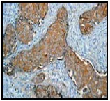
Immunohistochemistry (Formalin/PFA-fixed paraffin-embedded sections) - Anti-Hsc70 antibody [EP1531Y] - BSA and Azide free (AB220821)
Unpurified ab51052 (1/250) staining Hsc70 in paraffin embedded human breast carcinoma tissue by Immunohistochemistry.
This data was developed using the same antibody clone in a different buffer formulation containing PBS, BSA, glycerol, and sodium azide (ab51052).
- ICC/IF
Unknown
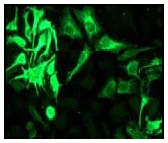
Immunocytochemistry/ Immunofluorescence - Anti-Hsc70 antibody [EP1531Y] - BSA and Azide free (AB220821)
Immunofluorescent staining of HeLa cells using unpurified ab51052 (1/100).
This data was developed using the same antibody clone in a different buffer formulation containing PBS, BSA, glycerol, and sodium azide (ab51052).
- IHC-P
Unknown
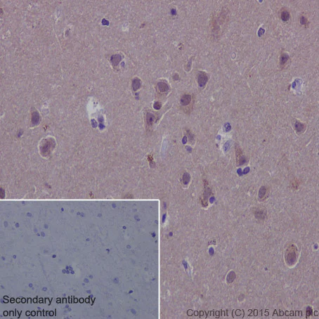
Immunohistochemistry (Formalin/PFA-fixed paraffin-embedded sections) - Anti-Hsc70 antibody [EP1531Y] - BSA and Azide free (AB220821)
Immunohistochemical staining of paraffin embedded human cerebral cortex with purified ab51052 at a working dilution of 1/500. The secondary antibody used is HRP goat anti-rabbit IgG H&L (ab97051) at 1/500. The sample is counter-stained with hematoxylin. Antigen retrieval was perfomed using Tris-EDTA buffer, pH 9.0. PBS was used instead of the primary antibody as the negative control, and is shown in the inset.This data was developed using the same antibody clone in a different buffer formulation containing PBS, BSA, glycerol, and sodium azide (ab51052).
- Flow Cyt (Intra)
Unknown
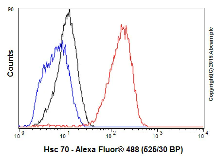
Flow Cytometry (Intracellular) - Anti-Hsc70 antibody [EP1531Y] - BSA and Azide free (AB220821)
Overlay histogram showing MCF7 cells fixed in 4% PFA and stained with purified ab51052 at a dilution of 1 in 70 (red line). The secondary antibody used was FITC goat anti-rabbit at a dilution of 1 in 500. Rabbit monoclonal IgG was used as an isotype control (black line) and cells incubated in the absence of both primary and secondary antibody were used as a negative control (blue line). This data was developed using the same antibody clone in a different buffer formulation containing PBS, BSA, glycerol, and sodium azide (ab51052).
- ICC/IF
Unknown
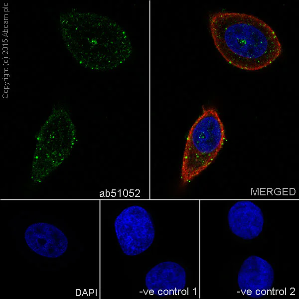
Immunocytochemistry/ Immunofluorescence - Anti-Hsc70 antibody [EP1531Y] - BSA and Azide free (AB220821)
Immunofluorescence staining of MCF7 cells with purified ab51052 at a working dilution of 1/250, counter-stained with DAPI. The secondary antibody was Alexa Fluor® 488 goat anti-rabbit (ab150077), used at a dilution of 1/1000. ab7291, a mouse anti-tubulin antibody (1/1000), was used to stain tubulin along with ab150120 (Alexa Fluor® 594 goat anti-mouse, 1/1000), shown in the top right hand panel. The cells were fixed in 4% PFA and permeabilized using 0.1% Triton X 100. The negative controls are shown in bottom middle and right hand panels - for negative control 1, purified ab51052 was used at a dilution of 1/500 followed by an Alexa Fluor® 594 goat anti-mouse antibody (ab150120) at a dilution of 1/500. For negative control 2, ab7291 (mouse anti-tubulin) was used at a dilution of 1/500 followed by an Alexa Fluor® 488 goat anti-rabbit antibody (ab150077) at a dilution of 1/400.
This data was developed using the same antibody clone in a different buffer formulation containing PBS, BSA, glycerol, and sodium azide (ab51052).
- IP
Unknown
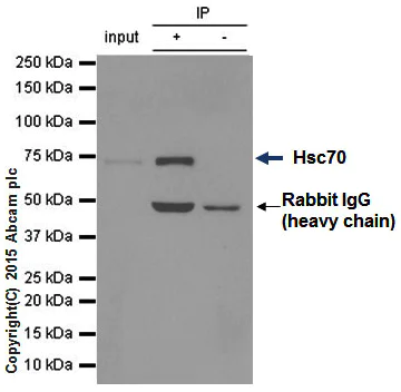
Immunoprecipitation - Anti-Hsc70 antibody [EP1531Y] - BSA and Azide free (AB220821)
ab51052 (purified) at 1/20 immunoprecipitating Hsc70 in 10 μg HeLa (Lanes 1 and 2, observed at 71 kDa). Lane 3 - PBS. For western blotting, HRP Veriblot for IP (ab131366) was used for detection at 1/1000 dilution. Blocking buffer and concentration : 5% NFDM/TBST Dilution buffer and concentration : 5% NFDM/TBST
This data was developed using the same antibody clone in a different buffer formulation containing PBS, BSA, glycerol, and sodium azide (ab51052).
All lanes:
Immunoprecipitation - Anti-Hsc70 antibody [EP1531Y] (<a href='/products/primary-antibodies/hsc70-antibody-ep1531y-ab51052'>ab51052</a>)
Predicted band size: 70 kDa
false
- WB
Lab
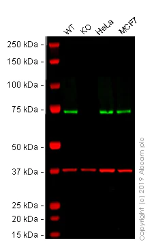
Western blot - Anti-Hsc70 antibody [EP1531Y] - BSA and Azide free (AB220821)
This data was developed using the same antibody clone in a different buffer formulation (ab51052).
Lanes 1 - 4 : Merged signal (red and green). Green - ab51052 observed at 73 kDa. Red - loading control, ab8245 (Mouse anti-GAPDH antibody [6C5]) observed at 37kDa.
ab51052 was shown to react with HSPA8 in A431 wild-type cells in Western blot. Loss of signal was observed when HSPA8 knockout sample was used. A431 wild-type and HSPA8 knockout cell lysates were subjected to SDS-PAGE. Membranes were blocked in 3% Milk in TBS-T (0.1% Tween®) before incubation with ab51052 and ab8245 (Mouse anti-GAPDH antibody [6C5]) overnight at 4°C at a 1 in 500 dilution and a 1 in 20000 dilution respectively. Blots were developed with Goat anti-Rabbit IgG H&L (IRDye® 800CW) preabsorbed (ab216773) and Goat anti-Mouse IgG H&L (IRDye® 680RD) preabsorbed (ab216776) secondary antibodies at 1 in 20000 dilution for 1 hour at room temperature before imaging.
All lanes:
Western blot - Anti-Hsc70 antibody [EP1531Y] - BSA and Azide free (ab220821) at 1/500 dilution
Lane 1:
Wild-type A431 whole cell lysate at 20 µg
Lane 2:
HSPA8 knockout A431 whole cell lysate at 20 µg
Lane 3:
HeLa whole cell lysate at 20 µg
Lane 4:
MCF7 whole cell lysate at 20 µg
Predicted band size: 70 kDa
Observed band size: 73 kDa
false
- WB
Lab
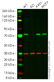
Western blot - Anti-Hsc70 antibody [EP1531Y] - BSA and Azide free (AB220821)
This data was developed using the same antibody clone in a different buffer formulation (ab51052).
Lanes 1- 4 : Merged signal (red and green). Green - ab51052 observed at 71 kDa. Red - Anti-GAPDH antibody [6C5] - Loading Control (ab8245) observed at 37 kDa.
ab51052 was shown to react with Hsc70 in wild-type HeLa cells in western blot. Loss of signal was observed when knockout cell line ab265664 (knockout cell lysate ab256944) was used. Wild-type HeLa and HSPA8 knockout HeLa cell lysates were subjected to SDS-PAGE. Membrane was blocked for 1 hour at room temperature in 0.1% TBST with 3% non-fat dried milk. ab51052 and Anti-GAPDH antibody [6C5] - Loading Control (ab8245) overnight at 4°C at a 1 in 500 dilution and a 1 in 20000 dilution respectively. Blots were developed with Goat anti-Rabbit IgG H&L (IRDye®800CW) preadsorbed (ab216773) and Goat anti-Mouse IgG H&L (IRDye®680RD) preadsorbed (ab216776) secondary antibodies at 1 in 20000 dilution for 1 hour at room temperature before imaging.
All lanes:
Western blot - Anti-Hsc70 antibody [EP1531Y] (<a href='/products/primary-antibodies/hsc70-antibody-ep1531y-ab51052'>ab51052</a>) at 1/500 dilution
Lane 1:
Wild-type HeLa cell lysate at 20 µg
Lane 2:
Western blot - Human HSPA8 (Hsc70) knockout HeLa cell lysate (<a href='/products/cell-lysates/human-hspa8-hsc70-knockout-hela-cell-lysate-ab256944'>ab256944</a>) at 20 µg
Lane 3:
A431 cell lysate at 20 µg
Lane 4:
MCF7 cell lysate at 20 µg
Predicted band size: 70 kDa
Observed band size: 71 kDa
false
関連する標識済み抗体及び組成の異なる製品 (8)
-
Anti-Hsc70 antibody [EP1531Y]
-
660 APC
APC Anti-Hsc70 antibody [EP1531Y]
-
519 Alexa Fluor® 488
Alexa Fluor® 488 Anti-Hsc70 antibody [EP1531Y]
-
565 Alexa Fluor® 555
Alexa Fluor® 555 Anti-Hsc70 antibody [EP1531Y]
-
617 Alexa Fluor® 594
Alexa Fluor® 594 Anti-Hsc70 antibody [EP1531Y]
-
665 Alexa Fluor® 647
Alexa Fluor® 647 Anti-Hsc70 antibody [EP1531Y]
-
775 Alexa Fluor® 750
Alexa Fluor® 750 Anti-Hsc70 antibody [EP1531Y]
-
578 PE
PE Anti-Hsc70 antibody [EP1531Y]
代替製品


組合せ製品


Primary Antibodies
AB125068
Anti-LAMP2A antibody [EPR4207(2)] - Lysosome Marker
20ul selling size|RabMAb|Recombinant|Lab Essentials|KO Validated

- 4
- 4

Primary Antibodies
AB125066
Anti-Glutathione Peroxidase 4 antibody [EPNCIR144]
BOND RX™ Validated|KO Validated|RabMAb|Recombinant|Lab Essentials

- 5
- 13
Primary Antibodies
AB192890
Anti-LC3B antibody [EPR18709] - Autophagosome Marker
BOND RX™ Validated|RabMAb|Recombinant|KO Validated|Lab Essentials|20ul selling size

- 5
- 19
Reactivity data
製品の詳細
ab220821 is the carrier-free version of ab51052.
Patented technology
Our RabMAb® technology is a patented hybridoma-based technology for making rabbit monoclonal antibodies. For details on our patents, please refer to RabMAb® patents.
What are the advantages of a recombinant monoclonal antibody?
This product is a recombinant monoclonal antibody, which offers several advantages including:
- - High batch-to-batch consistency and reproducibility
- - Improved sensitivity and specificity
- - Long-term security of supply
- - Animal-free batch production
For more information, read more on recombinant antibodies.
Conjugation ready
Our carrier-free antibodies are typically supplied in a PBS-only formulation, purified and free of BSA, sodium azide and glycerol. This conjugation-ready format is designed for use with fluorochromes, metal isotopes, oligonucleotides, and enzymes, which makes them ideal for antibody labelling, functional and cell-based assays, flow-based assays (e.g. mass cytometry) and Multiplex Imaging applications.
Use our conjugation kits for antibody conjugates that are ready-to-use in as little as 20 minutes with 1 minute hands-on-time and 100% antibody recovery: available for fluorescent dyes, HRP, biotin and gold.
Compatibility
This product is compatible with the Maxpar® Antibody Labeling Kit from Fluidigm, without the need for antibody preparation. Maxpar® is a trademark of Fluidigm Canada Inc.
出荷温度及び保存条件
製品の状態
精製方法
バッファー組成
出荷温度
短期保存温度
長期保存温度
保管に関する情報
補足情報
This supplementary information is collated from multiple sources and compiled automatically.
Biological function summary
Hsc70 participates in several processes beyond protein folding. This includes its involvement in the transport of proteins across cellular membranes and the degradation of misfolded proteins. Hsc70 often works in conjunction with co-chaperones and forms part of large protein complexes such as the chaperone machinery involved in clathrin-mediated endocytosis. It also plays a part in cellular stress responses supporting cell survival under various stress conditions.
Pathways
Hsc70 integrates into fundamental cellular pathways such as the ubiquitin-proteasome system and the ER-associated degradation pathway. These pathways are important for protein quality control where Hsc70 collaborates with other heat shock proteins like Hsp40 to facilitate the correct folding and clearance of proteins. In the context of autophagy Hsc70 mediates the recognition and trafficking of specific substrates to lysosomes for degradation demonstrating its importance in maintaining cellular health through these pathways.
製品プロトコール
- Visit the General protocols
- Visit the Troubleshooting
ターゲットの情報
文献 (6)
Recent publications for all applications. Explore the full list and refine your search
Laboratory investigation; a journal of technical m 96:1004-15 PubMed27501049
2016
Applications
Unspecified application
Species
Unspecified reactive species
The Journal of biological chemistry 288:27752-63 PubMed23921388
2013
Applications
Unspecified application
Species
Unspecified reactive species
Molecular oncology 7:484-96 PubMed23333597
2013
Applications
Unspecified application
Species
Unspecified reactive species
Molecular cancer research : MCR 9:936-47 PubMed21636681
2011
Applications
Unspecified application
Species
Unspecified reactive species
Archives of neurology 67:1464-72 PubMed20697033
2010
Applications
Unspecified application
Species
Unspecified reactive species
Molecular cell 36:15-27 PubMed19818706
2009
Applications
Unspecified application
Species
Unspecified reactive species
Abcam product promise
Please note: All products are 'FOR RESEARCH USE ONLY. NOT FOR USE IN DIAGNOSTIC OR THERAPEUTIC PROCEDURES'.
For licensing inquiries, please contact partnerships@abcam.com