Anti-HIKESHI 抗体 [EPR17761]
Anti-HIKESHI antibody [EPR17761]
- 20ul selling size
- RabMAb
- Recombinant
- 詳細を見る
Be the first to review this product! Submit a review
|
(1 Publication)
Rabbit Recombinant Monoclonal HIKESHI antibody. Suitable for IP, WB, ICC/IF, IHC-P and reacts with Human, Mouse samples. Cited in 1 publication.
別名を表示する
C11orf73, HSPC138, HSPC179, HSPC248, HIKESHI, Protein Hikeshi
- ICC/IF
Supplier Data
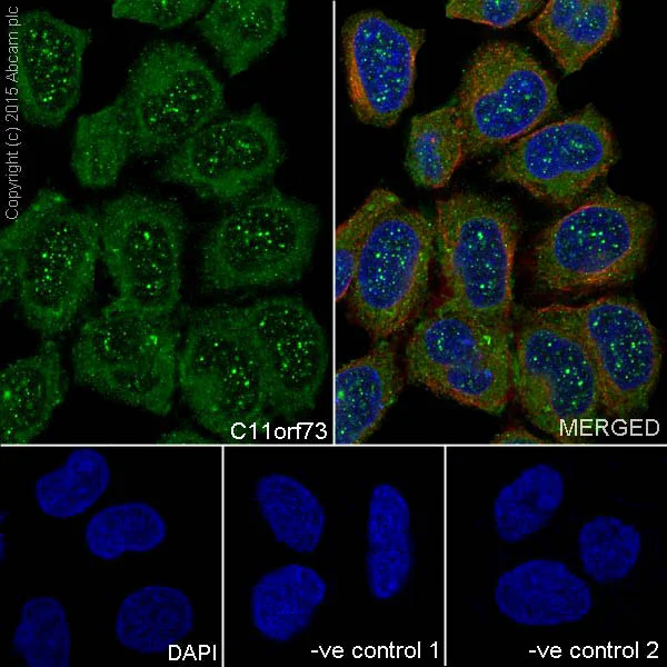
Immunocytochemistry/ Immunofluorescence - Anti-HIKESHI antibody [EPR17761] (AB202065)
Immunofluorescent analysis of 4% paraformaldehyde-fixed 0.1% Triton X-100 permeabilized HeLa (Human epithelial cells from cervix adenocarcinoma) cells labeling HIKESHI with ab202065 at 1/400 dilution followed by Goat anti-rabbit IgG (Alexa Fluor® 488) (ab150077) secondary antibody at 1/500 dilution (green).
Confocal image showing nuclear and cytoplasmic staining on HeLa cell line.
The nuclear counter stain is DAPI (blue).
Tubulin is detected with ab7291 (anti-Tubulin mouse mAb) at 1/1000 dilution and ab150120 (AlexaFluor®594 Goat anti-Mouse secondary) at 1/500 dilution (red).
The negative controls are as follows :
-ve control 1 : ab202065 at 1/400 dilution followed by ab150120 (AlexaFluor®594 Goat anti-Mouse secondary) at 1/500 dilution.
-ve control 2 : ab7291 (anti-Tubulin mouse mAb) at 1/1000 dilution followed by ab150077 (Alexa Fluor®488 Goat Anti-Rabbit IgG H&L) at 1/500 dilution.
- IHC-P
Supplier Data
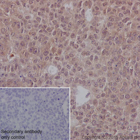
Immunohistochemistry (Formalin/PFA-fixed paraffin-embedded sections) - Anti-HIKESHI antibody [EPR17761] (AB202065)
Immunohistochemical analysis of paraffin-embedded Human cervix carcinoma tissue labeling HIKESHI with ab202065 at 1/100 dilution followed by Goat Anti-Rabbit IgG H&L (HRP) (ab97051) secondary antibody at 1/500 dilution.
Cytoplasmic and nuclear staining on Human cervix carcinoma tissue is observed.
Counter stained with Hematoxylin.
Secondary antibody only control : Used PBS instead of primary antibody secondary antibody is Goat Anti-Rabbit IgG H&L (HRP) (ab97051) at 1/500 dilution.
Perform heat mediated antigen retrieval with Tris/EDTA buffer pH 9.0 before commencing with IHC staining protocol.
- ICC/IF
Supplier Data
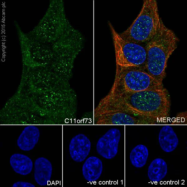
Immunocytochemistry/ Immunofluorescence - Anti-HIKESHI antibody [EPR17761] (AB202065)
Immunofluorescent analysis of 4% paraformaldehyde-fixed 0.1% Triton X-100 permeabilized MCF7 (Human breast adenocarcinoma cell line) cells labeling HIKESHI with ab202065 at 1/400 dilution followed by Goat anti-rabbit IgG (Alexa Fluor® 488) (ab150077) secondary antibody at 1/500 dilution (green).
Confocal image showing nuclear and cytoplasmic staining on MCF7 cell line.
The nuclear counter stain is DAPI (blue).
Tubulin is detected with ab7291 (anti-Tubulin mouse mAb) at 1/1000 dilution and ab150120 (AlexaFluor®594 Goat anti-Mouse secondary) at 1/500 dilution (red).
The negative controls are as follows :
-ve control 1 : ab202065 at 1/400 dilution followed by ab150120 (AlexaFluor®594 Goat anti-Mouse secondary) at 1/500 dilution.
-ve control 2 : ab7291 (anti-Tubulin mouse mAb) at 1/1000 dilution followed by ab150077 (Alexa Fluor®488 Goat Anti-Rabbit IgG H&L) at 1/500 dilution.
- IP
Supplier Data
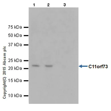
Immunoprecipitation - Anti-HIKESHI antibody [EPR17761] (AB202065)
HIKESHI was immunoprecipitated from 1mg of MCF7 (Human breast adenocarcinoma cell line) whole cell lysate with ab202065 at 1/50 dilution.
Western blot was performed from the immunoprecipitate using ab202065 at 1/1000 dilution.
Anti-Rabbit IgG (HRP), specific to the non-reduced form of IgG was used as secondary antibody at 1/1500 dilution.
Lane 1 : MCF7 whole cell lysate 10 μg (Input).
Lane 2 : ab202065 IP in MCF7 whole cell lysate.
Lane 3 : Rabbit monoclonal IgG (ab172730) instead of ab202065 in MCF7 whole cell lysate.
Blocking and dilution buffer and concentration : 5% NFDM/TBST.
Exposure time : 30 seconds.
All lanes:
Immunoprecipitation - Anti-HIKESHI antibody [EPR17761] (ab202065)
Predicted band size: 21 kDa,79 kDa
false
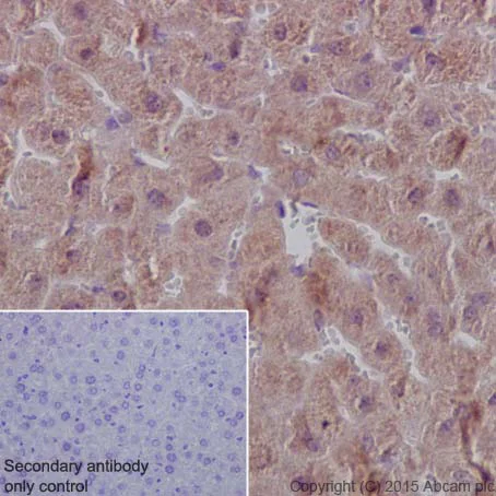
- IHC-P
Supplier Data
Immunohistochemistry (Formalin/PFA-fixed paraffin-embedded sections) - Anti-HIKESHI antibody [EPR17761] (AB202065)
Immunohistochemical analysis of paraffin-embedded Mouse liver tissue labeling HIKESHI with ab202065 at 1/100 dilution followed by Goat Anti-Rabbit IgG H&L (HRP) (ab97051) secondary antibody at 1/500 dilution.
Cytoplasmic and nuclear staining on mouse liver tissue is observed.
Counter stained with Hematoxylin.
Secondary antibody only control : Used PBS instead of primary antibody secondary antibody is Goat Anti-Rabbit IgG H&L (HRP) (ab97051) at 1/500 dilution.
Perform heat mediated antigen retrieval with Tris/EDTA buffer pH 9.0 before commencing with IHC staining protocol.
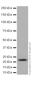
- WB
Supplier Data
Western blot - Anti-HIKESHI antibody [EPR17761] (AB202065)
Blocking/Dilution buffer : 5% NFDM/TBST.
All lanes:
Western blot - Anti-HIKESHI antibody [EPR17761] (ab202065) at 1/1000 dilution
All lanes:
Human fetal heart lysate at 10 µg
Secondary
All lanes:
Anti-Rabbit IgG (HRP), specific to the non-reduced form of IgG at 1/1000 dilution
Predicted band size: 21 kDa
Observed band size: 22 kDa
false
Exposure time: 3min
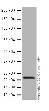
- WB
Supplier Data
Western blot - Anti-HIKESHI antibody [EPR17761] (AB202065)
Blocking/Dilution buffer : 5% NFDM/TBST.
All lanes:
Western blot - Anti-HIKESHI antibody [EPR17761] (ab202065) at 1/1000 dilution
All lanes:
MCF7 (Human breast adenocarcinoma cell line) cell lysate at 20 µg
Secondary
All lanes:
Goat Anti-Rabbit IgG, (H+L), Peroxidase conjugated at 1/1000 dilution
Predicted band size: 21 kDa
Observed band size: 22 kDa
false
Exposure time: 3min
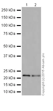
- WB
Unknown
Western blot - Anti-HIKESHI antibody [EPR17761] (AB202065)
Blocking/Dilution buffer : 5% NFDM/TBST.
All lanes:
Western blot - Anti-HIKESHI antibody [EPR17761] (ab202065) at 1/1000 dilution
Lane 1:
Mouse brain lysate at 10 µg
Lane 2:
Mouse kidney lysate at 10 µg
Secondary
All lanes:
Goat Anti-Rabbit IgG, (H+L), Peroxidase conjugated at 1/1000 dilution
Predicted band size: 21 kDa
Observed band size: 22 kDa
false
Exposure time: 3min
関連する標識済み抗体及び組成の異なる製品 (1)
-
Anti-HIKESHI antibody [EPR17761] - BSA and Azide free
組合せ製品
Primary Antibodies
AB172730
Rabbit IgG, monoclonal [EPR25A] - Isotype Control
RabMAb|Advanced Validation|Recombinant

- 0
- 0
Primary Antibodies
AB203826
Anti-p40 - DeltaNp63 antibody [EPR17863-47]
20ul selling size|BOND RX™ Validated|RabMAb|Recombinant
- 0
- 0
Primary Antibodies
AB281588
Anti-MAP2 antibody [RM1010] - Neuronal Marker
RabMAb|Recombinant|Lab Essentials|20ul selling size
- 0
- 0
Reactivity data
製品の詳細
Patented technology
Our RabMAb® technology is a patented hybridoma-based technology for making rabbit monoclonal antibodies. For details on our patents, please refer to RabMAb® patents.
What are the advantages of a recombinant monoclonal antibody?
This product is a recombinant monoclonal antibody, which offers several advantages including:
- - High batch-to-batch consistency and reproducibility
- - Improved sensitivity and specificity
- - Long-term security of supply
- - Animal-free batch production
For more information, read more on recombinant antibodies.
出荷温度及び保存条件
製品の状態
精製方法
バッファー組成
出荷温度
短期保存期間
短期保存温度
長期保存温度
分注に関する情報
保管に関する情報
補足情報
This supplementary information is collated from multiple sources and compiled automatically.
Biological function summary
HIKESHI engages in protecting cells from proteotoxic stress by ensuring the proper functioning of Hsp70 which is not bound to other co-chaperones. It does not heterodimerize but operates independently to import Hsp70 into the nucleus. This nuclear import contributes to cellular homeostasis and stress response mechanisms enhancing the cell's ability to manage damaged proteins and stress-induced protein aggregation.
Pathways
HIKESHI participates in cellular stress response and protein folding pathways. This action connects it with proteins involved in the importin beta pathway which contains various nucleocytoplasmic transport mechanisms. HIKESHI shares functional relationships with importin proteins emphasizing its specialized role in ensuring that only specific proteins like Hsp70 access the nuclear compartment during stress states.
製品プロトコール
- Visit the General protocols
- Visit the Troubleshooting
ターゲットの情報
文献 (1)
Recent publications for all applications. Explore the full list and refine your search
Nature cell biology 25:1575-1589 PubMed37770567
2023
Applications
Unspecified application
Species
Unspecified reactive species
Abcam product promise
Please note: All products are 'FOR RESEARCH USE ONLY. NOT FOR USE IN DIAGNOSTIC OR THERAPEUTIC PROCEDURES'.
For licensing inquiries, please contact partnerships@abcam.com