Anti-Glutaminase C 抗体 [EPR19525]
Anti-Glutaminase C antibody [EPR19525]
- 20ul selling size
- RabMAb
- Recombinant
- KO Validated
- 詳細を見る
0
(1 Review)
|
(6 Publications)
Rabbit Recombinant Monoclonal Glutaminase antibody. Suitable for IP, WB, ICC/IF, Flow Cyt (Intra) and reacts with Human, Mouse, Rat samples. Cited in 6 publications.
別名を表示する
GLS1, KIAA0838, GLS, K-glutaminase, L-glutamine amidohydrolase

- ICC/IF
Supplier Data
Immunocytochemistry/ Immunofluorescence - Anti-Glutaminase C antibody [EPR19525] (AB202027)
Immunofluorescent analysis of 100% methanol-fixed, 0.1% Triton X-100 permeabilized HeLa (Human epithelial cell line from cervix adenocarcinoma) cells labeling Glutaminase C with ab202027 at 1/500 dilution, followed by Goat anti-rabbit IgG (Alexa Fluor® 488) (ab150077) secondary antibody at 1/1000 dilution (green). Confocal image showing cytoplasmic staining on HeLa cell line.
The nuclear counter stain is DAPI (blue). Tubulin is detected with ab195889 (Anti-alpha Tubulin antibody [DM1A] - Microtubule Marker (Alexa Fluor® 594)) at 1/200 dilution (red).
Secondary antibody only control : Used PBS instead of primary antibody, secondary antibody is Goat anti-rabbit IgG (Alexa Fluor® 488) (ab150077) at 1/1000 dilution.

- Flow Cyt (Intra)
Supplier Data
Flow Cytometry (Intracellular) - Anti-Glutaminase C antibody [EPR19525] (AB202027)
Intracellular flow cytometric analysis of 4% paraformaldehyde-fixed HeLa (Human epithelial cell line from cervix adenocarcinoma) cells labeling Glutaminase Cwith ab202027 at 1/600 dilution (red) compared with a rabbit monoclonal IgG isotype control (ab172730; black) and an unlabelled control (cells without incubation with primary antibody and secondary antibody; blue). Goat anti rabbit IgG (Alexa Fluorr® 488) at 1/2000 dilution was used as the secondary antibody.

- ICC/IF
Supplier Data
Immunocytochemistry/ Immunofluorescence - Anti-Glutaminase C antibody [EPR19525] (AB202027)
Immunofluorescent analysis of 100% methanol-fixed, 0.1% Triton X-100 permeabilized HeLa (Human epithelial cell line from cervix adenocarcinoma) cells labeling Glutaminase C with ab202027 at 1/500 dilution, followed by Goat anti-rabbit IgG (Alexa Fluor® 488) (ab150077) secondary antibody at 1/1000 dilution (green). Confocal image showing mitochondrial staining on HeLa cell line.
The nuclear counter stain is DAPI (blue). COX IV is detected with ab33985 (anti-COX IV(mouse mAb)) at 1/1000 dilution and ab150120 (AlexaFluor®594 Goat anti-Mouse secondary) at 1/1000 dilution (red).
Secondary antibody only control : Used PBS instead of primary antibody, secondary antibody is Goat anti-rabbit IgG (Alexa Fluor® 488) (ab150077) at 1/1000 dilution.

- IP
Supplier Data
Immunoprecipitation - Anti-Glutaminase C antibody [EPR19525] (AB202027)
Glutaminase was immunoprecipitated from 0.35 mg of HeLa (Human epithelial cell line from cervix adenocarcinoma) whole cell lysate with ab202027 at 1/30 dilution. Western blot was performed from the immunoprecipitate using ab202027 at 1/1000 dilution. VeriBlot for IP Detection Reagent (HRP) (ab131366), was used for detection at 1/10000 dilution.
Lane 1 : HeLa whole cell lysate, 10 μg (Input).
Lane 2 : ab202027 IP in HeLa whole cell lysate.
Lane 3 : Rabbit monoclonal IgG (ab172730) instead of ab202027 in HeLa whole cell lysate.
Blocking and dilution buffer and concentration : 5% NFDM/TBST.
Exposure time : 10 seconds.
All lanes:
Immunoprecipitation - Anti-Glutaminase C antibody [EPR19525] (ab202027)
Observed band size: 65 kDa
false

- ICC/IF
Supplier Data
Immunocytochemistry/ Immunofluorescence - Anti-Glutaminase C antibody [EPR19525] (AB202027)
Immunofluorescent analysis of 100% methanol-fixed, 0.1% Triton X-100 permeabilized NIH/3T3 (Mouse embryonic fibroblast cell line) cells labeling Glutaminase C with ab202027 at 1/500 dilution, followed by Goat anti-rabbit IgG (Alexa Fluor® 488) (ab150077) secondary antibody at 1/1000 dilution (green). Confocal image showing cytoplasmic staining on NIH/3T3 cell line.
The nuclear counter stain is DAPI (blue). Tubulin is detected with ab195889 (Anti-alpha Tubulin antibody [DM1A] - Microtubule Marker (Alexa Fluor® 594)) at 1/200 dilution (red).
Secondary antibody only control : Used PBS instead of primary antibody, secondary antibody is Goat anti-rabbit IgG (Alexa Fluor® 488) (ab150077) at 1/1000 dilution.

- WB
Supplier Data
Western blot - Anti-Glutaminase C antibody [EPR19525] (AB202027)
Blocking/Dilution buffer : 5% NFDM/TBST.
All lanes:
Western blot - Anti-Glutaminase C antibody [EPR19525] (ab202027) at 1/1000 dilution
Lane 1:
HepG2 (Human liver hepatocellular carcinoma cell line) whole cell lysate at 20 µg
Lane 2:
HeLa (Human epithelial cell line from cervix adenocarcinoma) whole cell lysate at 20 µg
Lane 3:
K562 (Human chronic myelogenous leukemia cell line from bone marrow) whole cell lysate at 20 µg
Lane 4:
Jurkat (Human T cell leukemia cell line from peripheral blood) whole cell lysate at 20 µg
Lane 5:
Hepa1-6 (Mouse epithelial hepatoma cell line) whole cell lysate at 20 µg
Secondary
All lanes:
Western blot - Goat Anti-Rabbit IgG H&L (HRP) (<a href='/products/secondary-antibodies/goat-rabbit-igg-h-l-hrp-ab97051'>ab97051</a>) at 1/100000 dilution
Predicted band size: 65 kDa
Observed band size: 65 kDa
false
Exposure time: 15s

- WB
Supplier Data
Western blot - Anti-Glutaminase C antibody [EPR19525] (AB202027)
Blocking/Dilution buffer : 5% NFDM/TBST.
The observed expression profile is consistent with what has been described in UniProtKB (Glutaminase C is highly expressed in heart and pancreas, but is not detected in liver).
All lanes:
Western blot - Anti-Glutaminase C antibody [EPR19525] (ab202027) at 1/1000 dilution
Lane 1:
Human fetal heart lysate at 10 µg
Lane 2:
Human fetal kidney lysate at 10 µg
Lane 3:
Human fetal liver lysate at 10 µg
Secondary
All lanes:
Goat Anti-Rabbit IgG Peroxidase Conjugate, specific to the non-reduced form of IgG at 1/10000 dilution
Predicted band size: 65 kDa
Observed band size: 65 kDa
false
Exposure time: 15s
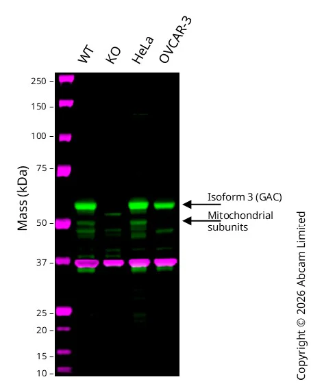
- WB
Lab
Western blot - Anti-Glutaminase C antibody [EPR19525] (AB202027)
Western blot : Anti-Glutaminase C antibody [EPR19525] ab202027 staining at 1/1000 dilution, shown in green; Mouse anti GAPDH ab8245 loading control staining at 1/20000 dilution, shown in magenta. A band was observed at 60 kDa in Wild-type MCF7 cell lysates with no signal observed at this size in GLS knockout MCF7 cell line. To generate this image, samples were run on an SDS-PAGE gel then transferred onto a nitrocellulose membrane. Membranes were blocked in 5 % Milk in TBS-0.1 % Tween® 20 (TBS-T) before incubation with primary antibodies overnight at 4 °C. Blots were washed four times in TBS-T, incubated with secondary antibodies for 1 h at room temperature, washed again four times then imaged. Secondary antibodies used were Goat anti-Rabbit 800CW and Goat anti-Mouse 680RD at 1/20,000 dilution.
All lanes:
Western blot - Anti-Glutaminase C antibody [EPR19525] (ab202027) at 1/1000 dilution
Lane 1:
Wild-type MCF7 cell lysates at 20 µg
Lane 2:
Western blot - Human GLS knockout MCF7 cell line (<a href='/products/cell-lines/human-gls-knockout-mcf7-cell-line-ab287444'>ab287444</a>) at 20 µg
Lane 3:
HeLa cell lysates at 20 µg
Lane 4:
OVCAR-3 cell lysates at 20 µg
Secondary
All lanes:
Goat anti-Rabbit 800CW & Goat anti-Mouse 680RD at 1/20000 dilution
Predicted band size: 73 kDa
Observed band size: 60 kDa
false

- WB
Supplier Data
Western blot - Anti-Glutaminase C antibody [EPR19525] (AB202027)
Blocking/Dilution buffer : 5% NFDM/TBST.
Exposure times : Lane 1 and 2 : 3 minutes; Lane 3 : 10 seconds; Lane 4, 5 and 6 : 3 minutes; Lane 7 : 30 seconds; Lane 8, 9 and 10 : 15 seconds.
All lanes:
Western blot - Anti-Glutaminase C antibody [EPR19525] (ab202027) at 1/5000 dilution
Lane 1:
Mouse brain lysate at 10 µg
Lane 2:
Mouse heart lysate at 10 µg
Lane 3:
Mouse spleen lysate at 10 µg
Lane 4:
Rat brain lysate at 10 µg
Lane 5:
Rat heart lysate at 10 µg
Lane 6:
Rat spleen lysate at 10 µg
Lane 7:
C6 (Rat glial tumor cell line) whole cell lysate at 10 µg
Lane 8:
RAW 264.7 (Mouse macrophage cell line transformed with Abelson murine leukemia virus) whole cell lysate at 10 µg
Lane 9:
PC-12 (Rat adrenal gland pheochromocytoma cell line) whole cell lysate at 10 µg
Lane 10:
NIH/3T3 (Mouse embryonic fibroblast cell line) whole cell lysate at 10 µg
Secondary
All lanes:
Western blot - Goat Anti-Rabbit IgG H&L (HRP) (<a href='/products/secondary-antibodies/goat-rabbit-igg-h-l-hrp-ab97051'>ab97051</a>) at 1/100000 dilution
Predicted band size: 65 kDa
Observed band size: 65 kDa
false
関連する標識済み抗体及び組成の異なる製品 (3)
-
Anti-Glutaminase C antibody [EPR19525] - BSA and Azide free
-
665 Alexa Fluor® 647
Alexa Fluor® 647 Anti-GLS/Glutaminase antibody [EPR19525]
-
428 SNv428
SNv428 Anti-GLS/Glutaminase antibody [EPR19525]
代替製品

組合せ製品

Primary Antibodies
AB272704
Anti-Acetyl Coenzyme A carboxylase alpha antibody [EPR23235-147] - BSA and Azide free
BOND RX™ Validated|RabMAb|Recombinant|KO Validated

- 5
- 2
Primary Antibodies
AB125066
Anti-Glutathione Peroxidase 4 antibody [EPNCIR144]
BOND RX™ Validated|KO Validated|RabMAb|Recombinant|Lab Essentials

- 5
- 13
Primary Antibodies
AB176562
Anti-Glutamine Synthetase antibody [EPR13022(B)]
BOND RX™ Validated|20ul selling size|Recombinant|KO Validated|Advanced Validation|RabMAb

- 5
- 4

Reactivity data
製品の詳細
Patented technology
Our RabMAb® technology is a patented hybridoma-based technology for making rabbit monoclonal antibodies. For details on our patents, please refer to RabMAb® patents.
What are the advantages of a recombinant monoclonal antibody?
This product is a recombinant monoclonal antibody, which offers several advantages including:
- - High batch-to-batch consistency and reproducibility
- - Improved sensitivity and specificity
- - Long-term security of supply
- - Animal-free batch production
For more information, read more on recombinant antibodies.
出荷温度及び保存条件
製品の状態
精製方法
バッファー組成
出荷温度
短期保存期間
短期保存温度
長期保存温度
分注に関する情報
保管に関する情報
補足情報
This supplementary information is collated from multiple sources and compiled automatically.
Biological function summary
Glutaminase C acts as an important player in nitrogen metabolism and cellular bioenergetics. It functions within the mitochondria facilitating the glutaminolysis pathway which provides intermediates for the tricarboxylic acid (TCA) cycle. Although glutaminase C mainly acts as a monomer in certain conditions it might participate in multiprotein complexes that aid its regulation and activity. This regulatory mechanism plays a vital role in energy homeostasis and cellular growth demands.
Pathways
Glutaminase C is involved in the glutaminolysis and TCA cycle pathways contributing significantly to energy production and biosynthesis in proliferating cells. It works in concert with related proteins like glutamate dehydrogenase which further converts the produced glutamate into α-ketoglutarate feeding into the TCA cycle. These combined actions support cellular adaptations during increased metabolic demand especially in cancerous cells where metabolic shifts are noticeable.
製品プロトコール
- Visit the General protocols
- Visit the Troubleshooting
ターゲットの情報
文献 (6)
Recent publications for all applications. Explore the full list and refine your search
Journal of traditional Chinese medicine = Chung i tsa chih ying wen pan 45:806-816 PubMed40810226
2025
Applications
Unspecified application
Species
Unspecified reactive species
Stem cell research & therapy 16:213 PubMed40301995
2025
Applications
Unspecified application
Species
Unspecified reactive species
Gut microbes 16:2428425 PubMed39567865
2024
Applications
Unspecified application
Species
Unspecified reactive species
PLoS pathogens 20:e1012552 PubMed39259763
2024
Applications
Unspecified application
Species
Unspecified reactive species
Cell metabolism 32:420-436.e12 PubMed32768387
2020
Applications
Unspecified application
Species
Unspecified reactive species
Cell metabolism 31:267-283.e12 PubMed31866442
2019
Applications
Unspecified application
Species
Unspecified reactive species
Abcam product promise
Please note: All products are 'FOR RESEARCH USE ONLY. NOT FOR USE IN DIAGNOSTIC OR THERAPEUTIC PROCEDURES'.
For licensing inquiries, please contact partnerships@abcam.com