Anti-Glucose Transporter GLUT4 抗体 [EPR26571-45]
Anti-Glucose Transporter GLUT4 antibody [EPR26571-45]
- BOND RX™ Validated
- 20ul selling size
- RabMAb
- Recombinant
- 詳細を見る
5
(1 Review)
|
(4 Publications)
Anti-Glucose Transporter GLUT4 antibody [EPR26571-45] (ab313775) is a rabbit monoclonal antibody detecting Glucose Transporter GLUT4 in Western Blot, IP, IHC-P, ICC/IF, Dot Blot. Suitable for Human, Mouse, Rat.
- Biophysical QC for unrivalled batch-batch consistency
別名を表示する
Glut4, Slc2a4, GT2, GLUT-4
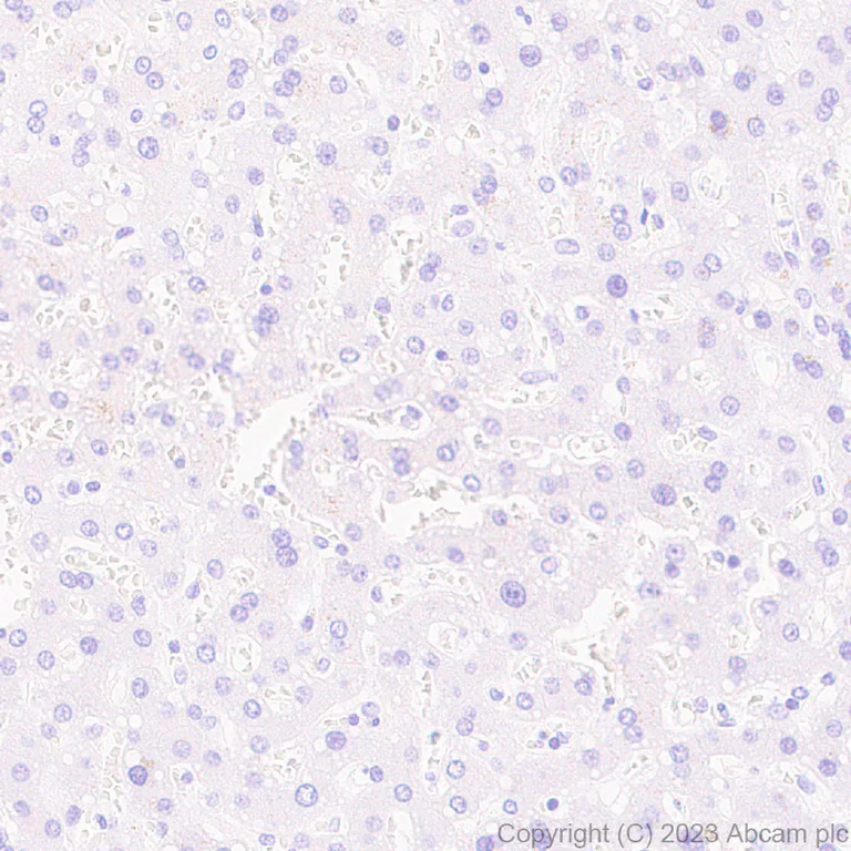
- IHC-P
Supplier Data
Immunohistochemistry (Formalin/PFA-fixed paraffin-embedded sections) - Anti-Glucose Transporter GLUT4 antibody [EPR26571-45] (AB313775)
Immunohistochemical analysis of paraffin-embedded Human liver tissue labeling SLC2A4 with ab313775 at 1/5000 (0.104 µg/ml) followed by a ready to use LeicaDS9800 (Bond™ Polymer Refine Detection). Negative control : No staining on human liver. The section was incubated with ab313775 for 30 mins at room temperature. The immunostaining was performed on a Leica Biosystems BOND® RX instrument Counterstained with Hematoxylin. Secondary antibody only control : Secondary antibody is a ready to use LeicaDS9800 (Bond™ Polymer Refine Detection).Heat mediated antigen retrieval was performed with Citrate buffer (pH 6.0, Epitope Retrieval Solution 1) for 20 mins
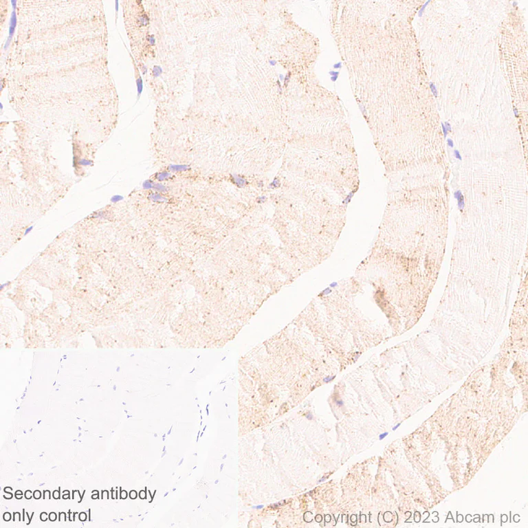
- IHC-P
Supplier Data
Immunohistochemistry (Formalin/PFA-fixed paraffin-embedded sections) - Anti-Glucose Transporter GLUT4 antibody [EPR26571-45] (AB313775)
Immunohistochemical analysis of paraffin-embedded Human skeletal muscle tissue labeling SLC2A4 with ab313775 at 1/5000 (0.104 µg/ml) followed by a ready to use LeicaDS9800 (Bond™ Polymer Refine Detection). Cytoplasmic staining on human skeletal muscle. The section was incubated with ab313775 for 30 mins at room temperature. The immunostaining was performed on a Leica Biosystems BOND® RX instrument Counterstained with Hematoxylin. Secondary antibody only control : Secondary antibody is a ready to use LeicaDS9800 (Bond™ Polymer Refine Detection).Heat mediated antigen retrieval was performed with Citrate buffer (pH 6.0, Epitope Retrieval Solution 1) for 20 mins
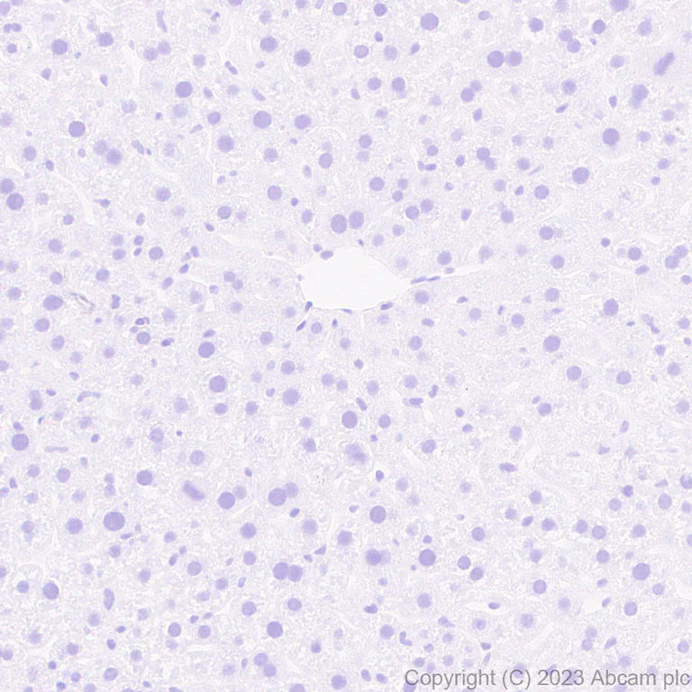
- IHC-P
Supplier Data
Immunohistochemistry (Formalin/PFA-fixed paraffin-embedded sections) - Anti-Glucose Transporter GLUT4 antibody [EPR26571-45] (AB313775)
Immunohistochemical analysis of paraffin-embedded Mouse liver tissue labeling SLC2A4 with ab313775 at 1/5000 (0.104 µg/ml) followed by a ready to use LeicaDS9800 (Bond™ Polymer Refine Detection). Negative control : No staining on mouse liver. The section was incubated with ab313775 for 30 mins at room temperature. The immunostaining was performed on a Leica Biosystems BOND® RX instrument Counterstained with Hematoxylin. Secondary antibody only control : Secondary antibody is a ready to use LeicaDS9800 (Bond™ Polymer Refine Detection).Heat mediated antigen retrieval was performed with Citrate buffer (pH 6.0, Epitope Retrieval Solution 1) for 20 mins
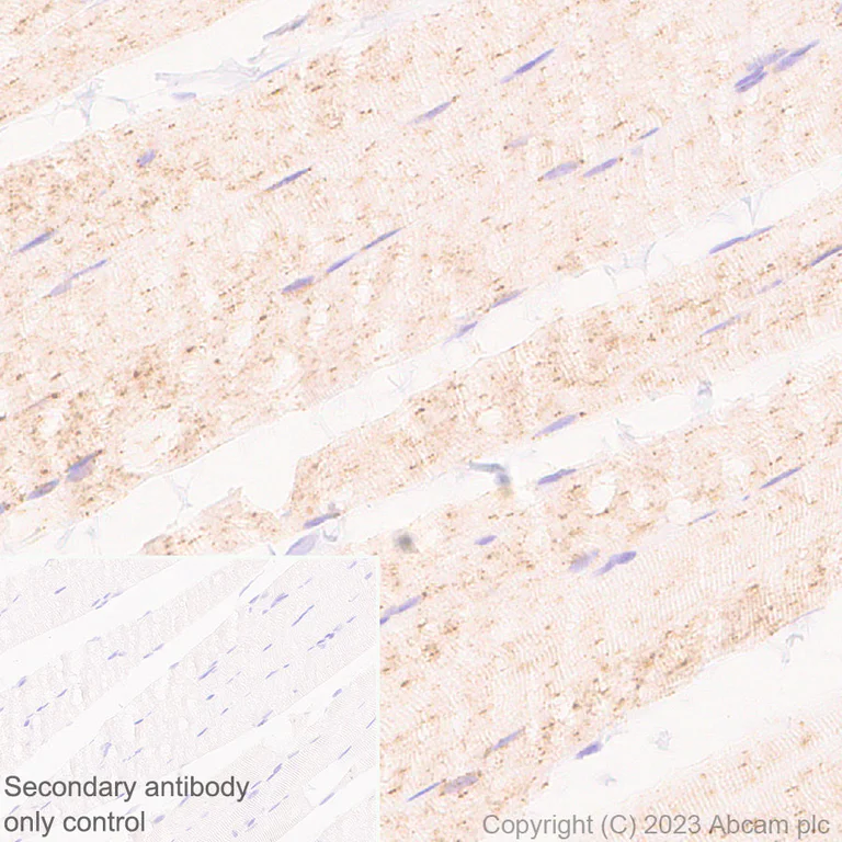
- IHC-P
Supplier Data
Immunohistochemistry (Formalin/PFA-fixed paraffin-embedded sections) - Anti-Glucose Transporter GLUT4 antibody [EPR26571-45] (AB313775)
Immunohistochemical analysis of paraffin-embedded Rat skeletal muscle tissue labeling SLC2A4 with ab313775 at 1/5000 (0.104 µg/ml) followed by a ready to use LeicaDS9800 (Bond™ Polymer Refine Detection). Cytoplasmic staining on rat skeletal muscle. The section was incubated with ab313775 for 30 mins at room temperature. The immunostaining was performed on a Leica Biosystems BOND® RX instrument Counterstained with Hematoxylin. Secondary antibody only control : Secondary antibody is a ready to use LeicaDS9800 (Bond™ Polymer Refine Detection).Heat mediated antigen retrieval was performed with Citrate buffer (pH 6.0, Epitope Retrieval Solution 1) for 20 mins
- IHC-P
Supplier Data
Immunohistochemistry (Formalin/PFA-fixed paraffin-embedded sections) - Anti-Glucose Transporter GLUT4 antibody [EPR26571-45] (AB313775)
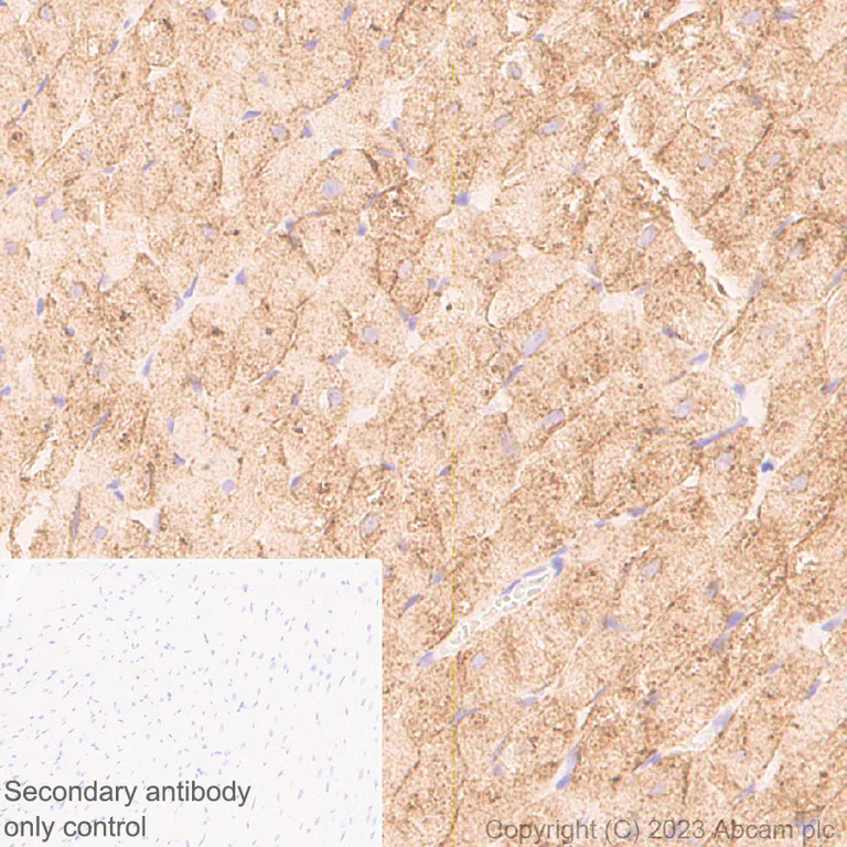
Immunohistochemical analysis of paraffin-embedded Rat cardiac muscle tissue labeling SLC2A4 with ab313775 at 1/5000 (0.104 µg/ml) followed by a ready to use LeicaDS9800 (Bond™ Polymer Refine Detection). Cytoplasmic staining on rat cardiac muscle. The section was incubated with ab313775 for 30 mins at room temperature. The immunostaining was performed on a Leica Biosystems BOND® RX instrument Counterstained with Hematoxylin. Secondary antibody only control : Secondary antibody is a ready to use LeicaDS9800 (Bond™ Polymer Refine Detection).Heat mediated antigen retrieval was performed with Citrate buffer (pH 6.0, Epitope Retrieval Solution 1) for 20 mins
- ICC/IF
Supplier Data
Immunocytochemistry/ Immunofluorescence - Anti-Glucose Transporter GLUT4 antibody [EPR26571-45] (AB313775)
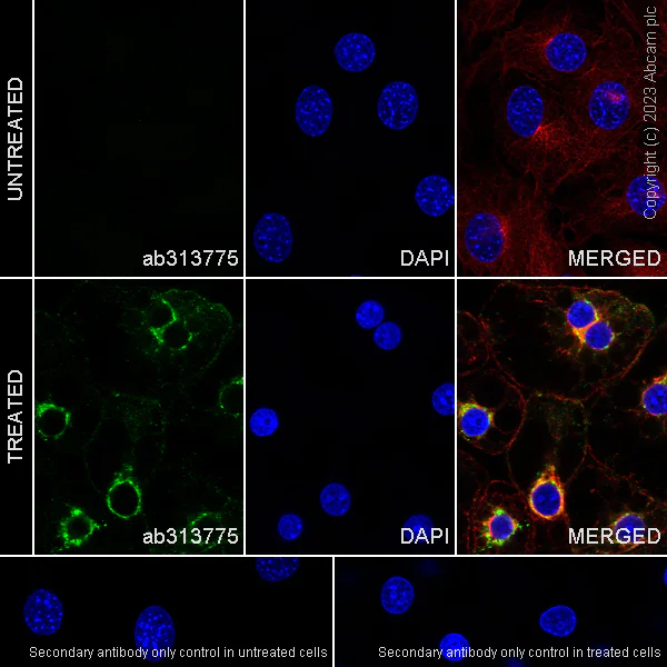
Immunofluorescent analysis of 4% Paraformaldehyde-fixed, 0.1% TritonX-100 permeabilized 3T3-L1 (mouse embryonic fibroblast) cells labelling SLC2A4 with ab313775 at 1/100 (5.18 µg/ml) dilution, followed by ab150081 Goat Anti-Rabbit IgG H&L (Alexa Fluor® 488) preadsorbed antibody at 1/1000 (2 µg/ml) dilution (Green). Confocal image showing cytoplasmic staining in differentiated 3T3-L1 cells, and no staining in 3T3-L1 cells. Image was taken with a confocal microscope(Leica-Microsystems, TCS SP8) ab195889 Anti-alpha Tubulin mouse monoclonal antibody - Microtubule Marker (Alexa Fluor® 594) was used to counterstain tubulin at 1/200 (2.5 µg/ml) dilution (Red). The Nuclear counterstain was DAPI (Blue). Secondary antibody only control : Secondary antibody is ab150081 Goat Anti-Rabbit IgG H&L (Alexa Fluor® 488) preadsorbed at 1/1000 (2 µg/ml) dilution.
- IHC-P
Supplier Data
Immunohistochemistry (Formalin/PFA-fixed paraffin-embedded sections) - Anti-Glucose Transporter GLUT4 antibody [EPR26571-45] (AB313775)
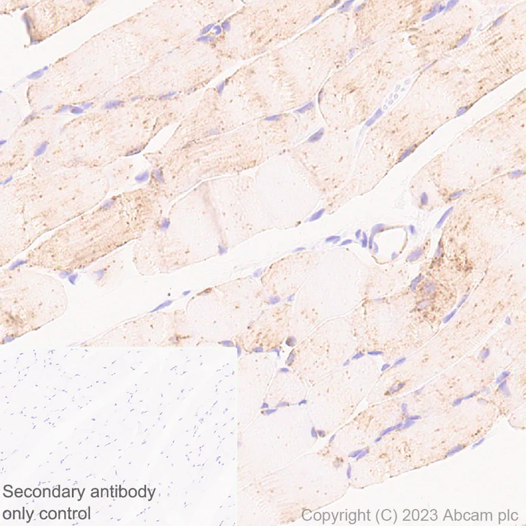
Immunohistochemical analysis of paraffin-embedded Mouse skeletal muscle tissue labeling SLC2A4 with ab313775 at 1/5000 (0.104 µg/ml) followed by a ready to use LeicaDS9800 (Bond™ Polymer Refine Detection). Cytoplasmic staining on mouse skeletal muscle (PMID : 2654938; PMID : 23877319; PMID : 33061888). The section was incubated with ab313775 for 30 mins at room temperature. The immunostaining was performed on a Leica Biosystems BOND® RX instrument Counterstained with Hematoxylin. Secondary antibody only control : Secondary antibody is a ready to use LeicaDS9800 (Bond™ Polymer Refine Detection).Heat mediated antigen retrieval was performed with Citrate buffer (pH 6.0, Epitope Retrieval Solution 1) for 20 mins
- IHC-P
Supplier Data
Immunohistochemistry (Formalin/PFA-fixed paraffin-embedded sections) - Anti-Glucose Transporter GLUT4 antibody [EPR26571-45] (AB313775)
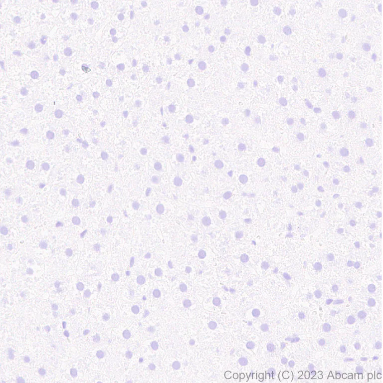
Immunohistochemical analysis of paraffin-embedded Rat liver tissue labeling SLC2A4 with ab313775 at 1/5000 (0.104 µg/ml) followed by a ready to use LeicaDS9800 (Bond™ Polymer Refine Detection). Negative control : No staining on rat liver. The section was incubated with ab313775 for 30 mins at room temperature. The immunostaining was performed on a Leica Biosystems BOND® RX instrument Counterstained with Hematoxylin. Secondary antibody only control : Secondary antibody is a ready to use LeicaDS9800 (Bond™ Polymer Refine Detection).Heat mediated antigen retrieval was performed with Citrate buffer (pH 6.0, Epitope Retrieval Solution 1) for 20 mins
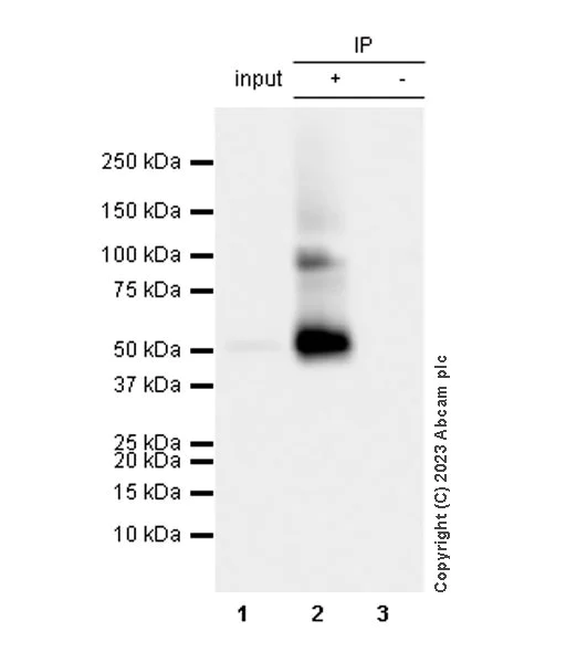
- IP
Supplier Data
Immunoprecipitation - Anti-Glucose Transporter GLUT4 antibody [EPR26571-45] (AB313775)
SLC2A4 was immunoprecipitated from 0.35 mg Mouse heart tissue lysate with ab313775 at 1/30 dilution (2µg in 0.35mg lysates). Western blot was performed on the immunoprecipitate using ab313775 at 1/1000 dilution. VeriBlot for IP secondary antibody (HRP) (ab131366) was used at 1/5000 dilution.
Lane 1 : Mouse heart tissue lysate 5 µg
Lane 2 : ab313775 IP in Mouse heart tissue lysate
Lane 3 : Rabbit monoclonal IgG (ab172730) instead of ab313775 in mouse heart tissue lysate.
Blocking and dilution buffer and concentration : 5% NFDM/TBST. Exposure time : 3 seconds The expression profile/ molecular weight observed is consistent with what has been described in the literature (PMID : 27302065).
All lanes:
Immunoprecipitation - Anti-Glucose Transporter GLUT4 antibody [EPR26571-45] (ab313775) at 1/30 dilution
All lanes:
Mouse heart tissue lysate
Secondary
All lanes:
Immunoprecipitation - VeriBlot for IP Detection Reagent (HRP) (<a href='/products/reagents/veriblot-for-ip-detection-reagent-hrp-ab131366'>ab131366</a>) at 1/5000 dilution
false
Exposure time: 3s
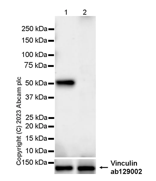
- WB
Supplier Data
Western blot - Anti-Glucose Transporter GLUT4 antibody [EPR26571-45] (AB313775)
Blocking and diluting buffer and concentration : 5% NFDM/TBST Negative control : liver (PMID : 2654938). Samples are non-boiled as boiling may cause protein aggregation. In Western blot, Anti-Vinculin antibody [EPR8185] (ab129002) staining at 1/10000 dilution. Exposure time : 48 seconds
All lanes:
Western blot - Anti-Glucose Transporter GLUT4 antibody [EPR26571-45] (ab313775) at 1/1000 dilution
Lane 1:
Human skeletal muscle tissue lysate at 20 µg
Lane 2:
Human liver tissue lysate at 20 µg
Secondary
All lanes:
Goat Anti-Rabbit IgG (HRP) with minimal cross-reactivity with human IgG at 1/1000 dilution
Observed band size: 51 kDa
false
Exposure time: 48s
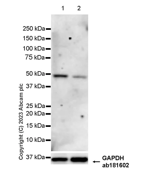
- WB
Supplier Data
Western blot - Anti-Glucose Transporter GLUT4 antibody [EPR26571-45] (AB313775)
Blocking and diluting buffer and concentration : 5% NFDM/TBST.
This blot was developed using a high sensitivity ECL substrate. The high-sensitivity ECL substrate used allows for the detection of proteins in the mid-femtogram range.
Samples are non-boiled as boiling may cause protein aggregation.
In Western blot, Anti-GAPDH antibody [EPR16891] - Loading Control (ab181602) staining at 1/200000 dilution.
All lanes:
Western blot - Anti-Glucose Transporter GLUT4 antibody [EPR26571-45] (ab313775) at 1/1000 dilution
Lane 1:
RD (Human muscle rhabdomyosarcoma) whole cell lysate at 20 µg
Lane 2:
A673 (human muscle Ewing's Sarcoma) whole cell lysate at 20 µg
Secondary
All lanes:
Western blot - Goat Anti-Rabbit IgG H&L (HRP) (<a href='/products/secondary-antibodies/goat-rabbit-igg-h-l-hrp-ab97051'>ab97051</a>) at 1/100000 dilution
Observed band size: 51 kDa
true
Exposure time: 180s
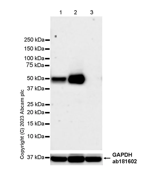
- WB
Supplier Data
Western blot - Anti-Glucose Transporter GLUT4 antibody [EPR26571-45] (AB313775)
Blocking and diluting buffer and concentration : 5% NFDM/TBST Low expression : kidney (PMID : 2654938). Samples are non-boiled as boiling may cause protein aggregation. In Western blot, Anti-GAPDH antibody [EPR16891] - Loading Control (ab181602) staining at 1/200000 dilution. Exposure time : 105 seconds
All lanes:
Western blot - Anti-Glucose Transporter GLUT4 antibody [EPR26571-45] (ab313775) at 1/1000 dilution
Lane 1:
Mouse skeletal muscle tissue lysate at 20 µg
Lane 2:
Mouse heart tissue lysate at 20 µg
Lane 3:
Mouse kidney tissue lysate at 20 µg
Secondary
All lanes:
Western blot - Goat Anti-Rabbit IgG H&L (HRP) (<a href='/products/secondary-antibodies/goat-rabbit-igg-h-l-hrp-ab97051'>ab97051</a>) at 1/100000 dilution
Observed band size: 51 kDa
false
Exposure time: 105s
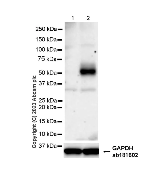
- WB
Supplier Data
Western blot - Anti-Glucose Transporter GLUT4 antibody [EPR26571-45] (AB313775)
Blocking and diluting buffer and concentration : 5% NFDM/TBST.
This blot was developed using a high sensitivity ECL substrate. The high-sensitivity ECL substrate used allows for the detection of proteins in the mid-femtogram range.
Samples are non-boiled as boiling may cause protein aggregation.
In Western blot, Anti-GAPDH antibody [EPR16891] - Loading Control (ab181602) staining at 1/200000 dilution.
All lanes:
Western blot - Anti-Glucose Transporter GLUT4 antibody [EPR26571-45] (ab313775) at 1/1000 dilution
Lane 1:
Untreated 3T3-L1 (mouse embryonic fibroblast) whole cell lysate at 20 µg
Lane 2:
3T3-L1 differentiation for 6 days whole cell lysate at 20 µg
Secondary
All lanes:
Western blot - Goat Anti-Rabbit IgG H&L (HRP) (<a href='/products/secondary-antibodies/goat-rabbit-igg-h-l-hrp-ab97051'>ab97051</a>) at 1/100000 dilution
Observed band size: 51 kDa
true
Exposure time: 158s
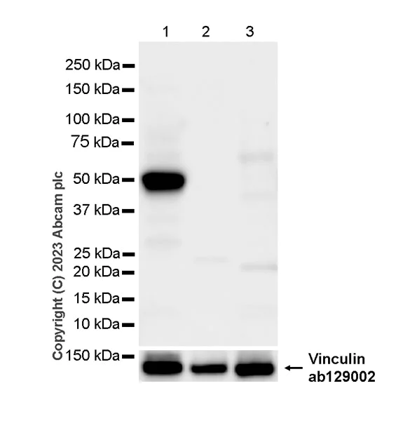
- WB
Supplier Data
Western blot - Anti-Glucose Transporter GLUT4 antibody [EPR26571-45] (AB313775)
Blocking and diluting buffer and concentration : 5% NFDM/TBST Negative control : liver (PMID : 2654938), lung (PMID : 2654938). Samples are non-boiled as boiling may cause protein aggregation. In Western blot, Anti-Vinculin antibody [EPR8185] (ab129002) staining at 1/10000 dilution. Exposure time : 15 seconds
All lanes:
Western blot - Anti-Glucose Transporter GLUT4 antibody [EPR26571-45] (ab313775) at 1/1000 dilution
Lane 1:
Rat heart tissue lysate at 20 µg
Lane 2:
Rat liver tissue lysate at 20 µg
Lane 3:
Rat lung tissue lysate at 20 µg
Secondary
All lanes:
Western blot - Goat Anti-Rabbit IgG H&L (HRP) (<a href='/products/secondary-antibodies/goat-rabbit-igg-h-l-hrp-ab97051'>ab97051</a>) at 1/100000 dilution
Observed band size: 51 kDa
false
Exposure time: 15s
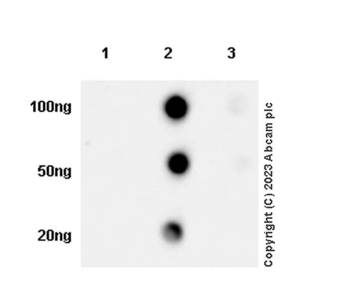
- Dot
Supplier Data
Dot Blot - Anti-Glucose Transporter GLUT4 antibody [EPR26571-45] (AB313775)
Dot blot analysis of SLC2A4 using ab313775 at 1 : 1000 (0.518 ug/ml) followed by a Goat Anti-Rabbit IgG, (H+L), Peroxidase conjugated (ab97051) at 1 : 100,000 dilution. Exposure time : 8 seconds. Blocking and diluting buffer and concentration : 5% NFDM/TBST
関連する標識済み抗体及び組成の異なる製品 (1)
-
Anti-Glucose Transporter GLUT4 antibody [EPR26571-45] - BSA and Azide free
組合せ製品
Primary Antibodies
AB115730
Anti-Glucose Transporter GLUT1 antibody [EPR3915]
BOND RX™ Validated|20ul selling size|RabMAb|Recombinant|KO Validated

- 4
- 20

Primary Antibodies
AB188317
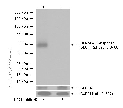
Anti-Glucose Transporter GLUT4 (phospho S488) antibody [EPR930(2)]
RabMAb|Recombinant|20ul selling size
- 5
- 3
Primary Antibodies
AB209847
Anti-Hexokinase II antibody [EPR20839]
RabMAb|Recombinant|KO Validated|20ul selling size

- 5
- 2
Reactivity data
製品の詳細
What is this antibody validated in?
Anti-Glucose Transporter GLUT4 antibody [EPR26571-45] (ab313775) is a rabbit recombinant monoclonal antibody and is validated for use in Western Blot (WB), Immunoprecipitation (IP), Immunohistochemistry (IHC-P), Immunocytochemistry/immunofluorescence (ICC/IF), Dot Blot in Human, Mouse, Rat samples.
What is the molecular weight of Glucose Transporter GLUT4?
Anti-Glucose Transporter GLUT4 [EPR26571-45] (ab313775) specifically detects a band for Glucose Transporter GLUT4 (UniProt: P14142) at a molecular weight of 54kDa.
Other related products
We have a range of other formats of antibody clone [EPR26571-45] also available for your convenience: ab313775, Carrier free - ab313776
Patented technology
Our RabMAb® technology is a patented hybridoma-based technology for making rabbit monoclonal antibodies. For details on our patents, please refer to RabMAb® patents.
What are the advantages of a recombinant monoclonal antibody?
This product is a recombinant monoclonal antibody, which offers several advantages including:
- - High batch-to-batch consistency and reproducibility
- - Improved sensitivity and specificity
- - Long-term security of supply
- - Animal-free batch production
For more information, read more on recombinant antibodies.
Trial sizes available!
Test your antibody or perform pre-screening before committing to a larger quantity. Sold in 20µl. Discover our selection of trial-size antibodies.
出荷温度及び保存条件
製品の状態
精製方法
バッファー組成
出荷温度
短期保存期間
短期保存温度
長期保存温度
分注に関する情報
保管に関する情報
補足情報
This supplementary information is collated from multiple sources and compiled automatically.
Biological function summary
GLUT4 helps regulate blood sugar levels through its control over glucose uptake in muscle and adipose tissues. As an important component of the insulin-regulated glucose transport system GLUT4 responds to increased insulin levels by moving to the cell's surface aiding in glucose transportation into the cell. This adaptive response reduces blood glucose concentration after food intake. GLUT4 operates largely independently but may interact with other glucose transporters like GLUT1 to maintain normal cellular glucose homeostasis.
Pathways
GLUT4 plays a significant role in the insulin signaling pathway and glucose homeostasis. Activation of the insulin receptor triggers a signaling cascade that includes enzymes like Akt (protein kinase B) which facilitates the translocation of GLUT4 vesicles to the plasma membrane. This translocation is important for glucose entry into the cells. Other key proteins in this pathway include IRS (insulin receptor substrate) proteins which act as signaling intermediates to link insulin receptor activation to downstream targets like GLUT4. Its activity is central to the maintenance of energy balance and storage in the body.
製品プロトコール
- Visit the General protocols
- Visit the Troubleshooting
ターゲットの情報
文献 (4)
Recent publications for all applications. Explore the full list and refine your search
Molecular medicine (Cambridge, Mass.) 31:180 PubMed40346484
2025
Applications
Unspecified application
Species
Unspecified reactive species
Journal of clinical biochemistry and nutrition 76:125-130 PubMed40151406
2025
Applications
Unspecified application
Species
Unspecified reactive species
Kidney international reports 10:227-246 PubMed39810759
2025
Applications
Unspecified application
Species
Unspecified reactive species
Diabetology & metabolic syndrome 16:33 PubMed38302999
2024
Applications
Unspecified application
Species
Unspecified reactive species
Abcam product promise
Please note: All products are 'FOR RESEARCH USE ONLY. NOT FOR USE IN DIAGNOSTIC OR THERAPEUTIC PROCEDURES'.
For licensing inquiries, please contact partnerships@abcam.com