Anti-GC-C 抗体 [EPR29752-507]
Anti-GC-C antibody [EPR29752-507]
- Advanced Validation
- BOND RX™ Validated
- RabMAb
- Recombinant
- 20ul selling size
- 詳細を見る
Be the first to review this product! Submit a review
|
(0 Publication)
Rabbit Recombinant Monoclonal GC-C antibody. Suitable for Dot, Flow Cyt, IHC-P, mIHC and reacts with Recombinant full length protein - Human, Human samples.
別名を表示する
GUC2C, STAR, GUCY2C, Guanylyl cyclase C, GC-C, Heat-stable enterotoxin receptor, Intestinal guanylate cyclase, STA receptor, hSTAR
- IHC-P
Supplier Data
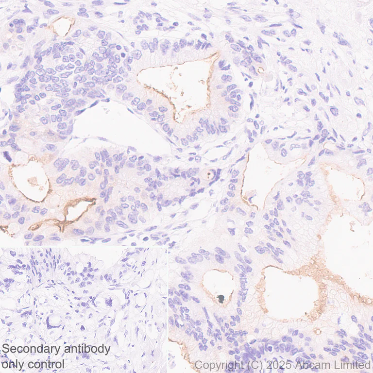
Immunohistochemistry (Formalin/PFA-fixed paraffin-embedded sections) - Anti-GC-C antibody [EPR29752-507] (AB323804)
Immunohistochemical analysis of paraffin-embedded Human pancreatic cancer tissue labeling GC-C with ab323804 at 1/100 (5.01 ug/ml) dilution, followed by a ready to use LeicaDS9800 (Bond™ Polymer Refine Detection).
Positive staining on human pancreatic cancer.
The primary antibody was incubated for 30 mins at room temperature.
The immunostaining was performed on a Leica Biosystems BOND® RX instrument
Counterstained with Hematoxylin.
Secondary antibody only control : Secondary antibody is a ready to use LeicaDS9800 (Bond™ Polymer Refine Detection).
Heat mediated antigen retrieval was performed with Tris-EDTA buffer (pH 9.0, Epitope Retrieval Solution2) for 20 mins
- mIHC
Lab
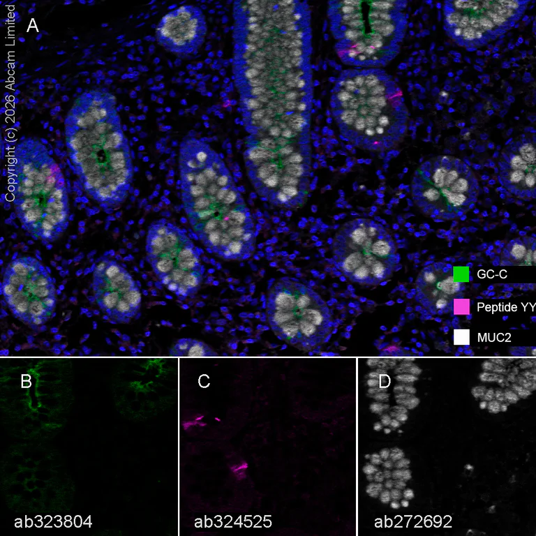
Multiplex immunohistochemistry - Anti-GC-C antibody [EPR29752-507] (AB323804)
Multiplex immunohistochemistry analysis of formalin/PFA-fixed paraffin-embedded human colon tissue staining GC-C with ab323804 at a 1/100 dilution, ab324525 anti-Peptide YY used at 1/2000 dilution and ab272692 anti-MUC2 used at a 1/2000 dilution.
Panel A : anti-GC-C (green; Opal™520), anti-Peptide YY (magenta; Opal™690) and anti-MUC2 (gray; Opal™570) on human colon.
Panel B : anti-GC-C staining apical membrane of epithelium in human colon.
Panel C : anti-Peptide YY staining enteroendocrine cells in human colon.
Panel D : anti-MUC2 staining goblet cells in human colon.
Nuclear DNA was labeled with DAPI (shown in blue).
The section was incubated in three rounds of staining : in the order of ab323804, ab324525 and ab272692 for 30 mins at room temperature. Each round was followed by a separate fluorescent tyramide signal amplification system.
The immunostaining was performed on a Leica Biosystems BOND® RX instrument with an Opal™ 4-color kit. Image acquisition was performed with Leica SP8 confocal microscope.
Heat mediated antigen retrieval was performed with Tris-EDTA buffer (pH 9.0, Epitope Retrieval Solution2) for 20 mins.
- IHC-P
Supplier Data
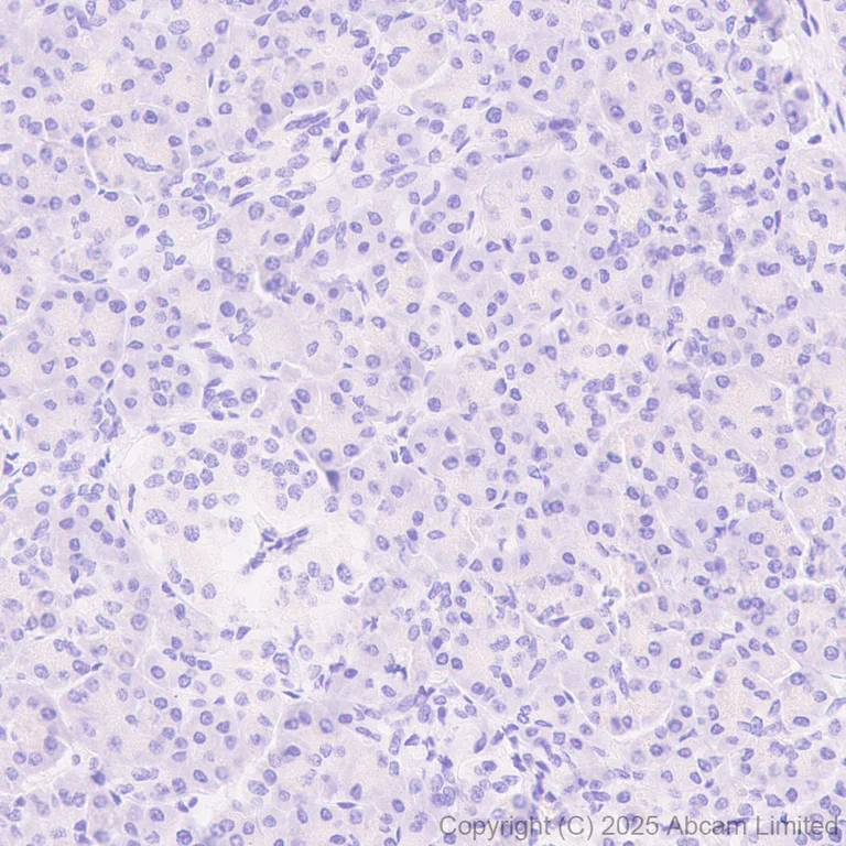
Immunohistochemistry (Formalin/PFA-fixed paraffin-embedded sections) - Anti-GC-C antibody [EPR29752-507] (AB323804)
Immunohistochemical analysis of paraffin-embedded Human pancreas tissue labeling GC-C with ab323804 at 1/100 (5.01 ug/ml) dilution, followed by a ready to use LeicaDS9800 (Bond™ Polymer Refine Detection).
Negative control : No staining on human pancreas.
The primary antibody was incubated for 30 mins at room temperature.
The immunostaining was performed on a Leica Biosystems BOND® RX instrument
Counterstained with Hematoxylin.
Secondary antibody only control : Secondary antibody is a ready to use LeicaDS9800 (Bond™ Polymer Refine Detection).
Heat mediated antigen retrieval was performed with Tris-EDTA buffer (pH 9.0, Epitope Retrieval Solution2) for 20 mins
- IHC-P
Supplier Data
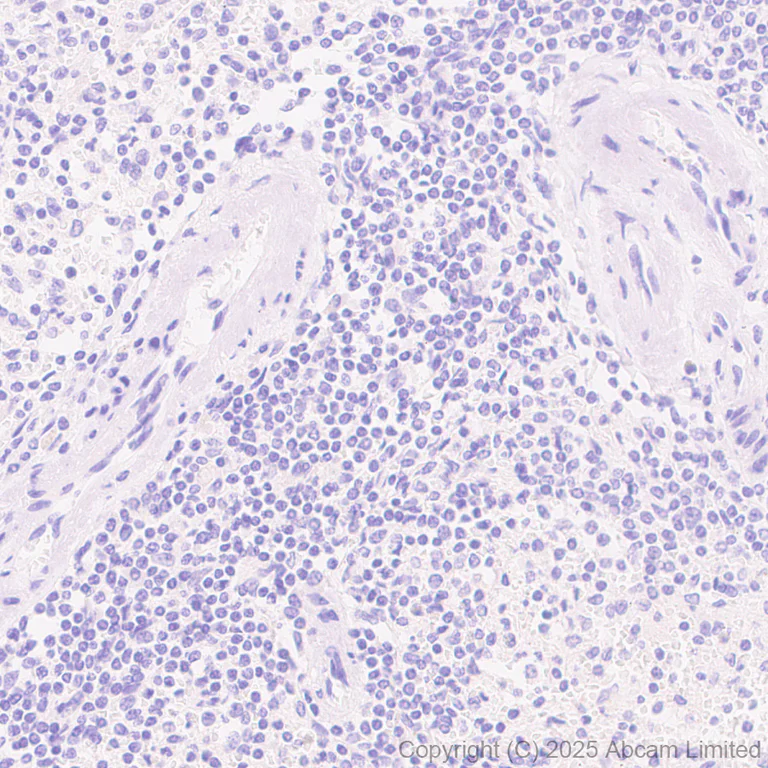
Immunohistochemistry (Formalin/PFA-fixed paraffin-embedded sections) - Anti-GC-C antibody [EPR29752-507] (AB323804)
Immunohistochemical analysis of paraffin-embedded Human spleen tissue labeling GC-C with ab323804 at 1/100 (5.01 ug/ml) dilution, followed by a ready to use LeicaDS9800 (Bond™ Polymer Refine Detection).
Negative control : No staining on human spleen.
The primary antibody was incubated for 30 mins at room temperature.
The immunostaining was performed on a Leica Biosystems BOND® RX instrument
Counterstained with Hematoxylin.
Secondary antibody only control : Secondary antibody is a ready to use LeicaDS9800 (Bond™ Polymer Refine Detection).
Heat mediated antigen retrieval was performed with Tris-EDTA buffer (pH 9.0, Epitope Retrieval Solution2) for 20 mins
- IHC-P
Supplier Data
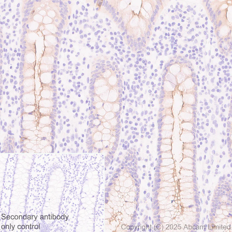
Immunohistochemistry (Formalin/PFA-fixed paraffin-embedded sections) - Anti-GC-C antibody [EPR29752-507] (AB323804)
Immunohistochemical analysis of paraffin-embedded Human colon tissue labeling GC-C with ab323804 at 1/100 (5.01 ug/ml) dilution, followed by a ready to use LeicaDS9800 (Bond™ Polymer Refine Detection).
Positive staining on human colon.
The primary antibody was incubated for 30 mins at room temperature.
The immunostaining was performed on a Leica Biosystems BOND® RX instrument
Counterstained with Hematoxylin.
Secondary antibody only control : Secondary antibody is a ready to use LeicaDS9800 (Bond™ Polymer Refine Detection).
Heat mediated antigen retrieval was performed with Tris-EDTA buffer (pH 9.0, Epitope Retrieval Solution2) for 20 mins
- IHC-P
Supplier Data
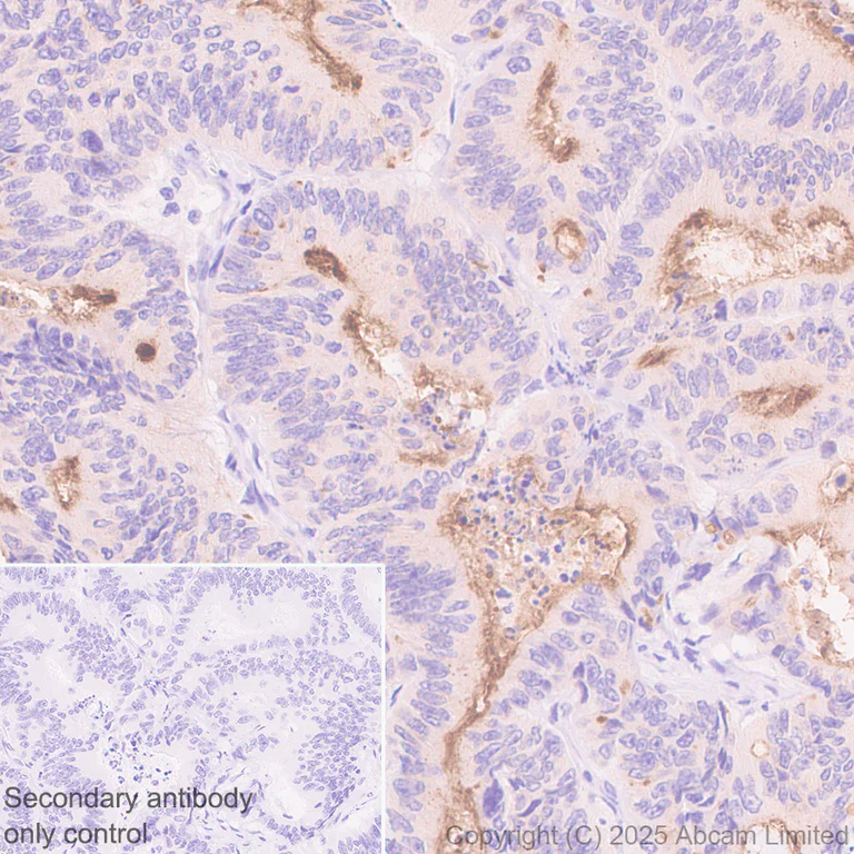
Immunohistochemistry (Formalin/PFA-fixed paraffin-embedded sections) - Anti-GC-C antibody [EPR29752-507] (AB323804)
Immunohistochemical analysis of paraffin-embedded Human colon carcinoma tissue labeling GC-C with ab323804 at 1/100 (5.01 ug/ml) dilution, followed by a ready to use LeicaDS9800 (Bond™ Polymer Refine Detection).
Positive staining on human colon carcinoma (PMID : 29261789).
The primary antibody was incubated for 30 mins at room temperature.
The immunostaining was performed on a Leica Biosystems BOND® RX instrument
Counterstained with Hematoxylin.
Secondary antibody only control : Secondary antibody is a ready to use LeicaDS9800 (Bond™ Polymer Refine Detection).
Heat mediated antigen retrieval was performed with Tris-EDTA buffer (pH 9.0, Epitope Retrieval Solution2) for 20 mins
- Flow Cyt
Supplier Data
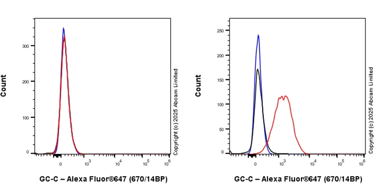
Flow Cytometry - Anti-GC-C antibody [EPR29752-507] (AB323804)
Flow cytometric analysis of HCT 116 (human colorectal carcinoma epithelial cell, Left) / T84 (human colon epithelial cell, Right) cells labeling GC-C with ab323804 at 1/500 dilution (0.1 ug) (Red) compared with a Rabbit monoclonal IgG (ab172730) (Black) isotype control and an unlabeled control (cells without incubation with primary antibody and secondary antibody).
Goat Anti-Rabbit IgG (Alexa Fluor® 647, ab150083) at 1/5000 dilution was used as the secondary antibody.
- IHC-P
Supplier Data
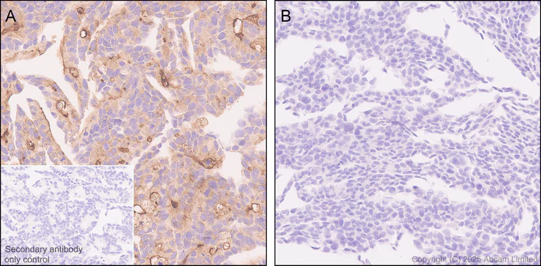
Immunohistochemistry (Formalin/PFA-fixed paraffin-embedded sections) - Anti-GC-C antibody [EPR29752-507] (AB323804)
Immunohistochemical analysis of paraffin-embedded (A) T84 (human colon epithelial cell) cell pellet. (B) HCT116 (Human colorectal carcinoma epithelial cell) cell pellet. tissue labeling GC-C with ab323804 at 1/100 (5.01 ug/ml) dilution, followed by a ready to use LeicaDS9800 (Bond™ Polymer Refine Detection).
Positive staining on (A) T84 cell pellet, no staining on (B) HCT116 cell pellet.
The primary antibody was incubated for 30 mins at room temperature.
The immunostaining was performed on a Leica Biosystems BOND® RX instrument
Counterstained with Hematoxylin.
Secondary antibody only control : Secondary antibody is a ready to use LeicaDS9800 (Bond™ Polymer Refine Detection).
Heat mediated antigen retrieval was performed with Tris-EDTA buffer (pH 9.0, Epitope Retrieval Solution2) for 20 mins
- Dot
Supplier Data
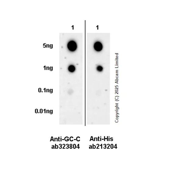
Dot Blot - Anti-GC-C antibody [EPR29752-507] (AB323804)
Dot blot analysis of GC-C using ab323804 at 1 : 1000 (0.501 ug/ml) followed by a Goat Anti-Rabbit IgG, (H+L), Peroxidase conjugated (ab97051) at 1 : 100,000 dilution.
Lane1 : His-tagged human GC-C recombinant protein
Exposure time : 180 seconds.
Blocking and diluting buffer and concentration : 5% NFDM/TBST.
In Dot Blot, Anti-6X His tag® antibody [EPR20547] - ChIP Grade (ab213204) staining at 1/5000 dilution.
All lanes:
Dot Blot - Anti-GC-C antibody [EPR29752-507] (ab323804) at 1/1000 dilution
All lanes:
His-tagged human GC-C recombinant protein
Secondary
All lanes:
Dot Blot - Goat Anti-Rabbit IgG H&L (HRP) (<a href='/products/secondary-antibodies/goat-rabbit-igg-h-l-hrp-ab97051'>ab97051</a>) at 1/100000 dilution
false
Exposure time: 180s
関連する標識済み抗体及び組成の異なる製品 (1)
-
Anti-GC-C antibody [EPR29752-507] - BSA and Azide free
組合せ製品

Primary Antibodies
AB172730
Rabbit IgG, monoclonal [EPR25A] - Isotype Control
RabMAb|Advanced Validation|Recombinant

- 0
- 0
Primary Antibodies
AB218810
RabMAb|Recombinant|KO Validated|20ul selling size

- 5
- 1
Primary Antibodies
AB227637
Anti-Met (c-Met) antibody [SP44] - C-terminal
RabMAb|Recombinant|KO Validated|20ul selling size

- 5
- 1
Primary Antibodies
AB78078
Anti-beta III Tubulin antibody [2G10] - Neuronal Marker
BOND RX™ Validated|KO Validated|Lab Essentials

- 5
- 41
Reactivity data
製品の詳細
Patented technology
Our RabMAb® technology is a patented hybridoma-based technology for making rabbit monoclonal antibodies. For details on our patents, please refer to RabMAb® patents.
What are the advantages of a recombinant monoclonal antibody?
This product is a recombinant monoclonal antibody, which offers several advantages including:
- - High batch-to-batch consistency and reproducibility
- - Improved sensitivity and specificity
- - Long-term security of supply
- - Animal-free batch production
For more information, read more on recombinant antibodies.
出荷温度及び保存条件
製品の状態
精製方法
バッファー組成
出荷温度
短期保存期間
短期保存温度
長期保存温度
分注に関する情報
保管に関する情報
製品プロトコール
- Visit the General protocols
- Visit the Troubleshooting
ターゲットの情報
Abcam product promise
Please note: All products are 'FOR RESEARCH USE ONLY. NOT FOR USE IN DIAGNOSTIC OR THERAPEUTIC PROCEDURES'.
For licensing inquiries, please contact partnerships@abcam.com