Anti-Cytokeratin 18 抗体 [RM1051]
Anti-Cytokeratin 18 antibody [RM1051]
- BOND RX™ Validated
- 20ul selling size
- RabMAb
- Recombinant
- KO Validated
- 詳細を見る
Be the first to review this product! Submit a review
|
(0 Publication)
Rabbit Recombinant Multiclonal Cytokeratin 18 antibody. Suitable for WB, IHC-P, ICC/IF, Flow Cyt (Intra), IP and reacts with Human, Mouse, Rat samples.
別名を表示する
CYK18, PIG46, KRT18, Cell proliferation-inducing gene 46 protein, Cytokeratin-18, Keratin-18, CK-18, K18
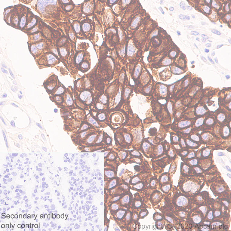
- IHC-P
Supplier Data
Immunohistochemistry (Formalin/PFA-fixed paraffin-embedded sections) - Anti-Cytokeratin 18 antibody [RM1051] (AB315096)
Immunohistochemical analysis of paraffin-embedded Human gastric cancer tissue labeling Cytokeratin 18 with ab315096 at 1/500 (0.948 ug/ml) dilution, followed by a ready to use LeicaDS9800 (Bond™ Polymer Refine Detection).
Positive staining on human gastric cancer. The section was incubated with ab315096 for 30 mins at room temperature. The immunostaining was performed on a Leica Biosystems BOND® RX instrument Counterstained with Hematoxylin.
Secondary antibody only control : Secondary antibody is a ready to use LeicaDS9800 (Bond™ Polymer Refine Detection).
Heat mediated antigen retrieval was performed with Citrate buffer (pH 6.0, Epitope Retrieval Solution 1) for 20 mins
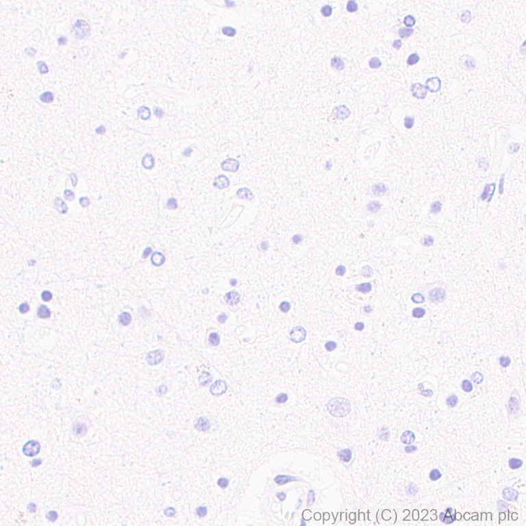
- IHC-P
Supplier Data
Immunohistochemistry (Formalin/PFA-fixed paraffin-embedded sections) - Anti-Cytokeratin 18 antibody [RM1051] (AB315096)
Immunohistochemical analysis of paraffin-embedded Human cerebrum tissue labeling Cytokeratin 18 with ab315096 at 1/500 (0.948 ug/ml) dilution, followed by a ready to use LeicaDS9800 (Bond™ Polymer Refine Detection).
Negative control : no staining on human cerebrum. The section was incubated with ab315096 for 30 mins at room temperature. The immunostaining was performed on a Leica Biosystems BOND® RX instrument Counterstained with Hematoxylin.
Secondary antibody only control : Secondary antibody is a ready to use LeicaDS9800 (Bond™ Polymer Refine Detection).
Heat mediated antigen retrieval was performed with Citrate buffer (pH 6.0, Epitope Retrieval Solution 1) for 20 mins
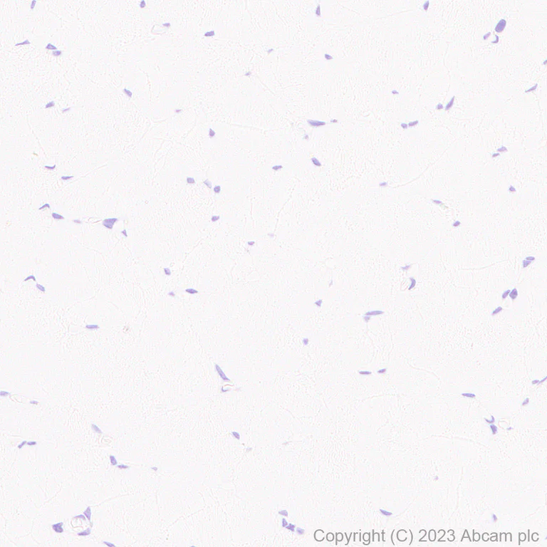
- IHC-P
Supplier Data
Immunohistochemistry (Formalin/PFA-fixed paraffin-embedded sections) - Anti-Cytokeratin 18 antibody [RM1051] (AB315096)
Immunohistochemical analysis of paraffin-embedded Human skeletal muscle tissue labeling Cytokeratin 18 with ab315096 at 1/500 (0.948 ug/ml) dilution, followed by a ready to use LeicaDS9800 (Bond™ Polymer Refine Detection).
Negative control : no staining on human skeletal muscle. The section was incubated with ab315096 for 30 mins at room temperature. The immunostaining was performed on a Leica Biosystems BOND® RX instrument Counterstained with Hematoxylin.
Secondary antibody only control : Secondary antibody is a ready to use LeicaDS9800 (Bond™ Polymer Refine Detection).
Heat mediated antigen retrieval was performed with Citrate buffer (pH 6.0, Epitope Retrieval Solution 1) for 20 mins
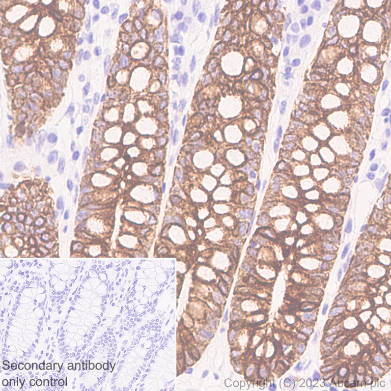
- IHC-P
Supplier Data
Immunohistochemistry (Formalin/PFA-fixed paraffin-embedded sections) - Anti-Cytokeratin 18 antibody [RM1051] (AB315096)
Immunohistochemical analysis of paraffin-embedded Human colon tissue labeling Cytokeratin 18 with ab315096 at 1/500 (0.948 ug/ml) dilution, followed by a ready to use LeicaDS9800 (Bond™ Polymer Refine Detection).
Positive staining on human colon.The section was incubated with ab315096 for 30 mins at room temperature. The immunostaining was performed on a Leica Biosystems BOND® RX instrument Counterstained with Hematoxylin.
Secondary antibody only control : Secondary antibody is a ready to use LeicaDS9800 (Bond™ Polymer Refine Detection).
Heat mediated antigen retrieval was performed with Citrate buffer (pH 6.0, Epitope Retrieval Solution 1) for 20 mins
- Flow Cyt (Intra)
Supplier Data
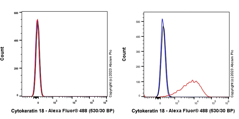
Flow Cytometry (Intracellular) - Anti-Cytokeratin 18 antibody [RM1051] (AB315096)
Flow cytometric analysis of 4% paraformaldehyde fixed 90% methanol permeabilized Molt-4 (human lymphoblastic leukemia T lymphoblast, Left) / A431 (human epidermoid carcinoma epithelial cell, Right) cells labelling Cytokeratin 18 with ab315096 at 1/500 dilution (0.1 ug, Red) compared with a Rabbit monoclonal IgG (ab172730) (Black) isotype control and an unlabelled control (cells without incubation with primary antibody and secondary antibody) (Blue).
Goat Anti-Rabbit IgG (Alexa Fluor® 488, ab150081) at 1/5000 dilution was used as the secondary antibody.
Negative control : Molt-4.
- IHC-P
Supplier Data
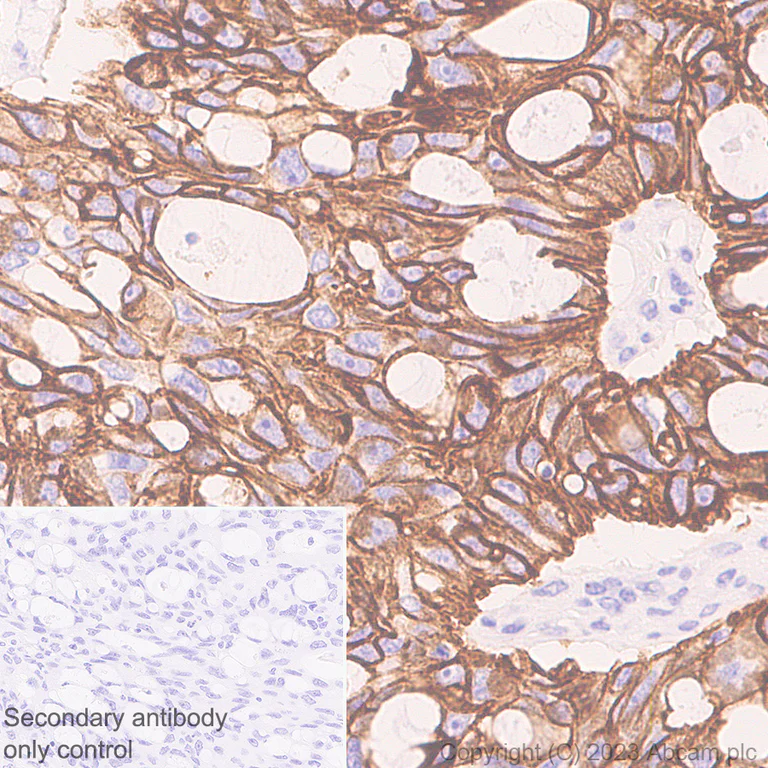
Immunohistochemistry (Formalin/PFA-fixed paraffin-embedded sections) - Anti-Cytokeratin 18 antibody [RM1051] (AB315096)
Immunohistochemical analysis of paraffin-embedded Human ovarian cancer tissue labeling Cytokeratin 18 with ab315096 at 1/500 (0.948 ug/ml) dilution, followed by a ready to use LeicaDS9800 (Bond™ Polymer Refine Detection).
Positive staining on human ovarian cancer.The section was incubated with ab315096 for 30 mins at room temperature.The immunostaining was performed on a Leica Biosystems BOND® RX instrument Counterstained with Hematoxylin.
Secondary antibody only control : Secondary antibody is a ready to use LeicaDS9800 (Bond™ Polymer Refine Detection).
Heat mediated antigen retrieval was performed with Citrate buffer (pH 6.0, Epitope Retrieval Solution 1) for 20 mins
- ICC/IF
Supplier Data
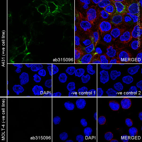
Immunocytochemistry/ Immunofluorescence - Anti-Cytokeratin 18 antibody [RM1051] (AB315096)
Immunofluorescent analysis of 4% Paraformaldehyde-fixed, 0.1% TritonX-100 permeabilized A431 (human epidermoid carcinoma epithelial cell) cells labelling Cytokeratin 18 with ab315096 at 1/50 (9.48 ug/ml) dilution, followed by ab150081 Goat Anti-Rabbit IgG H&L (Alexa Fluor® 488) preadsorbed antibody at 1/1000 (2 ug/ml) dilution (Green).
Confocal image showing cytoplasmic staining in A431 cell line. Negative control : MOLT-4.Image was taken with a confocal microscope(Leica-Microsystems, TCS SP8).
ab7291 Anti-alpha Tubulin mouse monoclonal antibody was used to counterstain tubulin at 1/1000 (1 ug/ml) dilution (Red). The Nuclear counterstain was DAPI (Blue).
Secondary antibody only control : Secondary antibody is ab150081 Goat Anti-Rabbit IgG H&L (Alexa Fluor® 488) preadsorbed at 1/1000 (2 ug/ml) dilution.
- IHC-P
Supplier Data
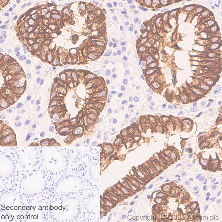
Immunohistochemistry (Formalin/PFA-fixed paraffin-embedded sections) - Anti-Cytokeratin 18 antibody [RM1051] (AB315096)
Immunohistochemical analysis of paraffin-embedded Human stomach tissue labeling Cytokeratin 18 with ab315096 at 1/500 (0.948 ug/ml) dilution, followed by a ready to use LeicaDS9800 (Bond™ Polymer Refine Detection).
Positive staining on human stomach. The section was incubated with ab315096 for 30 mins at room temperature. The immunostaining was performed on a Leica Biosystems BOND® RX instrument Counterstained with Hematoxylin.
Secondary antibody only control : Secondary antibody is a ready to use LeicaDS9800 (Bond™ Polymer Refine Detection).
Heat mediated antigen retrieval was performed with Citrate buffer (pH 6.0, Epitope Retrieval Solution 1) for 20 mins
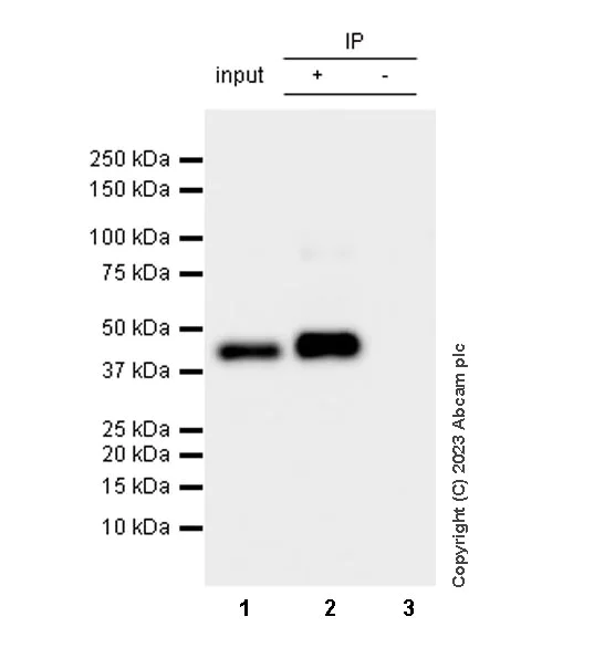
- IP
Supplier Data
Immunoprecipitation - Anti-Cytokeratin 18 antibody [RM1051] (AB315096)
Cytokeratin 18 was immunoprecipitated from 0.35 mg A431 (human epidermoid carcinoma epithelial cell) whole cell lysate with ab315096 at 1/30 dilution (2ug in 0.35mg lysates). Western blot was performed on the immunoprecipitate using ab315096 at 1/1000 dilution. VeriBlot for IP secondary antibody(HRP)(ab131366) was used at 1/5000 dilution.
Lane 1 : A431 (human epidermoid carcinoma epithelial cell) whole cell lysate
Lane 2 : ab315096 IP in A431 (human epidermoid carcinoma epithelial cell) whole cell lysate
Lane 3 : Rabbit monoclonal IgG (ab172730) instead of ab315096 in A431 whole cell lysate
Blocking and dilution buffer and concentration : 5% NFDM/TBST.
All lanes:
Immunoprecipitation - Anti-Cytokeratin 18 antibody [RM1051] (ab315096) at 1/30 dilution
All lanes:
A431 (human epidermoid carcinoma epithelial cell) whole cell lysate
Secondary
All lanes:
Immunoprecipitation - VeriBlot for IP Detection Reagent (HRP) (<a href='/products/reagents/veriblot-for-ip-detection-reagent-hrp-ab131366'>ab131366</a>) at 1/5000 dilution
false
Exposure time: 15s
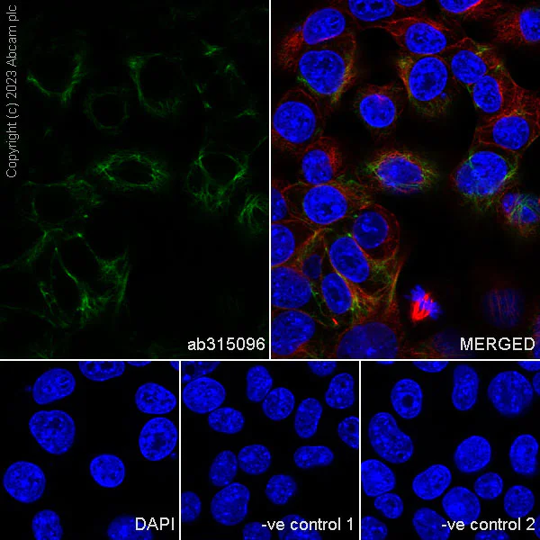
- ICC/IF
Supplier Data
Immunocytochemistry/ Immunofluorescence - Anti-Cytokeratin 18 antibody [RM1051] (AB315096)
Immunofluorescent analysis of 4% Paraformaldehyde-fixed, 0.1% TritonX-100 permeabilized Hepa1-6 (mouse hepatoma epithelial cell) cells labelling Cytokeratin 18 with ab315096 at 1/50 (9.48 ug/ml) dilution, followed by ab150081 Goat Anti-Rabbit IgG H&L (Alexa Fluor® 488) preadsorbed antibody at 1/1000 (2 ug/ml) dilution (Green).
Confocal image showing cytoplasmic staining in Hepa1-6 cell line. Image was taken with a confocal microscope(Leica-Microsystems, TCS SP8).
ab7291 Anti-alpha Tubulin mouse monoclonal antibody was used to counterstain tubulin at 1/1000 (1 ug/ml) dilution (Red). The Nuclear counterstain was DAPI (Blue).
Secondary antibody only control : Secondary antibody is ab150081 Goat Anti-Rabbit IgG H&L (Alexa Fluor® 488) preadsorbed at 1/1000 (2 ug/ml) dilution.
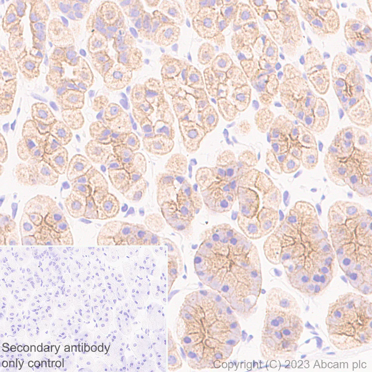
- IHC-P
Supplier Data
Immunohistochemistry (Formalin/PFA-fixed paraffin-embedded sections) - Anti-Cytokeratin 18 antibody [RM1051] (AB315096)
Immunohistochemical analysis of paraffin-embedded Mouse stomach tissue labeling Cytokeratin 18 with ab315096 at 1/500 (0.948 ug/ml) dilution, followed by a ready to use LeicaDS9800 (Bond™ Polymer Refine Detection).
Positive staining on mouse stomach. The section was incubated with ab315096 for 30 mins at room temperature. The immunostaining was performed on a Leica Biosystems BOND® RX instrument Counterstained with Hematoxylin.
Secondary antibody only control : Secondary antibody is a ready to use LeicaDS9800 (Bond™ Polymer Refine Detection).
Heat mediated antigen retrieval was performed with Citrate buffer (pH 6.0, Epitope Retrieval Solution 1) for 20 mins
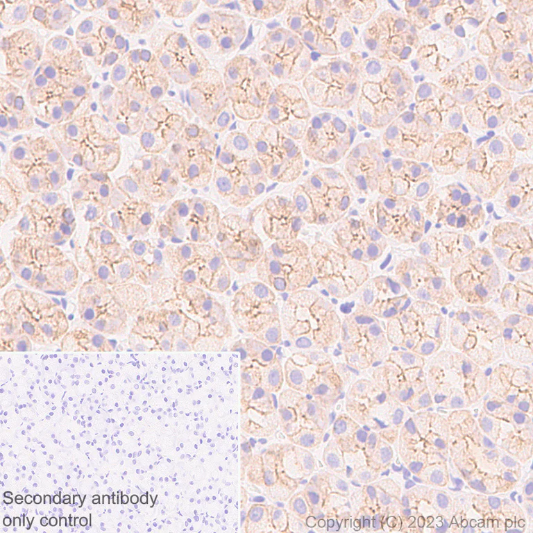
- IHC-P
Supplier Data
Immunohistochemistry (Formalin/PFA-fixed paraffin-embedded sections) - Anti-Cytokeratin 18 antibody [RM1051] (AB315096)
Immunohistochemical analysis of paraffin-embedded Rat stomach tissue labeling Cytokeratin 18 with ab315096 at 1/500 (0.948 ug/ml) dilution, followed by a ready to use LeicaDS9800 (Bond™ Polymer Refine Detection).
Positive staining on rat stomach. The section was incubated with ab315096 for 30 mins at room temperature. The immunostaining was performed on a Leica Biosystems BOND® RX instrument Counterstained with Hematoxylin.
Secondary antibody only control : Secondary antibody is a ready to use LeicaDS9800 (Bond™ Polymer Refine Detection).
Heat mediated antigen retrieval was performed with Citrate buffer (pH 6.0, Epitope Retrieval Solution 1) for 20 mins
- Flow Cyt (Intra)
Supplier Data
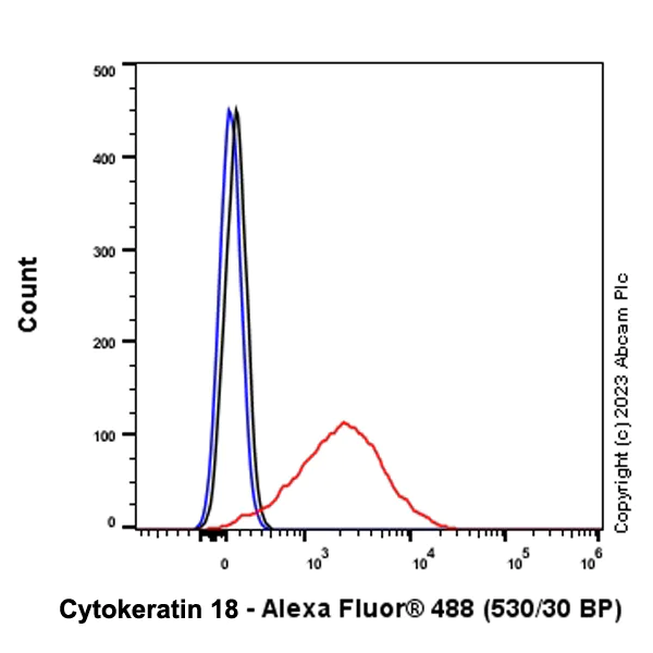
Flow Cytometry (Intracellular) - Anti-Cytokeratin 18 antibody [RM1051] (AB315096)
Flow cytometric analysis of 4% paraformaldehyde fixed 90% methanol permeabilized Hepa1-6 (mouse hepatoma epithelial cell) cells labelling Cytokeratin 18 with ab315096 at 1/500 dilution (0.1 ug, Red) compared with a Rabbit monoclonal IgG (ab172730) (Black) isotype control and an unlabelled control (cells without incubation with primary antibody and secondary antibody) (Blue).
Goat Anti-Rabbit IgG (Alexa Fluor® 488, ab150081) at 1/5000 dilution was used as the secondary antibody.
- IP
Supplier Data
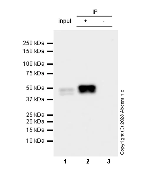
Immunoprecipitation - Anti-Cytokeratin 18 antibody [RM1051] (AB315096)
Cytokeratin 18 was immunoprecipitated from 0.35 mg Hepa1-6 (mouse hepatoma epithelial cell) whole cell lysate with ab315096 at 1/30 dilution (2ug in 0.35mg lysates). Western blot was performed on the immunoprecipitate using ab315096 at 1/1000 dilution. VeriBlot for IP secondary antibody(HRP)(ab131366) was used at 1/5000 dilution.
Lane 1 : Hepa1-6 (mouse hepatoma epithelial cell) whole cell lysate
Lane 2 : ab315096 IP in Hepa1-6 (mouse hepatoma epithelial cell) whole cell lysate
Lane 3 : Rabbit monoclonal IgG (ab172730) instead of ab315096 in Hepa1-6 whole cell lysate
Blocking and dilution buffer and concentration : 5% NFDM/TBST.
All lanes:
Immunoprecipitation - Anti-Cytokeratin 18 antibody [RM1051] (ab315096) at 1/30 dilution
All lanes:
Hepa1-6 (mouse hepatoma epithelial cell) whole cell lysate
Secondary
All lanes:
Immunoprecipitation - VeriBlot for IP Detection Reagent (HRP) (<a href='/products/reagents/veriblot-for-ip-detection-reagent-hrp-ab131366'>ab131366</a>) at 1/5000 dilution
false
Exposure time: 15s
- WB
Supplier Data
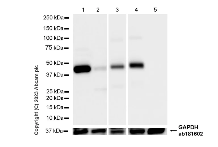
Western blot - Anti-Cytokeratin 18 antibody [RM1051] (AB315096)
Blocking and diluting buffer and concentration : 5% NFDM/TBST.
Low expression : Skeletal muscle.
Generation of the 22kDa fragment can be induced by staurosporine treatment (lane 1-2) (PMID : 34322741 and PMID : 9298992).
In Western blot, Anti-GAPDH antibody [EPR16891] - Loading Control (ab181602) staining at 1/200000 dilution.
Exposure time : Lanes 1-2 : 8 seconds; Lanes 3-5 : 1 second.
All lanes:
Western blot - Anti-Cytokeratin 18 antibody [RM1051] (ab315096) at 1/1000 dilution
Lane 1:
Untreated HeLa (human cervical adenocarcinoma epithelial cell) whole cell lysate at 20 µg
Lane 2:
HeLa treated with 2uM staurosporine for 24 hours whole cell lysate at 20 µg
Lane 3:
Human lung tissue lysate at 20 µg
Lane 4:
Human liver tissue lysate at 20 µg
Lane 5:
Human skeletal muscle tissue lysate at 20 µg
Secondary
All lanes:
Western blot - Goat Anti-Rabbit IgG H&L (HRP) (<a href='/products/secondary-antibodies/goat-rabbit-igg-h-l-hrp-ab97051'>ab97051</a>) at 1/20000 dilution
Observed band size: 22 kDa,48 kDa,36 kDa
false
- WB
Supplier Data
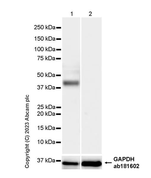
Western blot - Anti-Cytokeratin 18 antibody [RM1051] (AB315096)
Blocking and diluting buffer and concentration : 5% NFDM/TBST.
Negative control : MOLT-4 (PMID : 2473821)
In Western blot, Anti-GAPDH antibody [EPR16891] - Loading Control (ab181602) staining at 1/200000 dilution.
All lanes:
Western blot - Anti-Cytokeratin 18 antibody [RM1051] (ab315096) at 1/1000 dilution
Lane 1:
A431 (human epidermoid carcinoma epithelial cell) whole cell lysate at 7 µg
Lane 2:
MOLT-4 (human lymphoblastic leukemia T lymphoblast) whole cell lysate at 20 µg
Secondary
All lanes:
Western blot - Goat Anti-Rabbit IgG H&L (HRP) (<a href='/products/secondary-antibodies/goat-rabbit-igg-h-l-hrp-ab97051'>ab97051</a>) at 1/100000 dilution
Observed band size: 48 kDa,36 kDa
false
Exposure time: 4s
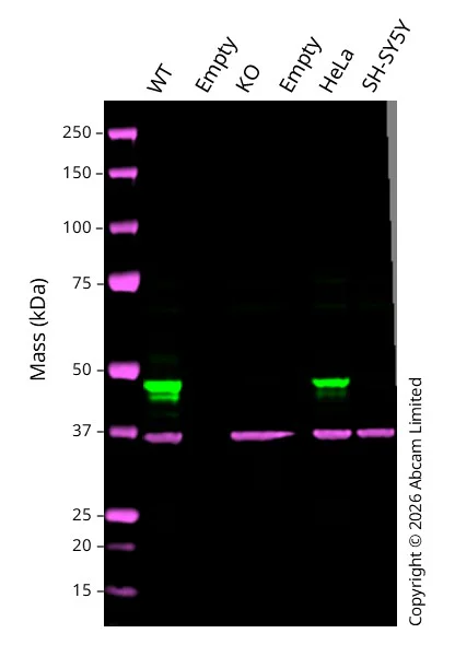
- WB
Lab
Western blot - Anti-Cytokeratin 18 antibody [RM1051] (AB315096)
Western blot : Anti-Cytokeratin 18 antibody [RM1051] ab315096 staining at 1/1000 dilution, shown in green; Mouse anti GAPDH ab8245 loading control staining at 1/20000 dilution, shown in magenta. A band was observed at 48 kDa in Wild-type A549 cell line with no signal observed at this size in KRT18 knockout A549 cell line. To generate this image, samples were run on an SDS-PAGE gel then transferred onto a nitrocellulose membrane. Membranes were blocked in 5% Milk in TBS-0.1 % Tween® 20 (TBS-T) before incubation with primary antibodies overnight at 4 °C. Blots were washed four times in TBS-T, incubated with secondary antibodies for 1 h at room temperature, washed again four times then imaged. Secondary antibodies used were Goat anti-Rabbit 800CW and Goat anti-Mouse 680RD at 1/20,000 dilution.
All lanes:
Western blot - Anti-Cytokeratin 18 antibody [RM1051] (ab315096) at 1/1000 dilution
Lane 1:
Wild-type A549 cell lysates at 20 µg
Lanes 2 and 4:
Empty
Lane 3:
KRT18 knockout A549 cell lysates at 20 µg
Lane 5:
HeLa cell lysates at 20 µg
Lane 6:
SH-SY5Y cell lysates at 20 µg
Secondary
All lanes:
Goat anti-Rabbit 800CW & Goat anti-Mouse 680RD at 1/20000 dilution
Predicted band size: 48 kDa
Observed band size: 48 kDa
false
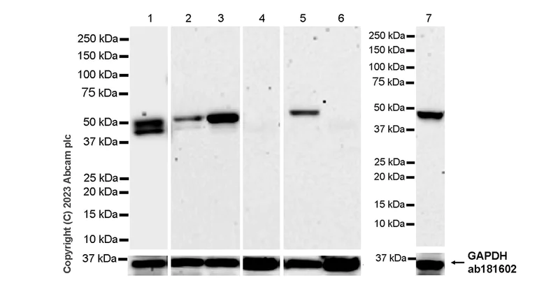
- WB
Supplier Data
Western blot - Anti-Cytokeratin 18 antibody [RM1051] (AB315096)
Blocking and diluting buffer and concentration : 5% NFDM/TBST.
Low expression : Skeletal muscle.
The observed MW is consistent with what has been described in the literature (PMID : 9298992).
In Western blot, Anti-GAPDH antibody [EPR16891] - Loading Control (ab181602) staining at 1/200000 dilution.
Exposure time : Lanes 1 : 26 seconds; Lanes 2-7 : 180 seconds.
All lanes:
Western blot - Anti-Cytokeratin 18 antibody [RM1051] (ab315096) at 1/1000 dilution
Lane 1:
Hepa1-6 (mouse hepatoma epithelial cell) whole cell lysate at 20 µg
Lane 2:
Mouse colon tissue lysate at 20 µg
Lane 3:
Mouse liver tissue lysate at 20 µg
Lane 4:
Mouse skeletal muscle tissue lysate at 20 µg
Lane 5:
Rat liver tissue lysate at 20 µg
Lane 6:
Rat skeletal muscle tissue lysate at 20 µg
Lane 7:
AR42J (rat pancreatic tumor epithelial cell) whole cell lysate at 20 µg
Secondary
All lanes:
Western blot - Goat Anti-Rabbit IgG H&L (HRP) (<a href='/products/secondary-antibodies/goat-rabbit-igg-h-l-hrp-ab97051'>ab97051</a>) at 1/20000 dilution
Observed band size: 45 kDa,48 kDa,36 kDa
false
関連する標識済み抗体及び組成の異なる製品 (1)
-
Anti-Cytokeratin 18 antibody [RM1051] - BSA and Azide free
組合せ製品

Primary Antibodies
AB53280
Anti-Cytokeratin 8 antibody [EP1628Y] - Cytoskeleton Marker
KO Validated|RabMAb|Recombinant|Lab Essentials|20ul selling size

- 5
- 9
Primary Antibodies
AB52625
Anti-Cytokeratin 19 antibody [EP1580Y] - Cytoskeleton Marker
BOND RX™ Validated|RabMAb|Recombinant|Lab Essentials|20ul selling size|Advanced Validation

- 5
- 12
Primary Antibodies
AB92547
Anti-Vimentin antibody [EPR3776] - Cytoskeleton Marker
BOND RX™ Validated|KO Validated|RabMAb|Recombinant|Lab Essentials|20ul selling size|Advanced Validation

- 5
- 92
Primary Antibodies
AB119695
Anti-Cytokeratin 14 antibody [SP53]
BOND RX™ Validated|20ul selling size|RabMAb|Advanced Validation|Recombinant|KO Validated

- 5
- 6
Reactivity data
製品の詳細
What are recombinant multiclonals?
Recombinant multiclonals are a mixture of recombinant antibodies co-expressed from a library of heavy and light chains. They offer several advantages including:
- - The sensitivity of polyclonal antibodies by recognising multiple epitopes
- - High batch-to-batch consistency and reproducibility
- - Improved sensitivity and specificity
- - Long-term security of supply
- - Animal-free batch production
View our range of recombinant multiclonal antibodies.
Patented technology
Our RabMAb® technology is a patented hybridoma-based technology for making rabbit monoclonal antibodies. For details on our patents, please refer to RabMAb® patents.
出荷温度及び保存条件
製品の状態
精製方法
バッファー組成
出荷温度
短期保存期間
短期保存温度
長期保存温度
分注に関する情報
保管に関する情報
補足情報
This supplementary information is collated from multiple sources and compiled automatically.
Biological function summary
CK18 maintains cellular integrity and mediates resistance to mechanical and non-mechanical stresses in epithelial cells. It forms heterodimers with KRT8 participating in a complex network of filaments important for maintaining cell shape and stability. This organization plays a role in managing cell survival during stress and apoptosis helping regulate cellular functions and responses in tissues where it is expressed.
Pathways
Cytokeratin 18 is involved in the apoptosis and cellular stress response pathways. It interacts with proteins such as caspases during apoptosis facilitating cytoskeletal reorganization and cell fragmentation. CK18 in coordination with KRT8 also participates in signaling pathways that regulate cell cycle and apoptotic processes underlining its importance in cellular turnover and tissue homeostasis.
製品プロトコール
- Visit the General protocols
- Visit the Troubleshooting
ターゲットの情報
Abcam product promise
Please note: All products are 'FOR RESEARCH USE ONLY. NOT FOR USE IN DIAGNOSTIC OR THERAPEUTIC PROCEDURES'.
For licensing inquiries, please contact partnerships@abcam.com