Anti-CTAG1B 抗体 [SP349]
Anti-CTAG1B antibody [SP349]
- RabMAb
- Recombinant
- 詳細を見る
Be the first to review this product! Submit a review
|
(2 Publications)
Rabbit Recombinant Monoclonal CTAG1B antibody. Suitable for Flow Cyt, WB, IHC-P and reacts with Human samples. Cited in 2 publications.
別名を表示する
CTAG, CTAG1, ESO1, LAGE2, LAGE2A, CTAG1B, LAGE2B, CTAG1A, Cancer/testis antigen 1, Autoimmunogenic cancer/testis antigen NY-ESO-1, Cancer/testis antigen 6.1, L antigen family member 2, CT6.1, LAGE-2

- IHC-P
Supplier Data
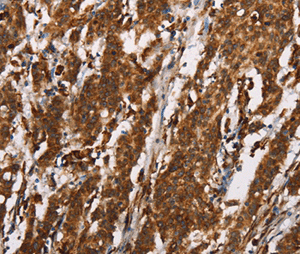
Immunohistochemistry (Formalin/PFA-fixed paraffin-embedded sections) - Anti-CTAG1B antibody [SP349] (AB223498)
Formalin-fixed paraffin-embedded human lung squamous cell carcinoma tissue stained for CTAG1B with ab223498 at 1/100 dilution in immunohistochemical analysis.
- IHC-P
Supplier Data
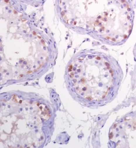
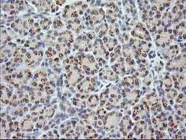
Immunohistochemistry (Formalin/PFA-fixed paraffin-embedded sections) - Anti-CTAG1B antibody [SP349] (AB223498)
Formalin-fixed paraffin-embedded human testis tissue stained for CTAG1B using ab223498 at 1/100 dilution in immunohistochemical analysis.
- Flow Cyt
Supplier Data
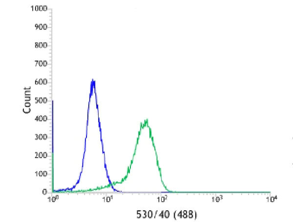
Flow Cytometry - Anti-CTAG1B antibody [SP349] (AB223498)
Flow cytometric analysis of ab223498 staining HeLa (human epithelial cell line from cervix adenocarcinoma) cells for CTAG1B (green) at 1/100 dilution compared to negative control of rabbit IgG (blue).
- IHC-P
Unknown
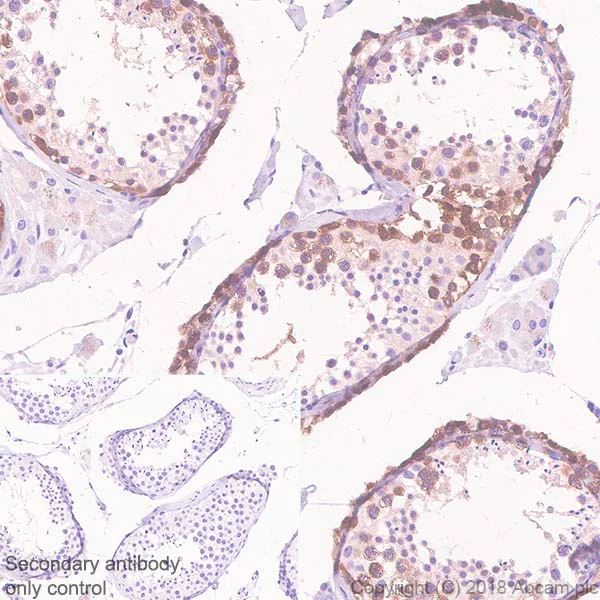
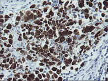
Immunohistochemistry (Formalin/PFA-fixed paraffin-embedded sections) - Anti-CTAG1B antibody [SP349] (AB223498)
Immunohistochemistry (Formalin/PFA-fixed paraffin-embedded sections) analysis of Human testis tissue sections labeling CTAG1B with Purified ab223498 at 1/100 dilution (3.83 µg/ml). Heat mediated antigen retrieval was performed Heat mediated antigen retrieval with sodium citrate buffer (pH 6.0, epitope retrieval solution
- for 10mins. Goat Anti-Rabbit & Mouse IgG (HRP) was used as the secondary antibody. Negative control : PBS instead of the primary antibody. Hematoxylin was used as a counterstain.

- Flow Cyt
Unknown
Flow Cytometry - Anti-CTAG1B antibody [SP349] (AB223498)
Flow Cytometry analysis of NCI-H1299 (human lung carcinoma epithelial cell) cells labeling CTAG1B with purified ab223498 at 1 : 380 dilution (1.01 µg/ml) - Red. Cells were fixed with 4% paraformaldehyde . A Goat anti rabbit IgG (Alexa Fluor® 488, ab150081) secondary antibody was used at 1 : 2000 dilution. Isotype control - Rabbit monoclonal IgG (ab172730) - Black. Unlabeled control - Blue.
- IHC-P
Unknown
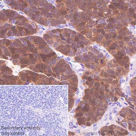
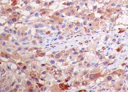
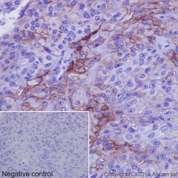
Immunohistochemistry (Formalin/PFA-fixed paraffin-embedded sections) - Anti-CTAG1B antibody [SP349] (AB223498)
Immunohistochemistry (Formalin/PFA-fixed paraffin-embedded sections) analysis of Human gastric carcinoma tissue sections labeling CTAG1B with Purified ab223498 at 1/100 dilution (3.83 µg/ml). Heat mediated antigen retrieval was performed Heat mediated antigen retrieval with sodium citrate buffer (pH 6.0, epitope retrieval solution 1) for 10mins. Goat Anti-Rabbit & Mouse IgG (HRP) was used as the secondary antibody. Negative control : PBS instead of the primary antibody. Hematoxylin was used as a counterstain.

- WB
Supplier Data
Western blot - Anti-CTAG1B antibody [SP349] (AB223498)
All lanes:
Western blot - Anti-CTAG1B antibody [SP349] (ab223498) at 1/100 dilution
Lane 1:
HEK-293 (human epithelial cell line from embryonic kidney) transfected with LAGE-1 cell lysate.
Lane 2:
HEK-293 (human epithelial cell line from embryonic kidney) CTAG1B isoform1a transfected cell lysate.
Predicted band size: 18 kDa
false
関連する標識済み抗体及び組成の異なる製品 (1)
-
Anti-CTAG1B antibody [SP349] - BSA and Azide free
代替製品
Primary Antibodies
AB183740
Anti-CTAG1B antibody [EPR13780]
20ul selling size|BOND RX™ Validated|RabMAb|Recombinant|Trial Size

- 3
- 1
Primary Antibodies
AB225536
Anti-CTAG1B antibody [EPR13780] - BSA and Azide free
BOND RX™ Validated|RabMAb|Recombinant
- 0
- 0
組合せ製品
Primary Antibodies
AB219650
Anti-PRAME antibody [EPR20330]
BOND RX™ Validated|RabMAb|Recombinant|20ul selling size

- 0
- 0
Primary Antibodies
AB172730
Rabbit IgG, monoclonal [EPR25A] - Isotype Control
RabMAb|Advanced Validation|Recombinant

- 0
- 0
Primary Antibodies
AB134162
RabMAb|Recombinant|KO Validated|20ul selling size
- 0
- 0
Reactivity data
出荷温度及び保存条件
製品の状態
精製方法
バッファー組成
出荷温度
短期保存期間
短期保存温度
長期保存温度
分注に関する情報
保管に関する情報
補足情報
This supplementary information is collated from multiple sources and compiled automatically.
Biological function summary
CTAG1B is involved in the immune response against tumors. It is part of the group called cancer/testis antigens due to its low expression in normal tissues except the testes. This restricted pattern allows CTAG1B to function in distinguishing between tumor and normal cells which is important for targeted therapies. It does not belong to a specific multiprotein complex but contributes to immune recognition by presenting as an antigenic determinant.
Pathways
CTAG1B engages predominantly in cellular processes associated with the immune system's adaptive arm. It plays a role in the antigen processing and presentation pathway where it functions to engage CD8+ T cells. The protein associates with other antigens expressed in cancer such as MAGE-A1 in enabling an immune response against tumor cells. By acting alongside these proteins CTAG1B supports pathways leading to the recognition and destruction of malignant cells.
製品プロトコール
- Visit the General protocols
- Visit the Troubleshooting
ターゲットの情報
文献 (2)
Recent publications for all applications. Explore the full list and refine your search
Frontiers in oncology 13:1106434 PubMed37081973
2023
Applications
Unspecified application
Species
Unspecified reactive species
Experimental biology and medicine (Maywood, N.J.) 246:2579-2588 PubMed34515546
2021
Applications
Unspecified application
Species
Unspecified reactive species
Abcam product promise
Please note: All products are 'FOR RESEARCH USE ONLY. NOT FOR USE IN DIAGNOSTIC OR THERAPEUTIC PROCEDURES'.
For licensing inquiries, please contact partnerships@abcam.com