Anti-CD2 抗体 [OX34] - BSA and Azide free
Anti-CD2 antibody [OX34] - BSA and Azide free
- Recombinant
- 詳細を見る
Be the first to review this product! Submit a review
|
(1 Publication)
Mouse Recombinant Monoclonal CD2 antibody. Carrier free. Suitable for Flow Cyt, IHC-P, IHC-Fr and reacts with Rat samples. Cited in 1 publication.
別名を表示する
CD2, SRBC, T-cell surface antigen CD2, Erythrocyte receptor, LFA-2, LFA-3 receptor, Rosette receptor, T-cell surface antigen T11/Leu-5
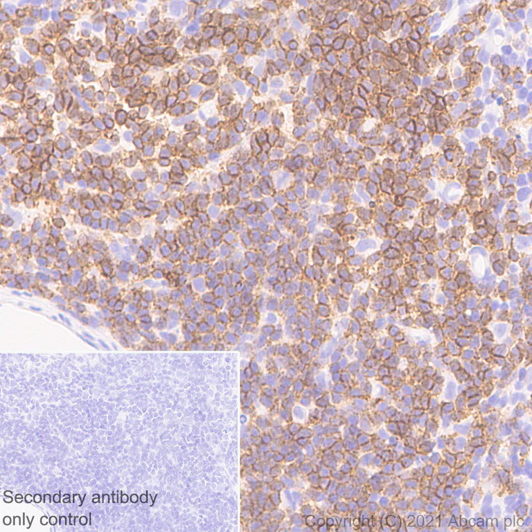
- IHC-P
Lab
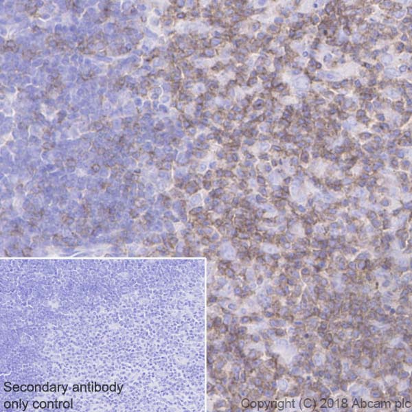
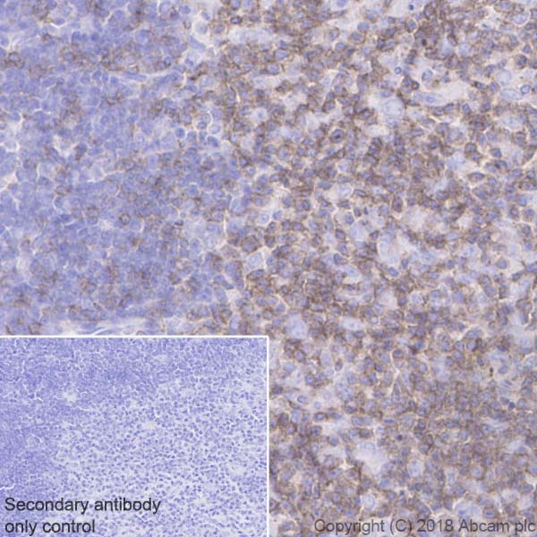
Immunohistochemistry (Formalin/PFA-fixed paraffin-embedded sections) - Anti-CD2 antibody [OX34] - BSA and Azide free (AB244591)
This data was developed using the same antibody clone in a different buffer formulation containing PBS and sodium azide (ab243837).
Immunohistochemical analysis of paraffin-embedded Rat spleen tissue labeling CD2 with ab243837 at 1/1000 (1.043 ug/ml) dilution, followed by a ready to use LeicaDS9800 (Bond™ Polymer Refine Detection). Counterstained with Hematoxylin.
Positive staining in rat spleen.
The section was incubated with ab243837 for 30 mins at room temperature, followed by anti-mouse IgG1 antibody (ab125913) for 8 mins during the LeicaDS9800 kit staining procedure.
Secondary antibody only control : Secondary antibody is a ready to use LeicaDS9800 (Bond™ Polymer Refine Detection).
Heat mediated antigen retrieval with Citrate buffer (pH 6.0, epitope retrieval solution 1) for 20 mins
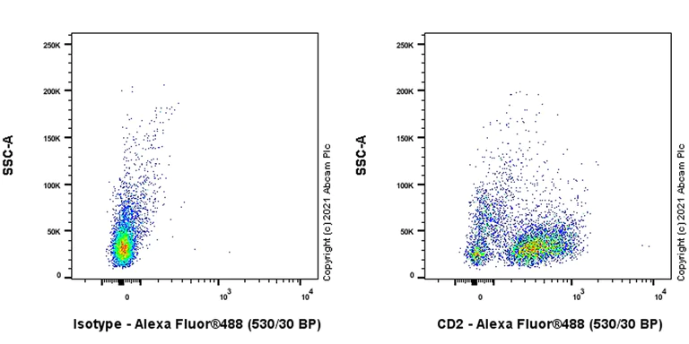
- Flow Cyt
Lab
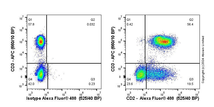
Flow Cytometry - Anti-CD2 antibody [OX34] - BSA and Azide free (AB244591)
This data was developed using the same antibody clone in a different buffer formulation containing PBS and sodium azide (ab243837).
Flow cytometric analysis of Rat splenocytes cells labelling CD2 with ab243837 at 1/1000 dilution (0.1 ug)/Right compared with a Mouse monoclonal IgG / Left isotype control.
Goat anti mouse IgG (Alexa Fluor® 488, ab150113) was used as a secondary antibody at 1/2000 dilution. Gated on viable cell.
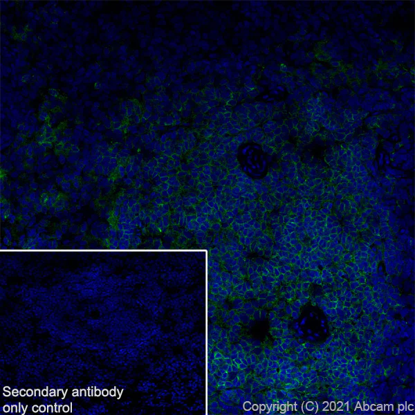
- IHC-Fr
Lab
Immunohistochemistry (Frozen sections) - Anti-CD2 antibody [OX34] - BSA and Azide free (AB244591)
This data was developed using the same antibody clone in a different buffer formulation containing PBS and sodium azide (ab243837).
Immunohistochemical analysis of 4% PFA-fixed, 0.2% Triton X-100 permeabilized frozen Rat spleen (fresh) tissue labeling CD2 with ab243837 at 1/100 (10.43 ug/ml) dilution followed by ab150113 Goat Anti-Mouse IgG H&L (Alexa Fluor® 488) at 1/1000 (2 ug/mL) dilution (Green).
Confocal image showing positive staining on rat spleen. The nuclear counterstain was DAPI (Blue).
Secondary antibody control : Secondary antibody is ab150113 Goat Anti-Mouse IgG H&L (Alexa Fluor® 488) at 1/1000 (2 ug/mL) dilution
関連する標識済み抗体及び組成の異なる製品 (4)
-
Anti-CD2 antibody [OX34]
-
660 APC
APC Anti-CD2 antibody [OX34]
-
519 Alexa Fluor® 488
Alexa Fluor® 488 Anti-CD2 antibody [OX34]
-
665 Alexa Fluor® 647
Alexa Fluor® 647 Anti-CD2 antibody [OX34]
代替製品
Primary Antibodies
AB131276
BOND RX™ Validated|20ul selling size|RabMAb|Recombinant
- 0
- 0
Primary Antibodies
AB248400
Anti-CD2 antibody [EPR6451] - BSA and Azide free
BOND RX™ Validated|RabMAb|Recombinant
- 0
- 0
Primary Antibodies
AB231059
Anti-CD2 antibody [EPR21825] - BSA and Azide free
RabMAb|Recombinant
- 0
- 0
組合せ製品
Primary Antibodies
AB16669
Anti-CD3 epsilon antibody [SP7]
BOND RX™ Validated|RabMAb|Advanced Validation|Recombinant|Lab Essentials|20ul selling size

- 4
- 80
Primary Antibodies
AB281586
BOND RX™ Validated|20ul selling size|RabMAb|Recombinant
- 5
- 3
Primary Antibodies
AB16667
BOND RX™ Validated|KO Validated|RabMAb|Advanced Validation|Recombinant|20ul selling size

- 5
- 133
Reactivity data
製品の詳細
This product has switched from a hybridoma to recombinant production method on 07 March 2024.
Want a custom formulation?
This antibody clone is manufactured by Abcam. If you require a custom buffer formulation or conjugation for your experiments, please contact orders@abcam.com
What are the advantages of a recombinant monoclonal antibody?
This product is a recombinant monoclonal antibody, which offers several advantages including:
- - High batch-to-batch consistency and reproducibility
- - Improved sensitivity and specificity
- - Long-term security of supply
- - Animal-free batch production
For more information, read more on recombinant antibodies.
Conjugation ready
Our carrier-free antibodies are typically supplied in a PBS-only formulation, purified and free of BSA, sodium azide and glycerol. This conjugation-ready format is designed for use with fluorochromes, metal isotopes, oligonucleotides, and enzymes, which makes them ideal for antibody labelling, functional and cell-based assays, flow-based assays (e.g. mass cytometry) and Multiplex Imaging applications.
Use our conjugation kits for antibody conjugates that are ready-to-use in as little as 20 minutes with 1 minute hands-on-time and 100% antibody recovery: available for fluorescent dyes, HRP, biotin and gold.
出荷温度及び保存条件
製品の状態
精製方法
バッファー組成
出荷温度
短期保存期間
短期保存温度
長期保存温度
分注に関する情報
保管に関する情報
補足情報
This supplementary information is collated from multiple sources and compiled automatically.
Biological function summary
The functioning of CD2 helps in the modulation of immune responses and T-cell activation. CD2 participates in signal transduction leading to cytokine production and proliferation. It is associated with the immunological synapse complex working closely with other surface molecules like Lck and CD3 to promote T-cell responses. This collaboration is vital for the development and function of immune cells driving adaptive immunity.
Pathways
CD2 engages in pathways integral to T-cell activation and signal transduction. A well-known pathway involving CD2 is the TCR (T-cell Receptor) signaling pathway where it partners with CD3 to initiate T-cell activation. Additionally CD2 influences the LAT (Linker for Activation of T-cells) pathway contributing to downstream signaling cascades that involve various signaling proteins such as Lck and ZAP-70 ensuring proper T-cell function.
製品プロトコール
- Visit the General protocols
- Visit the Troubleshooting
ターゲットの情報
文献 (1)
Recent publications for all applications. Explore the full list and refine your search
Journal of the National Cancer Institute 65:33-42 PubMed6993744
1980
Applications
Unspecified application
Species
Unspecified reactive species
Abcam product promise
Please note: All products are 'FOR RESEARCH USE ONLY. NOT FOR USE IN DIAGNOSTIC OR THERAPEUTIC PROCEDURES'.
For licensing inquiries, please contact partnerships@abcam.com