Anti-CD2 抗体 [EPR6451]
Anti-CD2 antibody [EPR6451]
- BOND RX™ Validated
- 20ul selling size
- RabMAb
- Recombinant
- 詳細を見る
Be the first to review this product! Submit a review
|
(4 Publications)
Rabbit Recombinant Monoclonal CD2 antibody. Suitable for IHC-P, IP, WB, Flow Cyt (Intra) and reacts with Human samples. Cited in 4 publications.
別名を表示する
CD2, SRBC, T-cell surface antigen CD2, Erythrocyte receptor, LFA-2, LFA-3 receptor, Rosette receptor, T-cell surface antigen T11/Leu-5

- Flow Cyt (Intra)
Lab
Flow Cytometry (Intracellular) - Anti-CD2 antibody [EPR6451] (AB131276)
Intracellular Flow Cytometry analysis of Jurkat (Human T cell leukemia T lymphocyte) cells labeling CD2 with purified ab131276 at 1 : 20 dilution (10 μg/ml) (Red). Cells were fixed with 4% Paraformaldehyde and permeabilised with 90% Methanol. A Goat anti rabbit IgG (Alexa Fluor® 488, ab150081) (1/2000 dilution) was used as the secondary antibody. Rabbit monoclonal IgG (Black) was used as a isotype control. Cell without incubation with primary antibody and secondary antibody (Blue) were used as unlabeled control.

- IHC-P
Unknown
Immunohistochemistry (Formalin/PFA-fixed paraffin-embedded sections) - Anti-CD2 antibody [EPR6451] (AB131276)
ab131276 showing positive staining in Hodgkin's lymphoma tissue.
Perform heat mediated antigen retrieval with citrate buffer pH 6 before commencing with IHC staining protocol.

- IHC-P
Lab
Immunohistochemistry (Formalin/PFA-fixed paraffin-embedded sections) - Anti-CD2 antibody [EPR6451] (AB131276)
Immunohistochemical analysis of Paraffin-embedded sections Human colon tissue labelling CD2 with ab131276 at 1/1000 dilution, followed by a ready to use secondary Rabbit specific IHC polymer detection kit HRP/DAB (ab209101). Staining on Human colon tissue is observed. Counter stained with Haematoxylin. Secondary antibody only control : Used PBS instead of primary antibody, secondary antibody is ready to use Rabbit specific IHC polymer detection kit HRP/DAB (ab209101). Heat mediated antigen retrieval using Bond™ Epitope Retrieval Solution 2 (pH 9.0). The immunostaining was performed on a Leica Biosystems BOND® RX instrument.

- Flow Cyt (Intra)
Lab
Flow Cytometry (Intracellular) - Anti-CD2 antibody [EPR6451] (AB131276)
Flow cytometry staining of human peripheral blood mononuclear cells (PBMCs) with ab131276 (right) or Recombinant Rabbit IgG, monoclonal [EPR25A] - Isotype Control (left).
PBMCs were incubated for 30 min on ice in 1x PBS containing 10 µg/ml human IgG and 10 % normal goat serum to block FC receptors and non-specific protein-protein interactions. Cells were fixed and permeabilised with BD Cytofix/Cytoperm™ for 20 min, followed by addition of the antibody ab131276 or Recombinant Rabbit IgG, monoclonal [EPR25A] - Isotype Control (1x 10⁶ in 100 µl at 0.04 μg/ml (1/12,500)) for 30 min at 22°C . The cells were simultaneously stained with CD3.
The secondary antibody Goat Anti-Rabbit IgG H&L (Alexa Fluor® 488) preadsorbed was incubated at 1/4000 for 30min at 22°C
Acquisition of >30000 events were collected using a 50 mW Blue laser (488nm) and 525/40 bandpass filter. Events were gated on live cells.

- IHC-P
Lab
Immunohistochemistry (Formalin/PFA-fixed paraffin-embedded sections) - Anti-CD2 antibody [EPR6451] (AB131276)
Immunohistochemical analysis of Paraffin-embedded sections Human tonsil tissue labelling CD2 with ab131276 at 1/1000 dilution, followed by a ready to use secondary Rabbit specific IHC polymer detection kit HRP/DAB (ab209101). Staining on Human tonsil tissue is observed. Counter stained with Haematoxylin. Secondary antibody only control : Used PBS instead of primary antibody, secondary antibody is ready to use Rabbit specific IHC polymer detection kit HRP/DAB (ab209101). Heat mediated antigen retrieval using Bond™ Epitope Retrieval Solution 2 (pH 9.0). The immunostaining was performed on a Leica Biosystems BOND® RX instrument.

- IHC-P
Lab
Immunohistochemistry (Formalin/PFA-fixed paraffin-embedded sections) - Anti-CD2 antibody [EPR6451] (AB131276)
Immunohistochemical analysis of Paraffin-embedded sections Human spleen tissue labelling CD2 with ab131276 at 1/1000 dilution, followed by a ready to use secondary Rabbit specific IHC polymer detection kit HRP/DAB (ab209101). Staining on Human spleen tissue is observed. Counter stained with Haematoxylin. Secondary antibody only control : Used PBS instead of primary antibody, secondary antibody is ready to use Rabbit specific IHC polymer detection kit HRP/DAB (ab209101). Heat mediated antigen retrieval using Bond™ Epitope Retrieval Solution 2 (pH 9.0). The immunostaining was performed on a Leica Biosystems BOND® RX instrument.

- IP
Lab
Immunoprecipitation - Anti-CD2 antibody [EPR6451] (AB131276)
CD2 was immunoprecipitated from 0.35 mg Jurkat (Human T cell leukemia T lymphocyte) whole cell lysate 10 μg with 131276 at 1/50 dilution (2μg). VeriBlot for IP Detection Reagent (HRP) (ab131366) was used at 1/5000 dilution.
Lane 1 : Jurkat (Human T cell leukemia T lymphocyte) whole cell lysate 10 μg
Lane 2 : ab131276 IP in Jurkat whole cell lysate
Lane 3 : Rabbit monoclonal IgG (ab172730) instead of ab131276 in Jurkat whole cell lysate
Blocking and dilution buffer and concentration : 5% NFDM/TBST.
All lanes:
Immunoprecipitation - Anti-CD2 antibody [EPR6451] (ab131276)
Predicted band size: 39 kDa
Observed band size: 50 kDa
false

- WB
Lab
Western blot - Anti-CD2 antibody [EPR6451] (AB131276)
Blocking and diluting buffer and concentration : 5% NFDM/TBST. The molecular weight observed is consistent with what has been described in the literature (PMID : 10510361).
All lanes:
Western blot - Anti-CD2 antibody [EPR6451] (ab131276) at 1/1000 dilution
All lanes:
Jurkat (Human T cell leukemia T lymphocyte) whole cell lysate at 15 µg
Secondary
All lanes:
Western blot - Goat Anti-Rabbit IgG H&L (HRP) (<a href='/products/secondary-antibodies/goat-rabbit-igg-h-l-hrp-ab97051'>ab97051</a>) at 1/20000 dilution
Predicted band size: 29 kDa
false
関連する標識済み抗体及び組成の異なる製品 (4)
-
Anti-CD2 antibody [EPR6451] - BSA and Azide free
-
519 Alexa Fluor® 488
Alexa Fluor® 488 Anti-CD2 antibody [EPR6451]
-
665 Alexa Fluor® 647
Alexa Fluor® 647 Anti-CD2 antibody [EPR6451]
-
578 PE
PE Anti-CD2 antibody [EPR6451]
代替製品


組合せ製品
Primary Antibodies
AB133616
BOND RX™ Validated|RabMAb|Recombinant|Lab Essentials|20ul selling size

- 5
- 20

Primary Antibodies
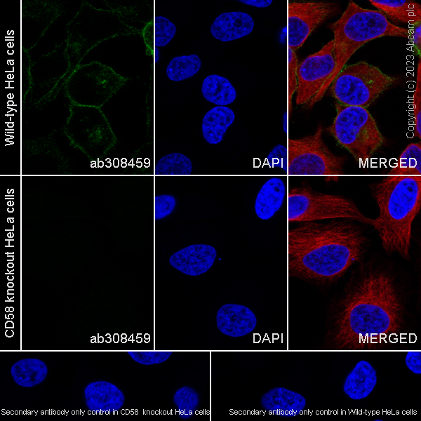
AB308459
Anti-CD58 antibody [EPR27435-85]
20ul selling size|RabMAb|Recombinant|KO Validated
- 0
- 0
Primary Antibodies
AB16667
BOND RX™ Validated|KO Validated|RabMAb|Advanced Validation|Recombinant|20ul selling size

- 5
- 133
Reactivity data
製品の詳細
Species reactivity
Mouse, Rat: We have preliminary internal testing data to indicate this antibody may not react with these species.
Please contact us for more information.
Patented technology
Our RabMAb® technology is a patented hybridoma-based technology for making rabbit monoclonal antibodies. For details on our patents, please refer to RabMAb® patents.
What are the advantages of a recombinant monoclonal antibody?
This product is a recombinant monoclonal antibody, which offers several advantages including:
- - High batch-to-batch consistency and reproducibility
- - Improved sensitivity and specificity
- - Long-term security of supply
- - Animal-free batch production
For more information, read more on recombinant antibodies.
出荷温度及び保存条件
製品の状態
精製方法
バッファー組成
出荷温度
短期保存期間
短期保存温度
長期保存温度
分注に関する情報
保管に関する情報
補足情報
This supplementary information is collated from multiple sources and compiled automatically.
Biological function summary
The functioning of CD2 helps in the modulation of immune responses and T-cell activation. CD2 participates in signal transduction leading to cytokine production and proliferation. It is associated with the immunological synapse complex working closely with other surface molecules like Lck and CD3 to promote T-cell responses. This collaboration is vital for the development and function of immune cells driving adaptive immunity.
Pathways
CD2 engages in pathways integral to T-cell activation and signal transduction. A well-known pathway involving CD2 is the TCR (T-cell Receptor) signaling pathway where it partners with CD3 to initiate T-cell activation. Additionally CD2 influences the LAT (Linker for Activation of T-cells) pathway contributing to downstream signaling cascades that involve various signaling proteins such as Lck and ZAP-70 ensuring proper T-cell function.
製品プロトコール
- Visit the General protocols
- Visit the Troubleshooting
ターゲットの情報
文献 (4)
Recent publications for all applications. Explore the full list and refine your search
Nature genetics 57:1142-1154 PubMed40229600
2025
Applications
Unspecified application
Species
Unspecified reactive species
European journal of haematology 109:513-518 PubMed35871391
2022
Applications
Unspecified application
Species
Unspecified reactive species
Breast cancer research : BCR 21:46 PubMed30922380
2019
Applications
Unspecified application
Species
Unspecified reactive species
Journal of gastroenterology 52:39-49 PubMed27032393
2016
Applications
Unspecified application
Species
Unspecified reactive species
Abcam product promise
Please note: All products are 'FOR RESEARCH USE ONLY. NOT FOR USE IN DIAGNOSTIC OR THERAPEUTIC PROCEDURES'.
For licensing inquiries, please contact partnerships@abcam.com