Anti-CCL3 / MIP-1 alpha + CCL3L1 抗体 [EPR23751-54]
Anti-CCL3 / MIP-1 alpha + CCL3L1 antibody [EPR23751-54]
- BOND RX™ Validated
- RabMAb
- Recombinant
- 20ul selling size
- 詳細を見る
Be the first to review this product! Submit a review
|
(9 Publications)
Rabbit Recombinant Monoclonal MIP-1 alpha/CCL3 antibody. Suitable for ICC/IF, IP, WB, Flow Cyt (Intra), IHC-P and reacts with Mouse, Human, Recombinant fragment - Human samples. Cited in 9 publications.
別名を表示する
G0S19-1, MIP1A, SCYA3, CCL3, C-C motif chemokine 3, G0/G1 switch regulatory protein 19-1, Macrophage inflammatory protein 1-alpha, PAT 464.1, SIS-beta, Small-inducible cytokine A3, Tonsillar lymphocyte LD78 alpha protein, MIP-1-alpha, D17S1718, G0S19-2, SCYA3L1, CCL3L3, CCL3L1, C-C motif chemokine 3-like 1, G0/G1 switch regulatory protein 19-2, LD78-beta(1-70), PAT 464.2, Small-inducible cytokine A3-like 1, Tonsillar lymphocyte LD78 beta protein
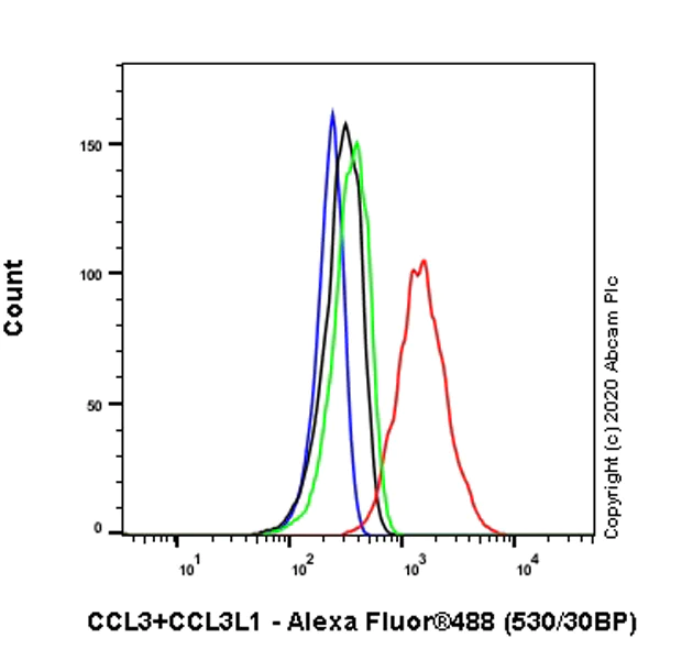
- Flow Cyt (Intra)
Supplier Data
Flow Cytometry (Intracellular) - Anti-CCL3 / MIP-1 alpha + CCL3L1 antibody [EPR23751-54] (AB259372)
Intracellular flow cytometric analysis of 5% paraformaldehyde-fixed, 90% methanol-permeabilized THP-1 (Human monocytic leukemia monocyte) (treated with 100nM phorbol 12-myristate 13-acetate (PMA) for 16 hours, then 100ng/ml lipopolysaccharide (LPS) for 4 hours, and add 1ug/ml Brefeldin A (BFA) for another 3 hours) (Red)/ Untreated control (Green) cells labeling Macrophage Inflammatory Protein 1 alpha / CCL3 + CCL3L1 with ab259372 at 1/500 dilution (Red / Green) compared with a Isotype control details (ab172730) (Black) and an unlabelled control (cells without incubation with primary antibody and secondary antibody) (Blue). Goat anti rabbit IgG (Alexa Fluor® 488, ab150077), at 1/2000 dilution was used as the secondary antibody.
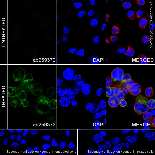
- ICC/IF
Supplier Data
Immunocytochemistry/ Immunofluorescence - Anti-CCL3 / MIP-1 alpha + CCL3L1 antibody [EPR23751-54] (AB259372)
Immunofluorescent analysis of 4% paraformaldehyde-fixed, 0.1% Triton X100 permeabilized THP-1 (human monocytic leukemia monocyte) cells labeling Macrophage Inflammatory Protein 1 alpha / CCL3 + CCL3L1 with ab259372 at 1/100 dilution, followed by ab150077 Goat Anti-Rabbit IgG H&L (Alexa Fluor® 488) secondary antibody at 1/1000 dilution (Green). Confocal image showing cytoplasmic staining in THP-1 cells treated with 12-myristate 13-acetate (100 nM) for 16 hours then with lipopolysaccharide (100 ng/ml) for 4 hours and with brefeldin A (1μg/ml) for another 3 hours. Tubulin is counterstained using ab195889 Anti-alpha Tubulin mouse monoclonal antibody - Microtubule Marker (Alexa Fluor® 594) at 1/200 dilution (Red). The nuclear counterstain is DAPI (Blue).
Secondary antibody only control : Used PBS instead of priary antibody, followed by ab150077 Goat Anti-Rabbit IgG H&L (Alexa Fluor® 488) secondary antibody at 1/1000 dilution.
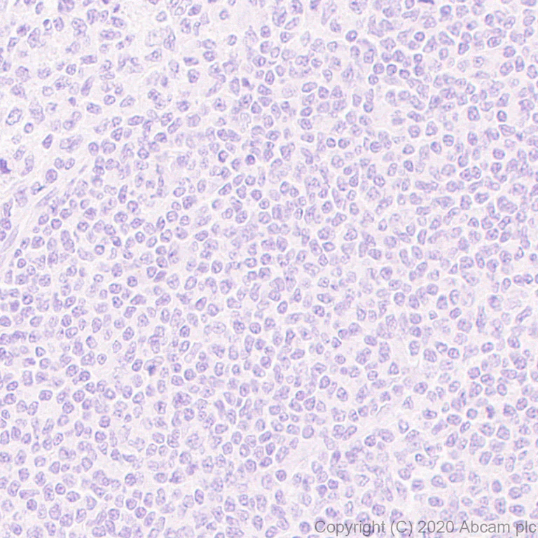
- IHC-P
Supplier Data
Immunohistochemistry (Formalin/PFA-fixed paraffin-embedded sections) - Anti-CCL3 / MIP-1 alpha + CCL3L1 antibody [EPR23751-54] (AB259372)
Immunohistochemical analysis of paraffin-embedded human tonsil tissue labeling Macrophage Inflammatory Protein 1 alpha / CCL3 + CCL3L1 with ab259372 at 1/1000 dilution, followed by a ready to use Rabbit specific IHC polymer detection kit HRP/DAB (ab209101). Negative control : No staining on human tonsil. The section was incubated with ab259372 for 30 mins at room temperature. The immunostaining was performed on a Leica Biosystems BOND® RX instrument. Counter stained with hematoxylin.
Secondary antibody only control : Used PBS instead of primary antibody, secondary antibody is a ready to use Rabbit specific IHC polymer detection kit HRP/DAB (ab209101).
Heat mediated antigen retrieval with Tris-EDTA buffer (pH 9.0, epitope retrieval solution2) for 20 mins.
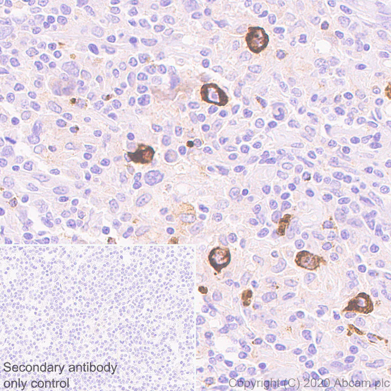
- IHC-P
Supplier Data
Immunohistochemistry (Formalin/PFA-fixed paraffin-embedded sections) - Anti-CCL3 / MIP-1 alpha + CCL3L1 antibody [EPR23751-54] (AB259372)
Immunohistochemical analysis of paraffin-embedded human Hodgkin lymphoma tissue labeling Macrophage Inflammatory Protein 1 alpha / CCL3 + CCL3L1 with ab259372 at 1/1000 dilution, followed by a ready to use Rabbit specific IHC polymer detection kit HRP/DAB (ab209101). Positive staining on human hodgkin Lymphoma (PMID : 31581676). The section was incubated with ab259372 for 30 mins at room temperature. The immunostaining was performed on a Leica Biosystems BOND® RX instrument. Counter stained with hematoxylin.
Secondary antibody only control : Used PBS instead of primary antibody, secondary antibody is a ready to use Rabbit specific IHC polymer detection kit HRP/DAB (ab209101).
Heat mediated antigen retrieval with Tris-EDTA buffer (pH 9.0, epitope retrieval solution2) for 20 mins.
- IHC-P
Supplier Data
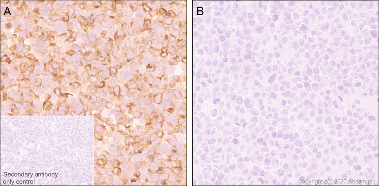
Immunohistochemistry (Formalin/PFA-fixed paraffin-embedded sections) - Anti-CCL3 / MIP-1 alpha + CCL3L1 antibody [EPR23751-54] (AB259372)
Immunohistochemical analysis of paraffin-embedded THP-1 cells labeling Macrophage Inflammatory Protein 1 alpha / CCL3 + CCL3L1 with ab259372 at 1/1000 dilution, followed by a ready to use Rabbit specific IHC polymer detection kit HRP/DAB (ab209101). Positive staining on THP-1 treated with 100nM PMA, LPS and Brefeldin treatment (image A), and negative staining on untreat THP-1 (image B). The section was incubated with ab259372 for 30 mins at room temperature. The immunostaining was performed on a Leica Biosystems BOND® RX instrument. Counter stained with hematoxylin.
Secondary antibody only control : Used PBS instead of primary antibody, secondary antibody is a ready to use Rabbit specific IHC polymer detection kit HRP/DAB (ab209101).
Heat mediated antigen retrieval with Tris-EDTA buffer (pH 9.0, epitope retrieval solution2) for 20 mins.
- IHC-P
Supplier Data
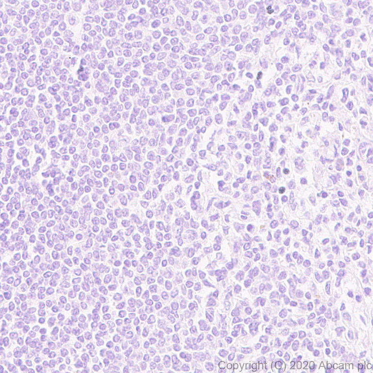
Immunohistochemistry (Formalin/PFA-fixed paraffin-embedded sections) - Anti-CCL3 / MIP-1 alpha + CCL3L1 antibody [EPR23751-54] (AB259372)
Immunohistochemical analysis of paraffin-embedded human spleen tissue labeling Macrophage Inflammatory Protein 1 alpha / CCL3 + CCL3L1 with ab259372 at 1/1000 dilution, followed by a ready to use Rabbit specific IHC polymer detection kit HRP/DAB (ab209101). Negative control : No staining on human spleen tissue. The section was incubated with ab259372 for 30 mins at room temperature. The immunostaining was performed on a Leica Biosystems BOND® RX instrument. Counter stained with hematoxylin.
Secondary antibody only control : Used PBS instead of primary antibody, secondary antibody is a ready to use Rabbit specific IHC polymer detection kit HRP/DAB (ab209101).
Heat mediated antigen retrieval with Tris-EDTA buffer (pH 9.0, epitope retrieval solution2) for 20 mins.

- IP
Supplier Data
Immunoprecipitation - Anti-CCL3 / MIP-1 alpha + CCL3L1 antibody [EPR23751-54] (AB259372)
Macrophage Inflammatory Protein 1 alpha / CCL3 + CCL3L1 was immunoprecipitated from 0.35 mg THP-1 (human monocytic leukemia monocyte) (treated with 100nM PMA for 16 hours. And then replace PMA with 100 ng/ml LPS (Lipopolysaccharide) for 7 hours, 1 μg/ml BFA was added for the last 3 hours) whole cell lysate whole cell lysate with ab259372 at 1/30 dilution (2ug in 0.35mg lysates). Western blot was performed on the immunoprecipitate using ab259372 at 1/1000 dilution. VeriBlot for IP Detection Reagent (HRP)(ab131366) was used at 1/5000 dilution.
Lane 1 : THP-1 (treated as above) whole cell lysate 3 ug
Lane 2 : ab259372 IP in THP-1 (treated as above) whole cell lysate
Lane 3 : Rabbit monoclonal IgG (ab172730) instead of ab259372 in THP-1 (treated as above) whole cell lysate
Blocking and dilution buffer and concentration : 5% NFDM/TBST.
Exposure time : 8 seconds.
All lanes:
Immunoprecipitation - Anti-CCL3 / MIP-1 alpha + CCL3L1 antibody [EPR23751-54] (ab259372)
Observed band size: 10 kDa
false

- ICC/IF
Supplier Data
Immunocytochemistry/ Immunofluorescence - Anti-CCL3 / MIP-1 alpha + CCL3L1 antibody [EPR23751-54] (AB259372)
Immunofluorescent analysis of 4% paraformaldehyde-fixed, 0.1% Triton X100 permeabilized RAW264.7 (mouse macrophage cell line transformed with Abelson murine leukemia virus) cells labeling Macrophage Inflammatory Protein 1 alpha / CCL3 + CCL3L1 with ab259372 at 1/100 dilution, followed by ab150077 Goat Anti-Rabbit IgG H&L (Alexa Fluor® 488) secondary antibody at 1/1000 dilution (Green). Confocal image showing cytoplasmic staining in RAW 264.7 cells treated with 12-myristate 13-acetate (100 nM) for 16 hours then with lipopolysaccharide (100 ng/ml) for 4 hours and with brefeldin A (1μg/ml) for another 3 hours. Tubulin is counterstained using ab195889 Anti-alpha Tubulin mouse monoclonal antibody - Microtubule Marker (Alexa Fluor® 594) at 1/200 dilution (Red). The nuclear counterstain is DAPI (Blue).
Secondary antibody only control : Used PBS instead of priary antibody, followed by ab150077 Goat Anti-Rabbit IgG H&L (Alexa Fluor® 488) secondary antibody at 1/1000 dilution.

- Flow Cyt (Intra)
Supplier Data
Flow Cytometry (Intracellular) - Anti-CCL3 / MIP-1 alpha + CCL3L1 antibody [EPR23751-54] (AB259372)
Intracellular flow cytometric analysis of5% paraformaldehyde-fixed, 90% methanol-permeabilized RAW264.7 (Mouse Abelson murine leukemia virus-induced tumor macrophage) (treated with 100ng/ml lipopolysaccharide (LPS) for 4 hours, and add Brefeldin A (BFA) for another 3 hours (Red)/ Untreated control (Green)) (Red)/ Untreated control (Green) cells labeling Macrophage Inflammatory Protein 1 alpha / CCL3 + CCL3L1 with ab259372at 1/500 dilution (Red / Green) compared with a Isotype control details (ab172730) (Black) and an unlabelled control (cells without incubation with primary antibody and secondary antibody) (Blue). Goat anti rabbit IgG (Alexa Fluor®488, ab150077), at 1/2000 dilution was used as the secondary antibody.
- WB
Supplier Data
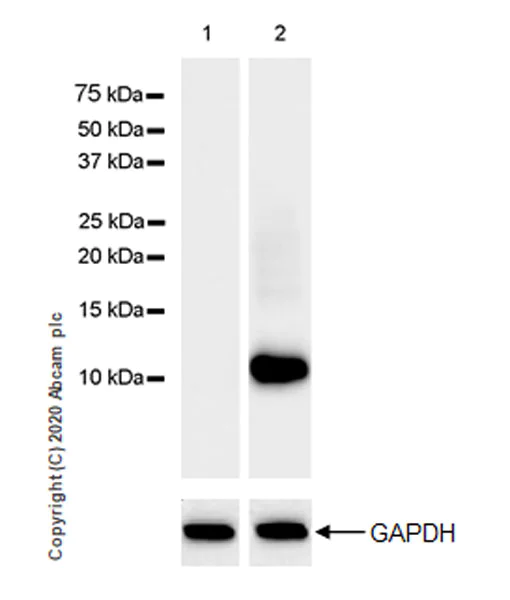
Western blot - Anti-CCL3 / MIP-1 alpha + CCL3L1 antibody [EPR23751-54] (AB259372)
Blocking and dilution buffer : 5% NFDM/TBST.
Exposure time : 8 seconds.
All lanes:
Western blot - Anti-CCL3 / MIP-1 alpha + CCL3L1 antibody [EPR23751-54] (ab259372) at 1/1000 dilution
Lane 1:
Untreated THP-1 (human monocytic leukemia monocyte) whole cell lysate at 20 µg
Lane 2:
THP-1 (treated with 100nM PMA for 16 hours. And then replace PMA with 100 ng/ml LPS (Lipopolysaccharide) for 4 hours, and then 1 μg/ml Brefeldin A was added for the last 3 hours), whole cell lysate at 20 µg
Secondary
All lanes:
Goat Anti-Rabbit IgG, (H+L), Peroxidase conjugated (<a href='/products/secondary-antibodies/goat-rabbit-igg-h-l-hrp-ab97051'>ab97051</a>) at 1/50000 dilution
Observed band size: 12 kDa
false
- WB
Supplier Data
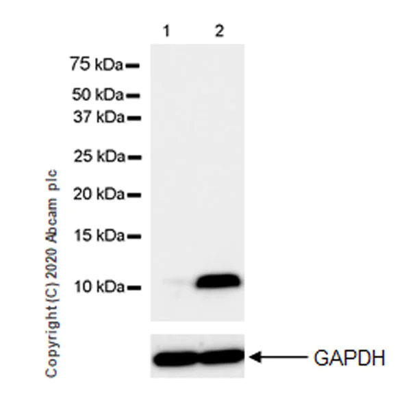
Western blot - Anti-CCL3 / MIP-1 alpha + CCL3L1 antibody [EPR23751-54] (AB259372)
Blocking and dilution buffer : 5% NFDM/TBST.
Exposure time : 3 seconds.
All lanes:
Western blot - Anti-CCL3 / MIP-1 alpha + CCL3L1 antibody [EPR23751-54] (ab259372) at 1/5000 dilution
Lane 1:
Untreated RAW 264.7 (mouse macrophage cell line transformed with Abelson murine leukemia virus) whole cell lysate at 10 µg
Lane 2:
RAW 264.7 (treated with 100 ng/ml LPS(Lipopolysaccharide) for 4 hours and then 1 μg/ml Brefeldin A was added for the last 3 hours), whole cell lysate at 10 µg
Secondary
All lanes:
Western blot - Goat Anti-Rabbit IgG H&L (HRP) (<a href='/products/secondary-antibodies/goat-rabbit-igg-h-l-hrp-ab97051'>ab97051</a>) at 1/20000 dilution
Observed band size: 12 kDa
false
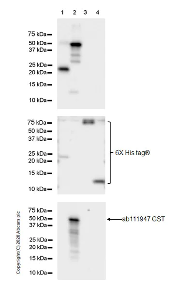
- WB
Supplier Data
Western blot - Anti-CCL3 / MIP-1 alpha + CCL3L1 antibody [EPR23751-54] (AB259372)
Blocking and dilution buffer : 5% NFDM/TBST.
Exposire time : 10 seconds.
All lanes:
Western blot - Anti-CCL3 / MIP-1 alpha + CCL3L1 antibody [EPR23751-54] (ab259372) at 1/1000 dilution
Lane 1:
His-tagged mouse CCL3 recombinant protein (aa24-92) * 2 ,10 ng
Lane 2:
GST/His-tagged human CCL3L1 recombinant protein (aa24-93), 10 ng
Lane 3:
LIF/His-tagged human CCL4 recombinant protein (aa24-92)*2, 10 ng
Lane 4:
His-tagged human CCL18 recombinant protein (aa21-89),10 ng
Secondary
All lanes:
Goat Anti-Rabbit IgG, (H+L), Peroxidase conjugated (<a href='/products/secondary-antibodies/goat-rabbit-igg-h-l-hrp-ab97051'>ab97051</a>) at 1/50000 dilution
Observed band size: 12 kDa
false
関連する標識済み抗体及び組成の異なる製品 (1)
-
Anti-CCL3 / MIP-1 alpha + CCL3L1 antibody [EPR23751-54] - BSA and Azide free
組合せ製品
Assay Kits
AB229400
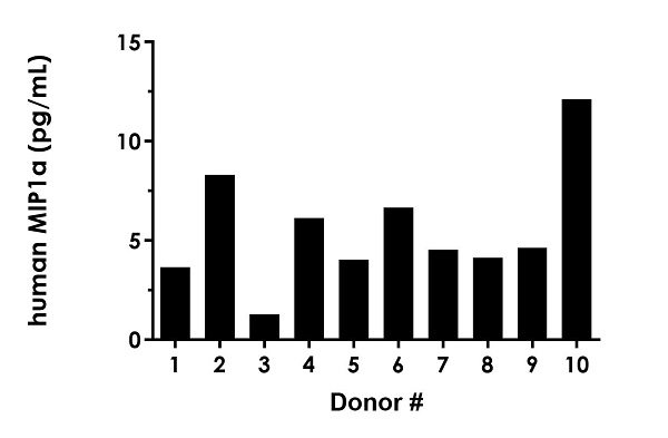
Human MIP-1 alpha/CCL3 ELISA Kit, Fluorescent
CatchPoint SimpleStep ELISA|Recombinant
- 0
- 0
Primary Antibodies
AB45690
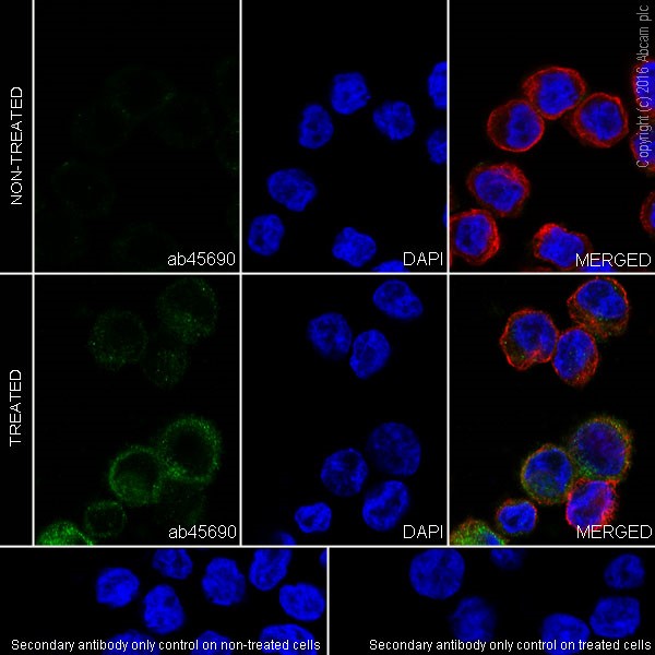
Anti-CCL4/MIP-1 beta antibody [EP521Y]
20ul selling size|RabMAb|Recombinant|KO Validated
- 1
- 1
Primary Antibodies
AB307712
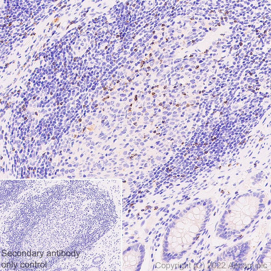
Anti-RANTES antibody [EPR26519-16]
BOND RX™ Validated|20ul selling size|RabMAb|Recombinant
- 0
- 0
Primary Antibodies
AB32536
Anti-NF-kB p65 antibody [E379]
BOND RX™ Validated|20ul selling size|RabMAb|Recombinant|KO Validated

- 4
- 15
Primary Antibodies
AB214819
20ul selling size|BOND RX™ Validated|RabMAb|Recombinant|KO Validated
- 0
- 0
Reactivity data
製品の詳細
Patented technology
Our RabMAb® technology is a patented hybridoma-based technology for making rabbit monoclonal antibodies. For details on our patents, please refer to RabMAb® patents.
What are the advantages of a recombinant monoclonal antibody?
This product is a recombinant monoclonal antibody, which offers several advantages including:
- - High batch-to-batch consistency and reproducibility
- - Improved sensitivity and specificity
- - Long-term security of supply
- - Animal-free batch production
For more information, read more on recombinant antibodies.
出荷温度及び保存条件
製品の状態
精製方法
バッファー組成
出荷温度
短期保存期間
短期保存温度
長期保存温度
分注に関する情報
保管に関する情報
補足情報
This supplementary information is collated from multiple sources and compiled automatically.
Biological function summary
The MIP-1 alpha functions as a chemotactic cytokine. It binds to receptors like CCR1 and CCR5 on the surface of target cells resulting in activation and migration toward areas of inflammation. MIP-1 alpha does not form a complex with other proteins; however its interactions with its receptors are vital for signal transduction. It also promotes the production of additional inflammatory cytokines contributing to amplifying immune responses. The chemotactic properties of MIP-1 alpha make it an important player in the body's defense against pathogens.
Pathways
MIP-1 alpha is involved in important immune pathways like the NF-kB signaling pathway. This pathway plays a significant role in immune cell activation and the inflammatory response. MIP-1 alpha interacts with other chemokines like CCL4 (MIP-1 beta) which shares similar functions and binds to CCR5. The coordinated activities of these chemokines enhance the recruitment and activation of various leukocyte populations facilitating an effective immune response.
製品プロトコール
- Visit the General protocols
- Visit the Troubleshooting
ターゲットの情報
追加のターゲット
文献 (9)
Recent publications for all applications. Explore the full list and refine your search
Frontiers in molecular biosciences 12:1571593 PubMed40656894
2025
Applications
Unspecified application
Species
Unspecified reactive species
Cell biology and toxicology 41:47 PubMed39976849
2025
Applications
Unspecified application
Species
Unspecified reactive species
Nature communications 15:47 PubMed38167807
2024
Applications
Unspecified application
Species
Unspecified reactive species
Advanced science (Weinheim, Baden-Wurttemberg, Germany) 10:e2302527 PubMed37867250
2023
Applications
Unspecified application
Species
Unspecified reactive species
Fundamental research 5:1804-1821 PubMed40777809
2023
Applications
Unspecified application
Species
Unspecified reactive species
Scientific reports 13:2864 PubMed36806315
2023
Applications
Unspecified application
Species
Unspecified reactive species
International journal of molecular sciences 24: PubMed36768133
2023
Applications
Unspecified application
Species
Unspecified reactive species
Nature chemical biology 19:275-283 PubMed36175661
2022
Applications
Unspecified application
Species
Unspecified reactive species
Journal of experimental & clinical cancer research : CR 41:60 PubMed35148799
2022
Applications
Unspecified application
Species
Unspecified reactive species
Abcam product promise
Please note: All products are 'FOR RESEARCH USE ONLY. NOT FOR USE IN DIAGNOSTIC OR THERAPEUTIC PROCEDURES'.
For licensing inquiries, please contact partnerships@abcam.com