Anti-Catalase 抗体 [EP1929Y] - Peroxisome Marker
Anti-Catalase antibody [EP1929Y] - Peroxisome Marker
- 20ul selling size
- KO Validated
- RabMAb
- Recombinant
- Lab Essentials
- 詳細を見る
5
(1 Review)
|
(26 Publications)
Rabbit Recombinant Monoclonal Catalase antibody. Peroxisome marker. Suitable for IHC-P, WB, ICC/IF and reacts with Human samples. Cited in 26 publications.
別名を表示する
Catalase, CAT
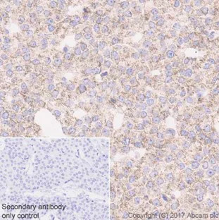
- IHC-P
Lab
Immunohistochemistry (Formalin/PFA-fixed paraffin-embedded sections) - Anti-Catalase antibody [EP1929Y] - Peroxisome Marker (AB76024)
Immunohistochemical analysis of Paraffin-embedded human bladder cancer tissue sections labeling Catalase with ab76024 at 1/1000. Goat Anti-Rabbit IgG H&L (HRP) was used as the secondary antibody. Sections were counterstained with Hematoxylin. Antigen retrieval was heat mediated using ab93684 (Tris/EDTA buffer, pH 9.0).
Granular cytoplasmic staining on human bladder cancer.
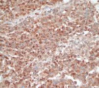
- IHC-P
Unknown
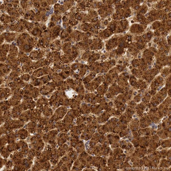
Immunohistochemistry (Formalin/PFA-fixed paraffin-embedded sections) - Anti-Catalase antibody [EP1929Y] - Peroxisome Marker (AB76024)
Immunohistochemical staining of Catalase in paraffin embedded human normal brain tissue using ab76024 at a 1/100 dilution.
Perform heat mediated antigen retrieval before commencing with IHC staining protocol.
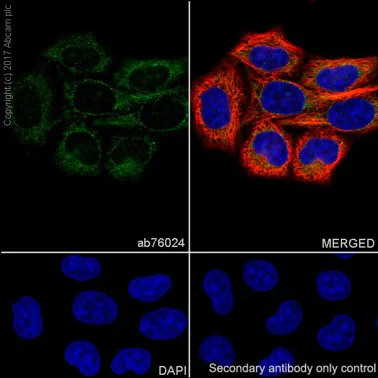
- ICC/IF
Lab
Immunocytochemistry/ Immunofluorescence - Anti-Catalase antibody [EP1929Y] - Peroxisome Marker (AB76024)
Immunocytochemistry/Immunofluorescence analysis of HeLa (human cervix adenocarcinoma epithelial cell) labeling Catalase ab76024 at 1/100. Cells were fixed with 100% Methanol. ab150077, an Alexa Fluor® 488-conjugated goat anti-rabbit IgG (1/1000) was used as the secondary antibody. DAPI (blue) was used as the nuclear counterstain.
ab195889, Anti-alpha Tubulin antibody [DM1A] - Microtubule Marker (Alexa Fluor® 594) 1/100 was used as counterstain antibody.
Confocal image showing membranous staining in HeLa cells.
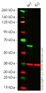
- WB
Lab
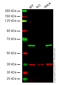
Western blot - Anti-Catalase antibody [EP1929Y] - Peroxisome Marker (AB76024)
Lanes 1-2 : Merged signal (red and green). Green - ab76024 observed at 60 kDa. Red - loading control ab8245 observed at 37 kDa.
ab76024 Anti-Catalase antibody [EP1929Y] - Peroxisome Marker was shown to specifically react with Catalase in wild-type HeLa cells. Loss of signal was observed when knockout cell line ab265250 (knockout cell lysate ab256859) was used. Wild-type and Catalase knockout samples were subjected to SDS-PAGE. ab76024 and Anti-GAPDH antibody [6C5] - Loading Control (ab8245) were incubated overnight at 4°C at 1 in 10000 and 1 in 20000 dilution respectively. Blots were developed with Goat anti-Rabbit IgG H&L (IRDye® 800CW) preadsorbed (ab216773) and Goat anti-Mouse IgG H&L (IRDye® 680RD) preadsorbed (ab216776) secondary antibodies at 1 in 20000 dilution for 1 hour at room temperature before imaging.
All lanes:
Western blot - Anti-Catalase antibody [EP1929Y] - Peroxisome Marker (ab76024) at 1/10000 dilution
Lane 1:
Wild-type HeLa cell lysate at 20 µg
Lane 2:
CAT knockout HeLa cell lysate at 20 µg
Lane 2:
Western blot - Human CAT (Catalase) knockout HeLa cell line (<a href='/products/cell-lines/human-cat-catalase-knockout-hela-cell-line-ab265250'>ab265250</a>)
Predicted band size: 60 kDa
Observed band size: 60 kDa
false
- WB
Unknown
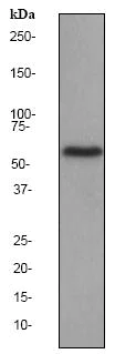
Western blot - Anti-Catalase antibody [EP1929Y] - Peroxisome Marker (AB76024)
Lanes 1 - 3 : Merged signal (red and green). Green - ab76024 observed at 60 kDa. Red - loading control, ab9484, observed at 37 kDa.
ab76024 was shown to specifically react with CAT when CAT knockout samples were used. Wild-type and CAT knockout samples were subjected to SDS-PAGE. ab76024 and ab9484 (Mouse anti GAPDH loading control) were incubated overnight at 4°C at 1/5000 dilution and 1/20000 dilution respectively. Blots were developed with Goat anti-Rabbit IgG H&L (IRDye® 800CW) preabsorbed ab216773 and Goat anti-Mouse IgG H&L (IRDye® 680RD) preabsorbed ab216776 secondary antibodies at 1/20000 dilution for 1 hour at room temperature before imaging.
All lanes:
Western blot - Anti-Catalase antibody [EP1929Y] - Peroxisome Marker (ab76024)
Lane 1:
Wild-type HAP1 whole cell lysate at 20 µg
Lane 2:
CAT knockout HAP1 whole cell lysate at 20 µg
Lane 3:
HeLa whole cell lysate at 20 µg
Predicted band size: 60 kDa
false
- WB
Unknown
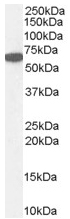
Western blot - Anti-Catalase antibody [EP1929Y] - Peroxisome Marker (AB76024)
All lanes:
Western blot - Anti-Catalase antibody [EP1929Y] - Peroxisome Marker (ab76024) at 1/20000 dilution
All lanes:
HeLa cell lysate at 10 µg
Secondary
All lanes:
HRP labelled goat anti-rabbit at 1/2000 dilution
Predicted band size: 60 kDa
Observed band size: 60 kDa
false
関連する標識済み抗体及び組成の異なる製品 (1)
-
Anti-Catalase antibody [EP1929Y] - BSA and Azide free
代替製品

Primary Antibodies
AB52477
Anti-Catalase antibody - Peroxisome Marker
BOND RX™ Validated|KO Validated|Lab Essentials
- 5
- 8
Primary Antibodies
AB209211
Anti-Catalase antibody [EPR20198] - Peroxisome Marker
KO Validated|RabMAb|Recombinant|Lab Essentials|20ul selling size

- 5
- 3
Primary Antibodies
AB76110
Anti-Catalase antibody [EPR1928Y]
20ul selling size|RabMAb|Recombinant|KO Validated

- 5
- 1
Primary Antibodies
AB223793
Anti-Catalase antibody [EPR20198] - BSA and Azide free
RabMAb|Recombinant|KO Validated
- 0
- 0
Primary Antibodies
AB110292
Anti-Catalase antibody [12C2DB9] - Peroxisome Marker
KO Validated|Lab Essentials
- 5
- 3
組合せ製品

Primary Antibodies
AB68155
Anti-SOD2/MnSOD antibody [EPR2560Y]
RabMAb|Recombinant|KO Validated|20ul selling size

- 5
- 2

Primary Antibodies
AB125066
Anti-Glutathione Peroxidase 4 antibody [EPNCIR144]
BOND RX™ Validated|KO Validated|RabMAb|Recombinant|Lab Essentials

- 5
- 13
Reactivity data
製品の詳細
Patented technology
Our RabMAb® technology is a patented hybridoma-based technology for making rabbit monoclonal antibodies. For details on our patents, please refer to RabMAb® patents.
Species reactivity
Rat: We have preliminary internal testing data to indicate this antibody may not react with this species.
Please contact us for more information.
出荷温度及び保存条件
製品の状態
精製度
バッファー組成
出荷温度
短期保存温度
長期保存温度
分注に関する情報
保管に関する情報
補足情報
This supplementary information is collated from multiple sources and compiled automatically.
Biological function summary
Catalase contributes to antioxidant defense by breaking down hydrogen peroxide preventing cellular damage. In peroxisomes it works alongside other peroxisomal enzymes to maintain cell health and metabolic regulation. Catalase does not form a complex but interacts closely with other enzymes like superoxide dismutase which dismutates superoxide radicals into less harmful substances. Increased Catalase activity levels can be measured using Catalase activity kits and activity assays allowing us to learn about peroxisome function.
Pathways
Hydrogen peroxide removal by Catalase is vital in the reactive oxygen species (ROS) metabolic process and plays a part in the cellular response to oxidative stress. Catalase interacts with the glutathione peroxidase pathway safeguarding cells from oxidative stress-related damage. Superoxide dismutase works synergistically with Catalase transforming superoxide anions into hydrogen peroxide before its decomposition by Catalase. These activities highlight the essential role Catalase plays in protecting cells from oxidative stress damage.
製品プロトコール
- Visit the General protocols
- Visit the Troubleshooting
ターゲットの情報
文献 (26)
Recent publications for all applications. Explore the full list and refine your search
Proceedings of the National Academy of Sciences of the United States of America 121:e2321972121 PubMed39008677
2024
Applications
Unspecified application
Species
Unspecified reactive species
Nature communications 15:5827 PubMed38992008
2024
Applications
Unspecified application
Species
Unspecified reactive species
Brain : a journal of neurology 147:3727-3741 PubMed38696728
2024
Applications
Unspecified application
Species
Unspecified reactive species
Frontiers in immunology 14:1126217 PubMed37313408
2023
Applications
Unspecified application
Species
Unspecified reactive species
Acta pharmacologica Sinica 44:1906-1919 PubMed37186123
2023
Applications
Unspecified application
Species
Unspecified reactive species
International journal of molecular sciences 23: PubMed36233157
2022
Applications
Unspecified application
Species
Unspecified reactive species
The Journal of physiology 600:2897-2917 PubMed35556249
2022
Applications
Unspecified application
Species
Unspecified reactive species
Clinical & experimental metastasis 39:641-659 PubMed35604506
2022
Applications
Unspecified application
Species
Unspecified reactive species
BioFactors (Oxford, England) 48:897-907 PubMed35170815
2022
Applications
Unspecified application
Species
Unspecified reactive species
Frontiers in cell and developmental biology 9:734818 PubMed34692691
2021
Applications
Unspecified application
Species
Unspecified reactive species
Abcam product promise
Please note: All products are 'FOR RESEARCH USE ONLY. NOT FOR USE IN DIAGNOSTIC OR THERAPEUTIC PROCEDURES'.
For licensing inquiries, please contact partnerships@abcam.com