Alexa Fluor® 647 Anti-PD1 抗体 [NAT105]
Alexa Fluor® 647 Anti-PD1 antibody [NAT105]
- Recombinant
- 詳細を見る
Be the first to review this product! Submit a review
|
(0 Publication)
Mouse Recombinant Monoclonal PD1 antibody - conjugated to Alexa Fluor® 647. Suitable for IHC-P, Flow Cyt, Flow Cyt (Intra), ICC/IF and reacts with Human samples.
別名を表示する
CD279, PD1, PDCD1, Programmed cell death protein 1, Protein PD-1, hPD-1
- Flow Cyt
Lab
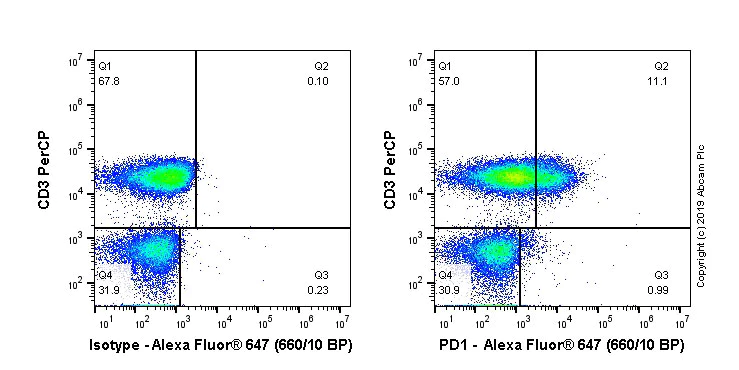
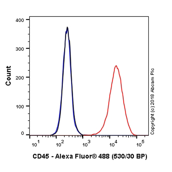
Flow Cytometry - Alexa Fluor® 647 Anti-PD1 antibody [NAT105] (AB279695)
Flow cytometry staining of human peripheral blood mononuclear cells (PBMCs) with ab279695 (right) or mouse IgG1κ Alexa Fluor® 647 isotype (left). PBMCs were incubated for 30 min on ice in 1x PBS containing 10μg/ml human IgG and 10% normal goat serum to block FC receptors and non-specific protein-protein interaction followed by the antibody (ab279695) or mouse IgG1κ Alexa Fluor® 647 isotype (100 μl at 1μg/ml (1/500)) for 30 min on ice. The cells were simultaneously stained with CD3.
Acquisition of >30000 events were collected using a 40 mW Red laser (638nm) and 660/10 bandpass filter. Events were gated on live lymphocytes.
関連する標識済み抗体及び組成の異なる製品 (6)
-
Anti-PD1 antibody [NAT105]
-
Anti-PD1 antibody [NAT105] - Rabbit IgG (Chimeric) - BSA and Azide free
-
Anti-PD1 antibody [NAT105] - Rabbit IgG (Chimeric)
-
Anti-PD1 antibody [NAT105] - BSA and Azide free
-
519 Alexa Fluor® 488
Alexa Fluor® 488 Anti-PD1 antibody [NAT105]
-
565 Alexa Fluor® 555
Alexa Fluor® 555 Anti-PD1 antibody [NAT105]
代替製品
組合せ製品
Primary Antibodies
AB280849
Alexa Fluor® 555 Anti-CD4 antibody [EPR6855]
BOND RX™ Validated|RabMAb|Recombinant|Trial Size
- 5
- 2
Primary Antibodies
AB274896
Alexa Fluor® 555 Anti-PD-L1 antibody [73-10]
BOND RX™ Validated|RabMAb|Recombinant|Trial Size
- 4
- 1
Reactivity data
製品の詳細
What are the advantages of a recombinant monoclonal antibody?
This product is a recombinant monoclonal antibody, which offers several advantages including:
- - High batch-to-batch consistency and reproducibility
- - Improved sensitivity and specificity
- - Long-term security of supply
- - Animal-free batch production
For more information, read more on recombinant antibodies.
Alexa Fluor® is a registered trademark of Molecular Probes, Inc, a Thermo Fisher Scientific Company. The Alexa Fluor® dye included in this product is provided under an intellectual property license from Life Technologies Corporation. As this product contains the Alexa Fluor® dye, the purchase of this product conveys to the buyer the non-transferable right to use the purchased product and components of the product only in research conducted by the buyer (whether the buyer is an academic or for-profit entity). As this product contains the Alexa Fluor® dye the sale of this product is expressly conditioned on the buyer not using the product or its components, or any materials made using the product or its components, in any activity to generate revenue, which may include, but is not limited to use of the product or its components: in manufacturing; (ii) to provide a service, information, or data in return for payment (iii) for therapeutic, diagnostic or prophylactic purposes; or (iv) for resale, regardless of whether they are sold for use in research. For information on purchasing a license to this product for purposes other than research, contact Life Technologies Corporation, 5781 Van Allen Way, Carlsbad, CA 92008 USA or outlicensing@thermofisher.com.
出荷温度及び保存条件
製品の状態
精製方法
バッファー組成
出荷温度
短期保存期間
短期保存温度
長期保存温度
分注に関する情報
保管に関する情報
補足情報
This supplementary information is collated from multiple sources and compiled automatically.
Biological function summary
PD1 serves as an inhibitory receptor acting as a checkpoint in the immune system. It becomes part of an immune-suppressive complex when it binds with its ligands PD-L1 or PD-L2 which are expressed on various cell types including some tumor cells. This interaction suppresses the proliferation of T cells and cytokine production contributing to immune homeostasis. By controlling T cell activity PD1 limits autoimmunity but can also reduce the immune system's capability to attack cancer cells.
Pathways
PD1 functions in the immune checkpoint pathway a critical regulatory circuit in immune regulation. The engagement of PD1 with its ligands initiates a cascade that inhibits the function and proliferation of T cells through downstream SHP-2 phosphatase activity. This pathway frequently involves other regulatory proteins like CTLA-4 and is an important mechanism by which the body modulates immune responses. Related pathways often intersect with those involving T cell receptor signaling and contribute to the overall modulation of immune activity.
製品プロトコール
- Visit the General protocols
- Visit the Troubleshooting
ターゲットの情報
Abcam product promise
Please note: All products are 'FOR RESEARCH USE ONLY. NOT FOR USE IN DIAGNOSTIC OR THERAPEUTIC PROCEDURES'.
For licensing inquiries, please contact partnerships@abcam.com