Alexa Fluor® 647 Anti-Indoleamine 2, 3-dioxygenase 抗体 [EPR20374]
Alexa Fluor® 647 Anti-Indoleamine 2, 3-dioxygenase antibody [EPR20374]
- RabMAb
- Recombinant
- 詳細を見る
Be the first to review this product! Submit a review
|
(0 Publication)
Rabbit Recombinant Monoclonal Indoleamine 2, 3-dioxygenase antibody - conjugated to Alexa Fluor® 647. Suitable for ICC/IF, IHC-P, Flow Cyt (Intra) and reacts with Human samples.
別名を表示する
IDO, INDO, IDO1, IDO-1
- IHC-P
Supplier Data
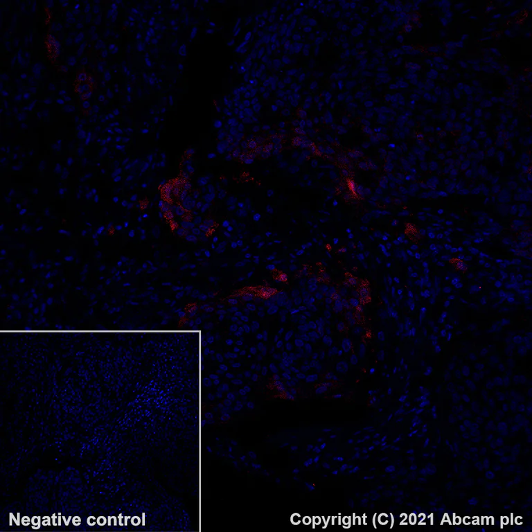
Immunohistochemistry (Formalin/PFA-fixed paraffin-embedded sections) - Alexa Fluor® 647 Anti-Indoleamine 2, 3-dioxygenase antibody [EPR20374] (AB281580)
Immunohistochemistry analysis of paraffin-embedded human cervix carcinoma tissue staining Indoleamine 2, 3-dioxygenase using ab281580 at a 1/50 diluiton (10.0 μg/ml).
Positive staining on human cervix carcinoma.
The section was then incubated overnight at +4°C in TBS containing 0.025% (v/v) Triton X-100 and 1% (w/v) BSA with ab281580 at 1/50 dilution (shown in Red). Nuclear DNA was labeled with DAPI (shown in blue). The section was then mounted using Fluoromount®.
Non-specific protein-protein interactions were then blocked in TBS containing 0.025% (v/v) Triton X-100, 0.3M (w/v) glycine and 1% (w/v) BSA for 1h at room temperature.
Image was taken with a confocal microscope (Leica-Microsystems, TCS SP8).
Heat mediated antigen retrieval using ab93684 (Tris/EDTA buffer, pH 9.0).
- ICC/IF
Supplier Data
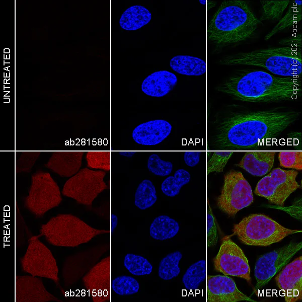
Immunocytochemistry/ Immunofluorescence - Alexa Fluor® 647 Anti-Indoleamine 2, 3-dioxygenase antibody [EPR20374] (AB281580)
Immunocytochemistry analysis of 4% Paraformaldehyde-fixed, 0.1% Triton X-100 permeabilized HeLa (human epithelial cell line from cervix adenocarcinoma) cells treated with human Interferon-γ (50 ng/ml) for 16 hours, labeling Indoleamine 2, 3-dioxygenase with ab281580 at 1/50 (10.0 μg/ml) dilution (Red).
Confocal image showing cytoplasmic and nuclear staining in Interferon-γ treated HeLa cells.
ab195887 Anti-alpha Tubulin mouse monoclonal antibody - Microtubule Marker (Alexa Fluor® 488) was used to counterstain tubulin at 1/200 dilution (Green). The nuclear counterstain was DAPI (Blue).
- Flow Cyt (Intra)
Supplier Data
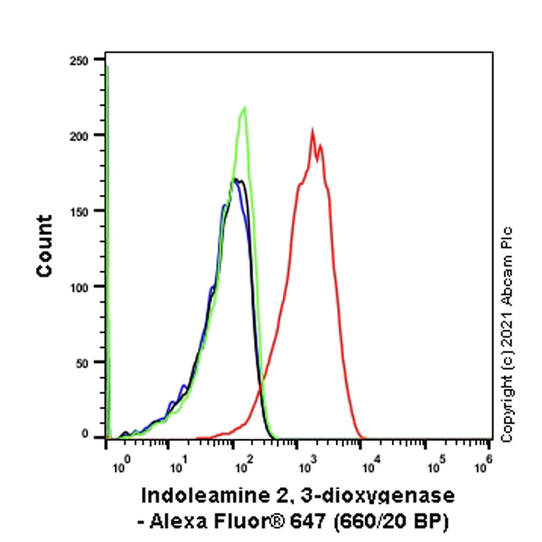
Flow Cytometry (Intracellular) - Alexa Fluor® 647 Anti-Indoleamine 2, 3-dioxygenase antibody [EPR20374] (AB281580)
Flow Cytometry analysis of 4% paraformaldehyde-fixed, 90% methanol-permeabilized HeLa (human cervix adenocarcinoma epithelial cell) treated with 50ng/ml Human Interferon-γ for 16 hours (Red)/ Untreated control (Green) cells stained for Indoleamine 2, 3-dioxygenase using ab281580 at a 1/500 dilution (0.1μg) (red), compared to Rabbit monoclonal isotype control - Alexa Fluor® 647 (Black) isotype control and an unlabeled control (cells without incubation with primary antibody and secondary antibody) (Blue).
The level of Indoleamine 2, 3-dioxygenase expression can be induced by IFN-γ treatment (PMID : 16368976).
- IHC-P
Supplier Data
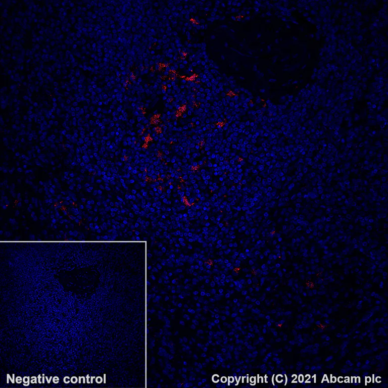
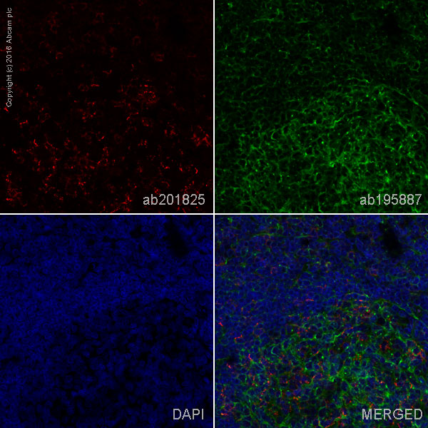
Immunohistochemistry (Formalin/PFA-fixed paraffin-embedded sections) - Alexa Fluor® 647 Anti-Indoleamine 2, 3-dioxygenase antibody [EPR20374] (AB281580)
Immunohistochemistry analysis of paraffin-embedded human spleen tissue staining Indoleamine 2, 3-dioxygenase using ab281580 at a 1/50 diluiton (10.0 μg/ml).
Positive staining on human spleen.
The section was then incubated overnight at +4°C in TBS containing 0.025% (v/v) Triton X-100 and 1% (w/v) BSA with ab281580 at 1/50 dilution (shown in Red). Nuclear DNA was labeled with DAPI (shown in blue). The section was then mounted using Fluoromount®.
Non-specific protein-protein interactions were then blocked in TBS containing 0.025% (v/v) Triton X-100, 0.3M (w/v) glycine and 1% (w/v) BSA for 1h at room temperature.
Image was taken with a confocal microscope (Leica-Microsystems, TCS SP8).
Heat mediated antigen retrieval using ab93684 (Tris/EDTA buffer, pH 9.0).
関連する標識済み抗体及び組成の異なる製品 (7)
-
Anti-Indoleamine 2, 3-dioxygenase antibody [EPR20374]
-
Anti-Indoleamine 2, 3-dioxygenase antibody [EPR20374] - BSA and Azide free (Capture)
-
Anti-Indoleamine 2, 3-dioxygenase antibody [EPR20374] - Low endotoxin, Azide free
-
Anti-Indoleamine 2, 3-dioxygenase antibody [EPR20374] - BSA and Azide free
-
519 Alexa Fluor® 488
Alexa Fluor® 488 Anti-Indoleamine 2, 3-dioxygenase antibody [EPR20374]
-
565 Alexa Fluor® 555
Alexa Fluor® 555 Anti-Indoleamine 2, 3-dioxygenase antibody [EPR20374]
-
617 Alexa Fluor® 594
Alexa Fluor® 594 Anti-Indoleamine 2, 3-dioxygenase antibody [EPR20374]
組合せ製品
Primary Antibodies
AB270127
Alexa Fluor® 647 Anti-CTLA4 antibody [CAL49]
BOND RX™ Validated|RabMAb|Recombinant|Advanced Validation

- 0
- 0
Primary Antibodies
AB313665
Alexa Fluor® 488 Anti-CD163 antibody [EPR19518]
BOND RX™ Validated|RabMAb|Recombinant
- 0
- 0
Reactivity data
製品の詳細
Patented technology
Our RabMAb® technology is a patented hybridoma-based technology for making rabbit monoclonal antibodies. For details on our patents, please refer to RabMAb® patents.
What are the advantages of a recombinant monoclonal antibody?
This product is a recombinant monoclonal antibody, which offers several advantages including:
- - High batch-to-batch consistency and reproducibility
- - Improved sensitivity and specificity
- - Long-term security of supply
- - Animal-free batch production
For more information, read more on recombinant antibodies.
Alexa Fluor® is a registered trademark of Molecular Probes, Inc, a Thermo Fisher Scientific Company. The Alexa Fluor® dye included in this product is provided under an intellectual property license from Life Technologies Corporation. As this product contains the Alexa Fluor® dye, the purchase of this product conveys to the buyer the non-transferable right to use the purchased product and components of the product only in research conducted by the buyer (whether the buyer is an academic or for-profit entity). As this product contains the Alexa Fluor® dye the sale of this product is expressly conditioned on the buyer not using the product or its components, or any materials made using the product or its components, in any activity to generate revenue, which may include, but is not limited to use of the product or its components: in manufacturing; (ii) to provide a service, information, or data in return for payment (iii) for therapeutic, diagnostic or prophylactic purposes; or (iv) for resale, regardless of whether they are sold for use in research. For information on purchasing a license to this product for purposes other than research, contact Life Technologies Corporation, 5781 Van Allen Way, Carlsbad, CA 92008 USA or outlicensing@thermofisher.com.
出荷温度及び保存条件
製品の状態
精製方法
バッファー組成
出荷温度
短期保存期間
短期保存温度
長期保存温度
分注に関する情報
保管に関する情報
補足情報
This supplementary information is collated from multiple sources and compiled automatically.
Biological function summary
IDO plays a role in immune response regulation by degrading tryptophan an essential amino acid needed for T-cell proliferation. The depletion of tryptophan and the accumulation of its metabolites such as kynurenine cause immunosuppressive effects within the tissue microenvironment. While IDO functions mostly as a standalone enzyme its activity influences the cellular surroundings by altering the balance of local immune reactions.
Pathways
IDO integrates into the tryptophan metabolism pathway and is important in the kynurenine pathway. Through this pathway it maintains immune homeostasis and cell defense mechanisms. Proteins like kynureninase and kynurenine 3-monooxygenase also participate in the same metabolic processes which further extend the effects of tryptophan metabolism on immune cell behavior.
製品プロトコール
- Visit the General protocols
- Visit the Troubleshooting
ターゲットの情報
Abcam product promise
Please note: All products are 'FOR RESEARCH USE ONLY. NOT FOR USE IN DIAGNOSTIC OR THERAPEUTIC PROCEDURES'.
For licensing inquiries, please contact partnerships@abcam.com