Anti-5-methylcytosine (5-mC) 抗体 [33D3]
Anti-5-methylcytosine (5-mC) antibody [33D3]
5
(12 Reviews)
|
(228 Publications)
Anti-5-methylcytosine (5-mC) antibody [33D3] (ab10805) is a mouse monoclonal antibody detecting 5-methylcytosine (5-mC) in Flow Cytometry, IP, IHC-P, IHC-Fr.
- Over 170 publications
- Trusted since 2005
別名を表示する
5 Methycytosine, 5-Me Cytidine, 5-Me Cytosine, 5-Methyl-Cytidine, 5-Methyl-Cytosine, 5-MethylCytidine, 5-MethylCytosine, 5-mC, 5-meC, methyl CpG

- IHC-P
AbReview13478****
Immunohistochemistry (Formalin/PFA-fixed paraffin-embedded sections) - Anti-5-methylcytosine (5-mC) antibody [33D3] (AB10805)
ab10805 staining 5-Methyl Cytidine in pig embryo (15 to 17 days) tissue section by Immunohistochemistry (Formalin/PFA-fixed paraffin-embedded sections). Tissue underwent fixation in paraformaldehyde, heat mediated antigen retrieval in Tris-EDTA buffer, permeabilization in Triton X-100 and blocking in 2% BSA for 10 minutes at 25°C. The primary antibody was diluted, 1/100 (PBS + 2% BSA) and incubated with sample for 1 hour at 25°C. An Alexa Fluor® 488 conjugated donkey polyclonal to mouse at 1/250 dilution, was used as secondary.
This image is courtesy of an Abreview submitted by Ms Sara Maj Wätjen Hyldig

- IHC-P
CiteAb
Immunohistochemistry (Formalin/PFA-fixed paraffin-embedded sections) - Anti-5-methylcytosine (5-mC) antibody [33D3] (AB10805)
5-methylcytosine immunohistochemistry using Anti-5-methylcytosine (5-mC) antibody [33D3] ab10805. Publication image and figure legend from Wu, T., Zhang, Z. W., et al., 2020, Clin Epigenetics, PubMed : 31992357.
5hmC, 5mC, and Ki-67 index IHC staining in EPN_PF samples and normal cerebellum. a Representative image of 5hmC, 5mC, and Ki-67 staining in EPN_PF and cerebellum. b The Pearson correlation between nuclear positive cells of 5hmC and global 5hmC levels. c The Pearson correlation between Ki-67 index cells and global 5hmC levels. d The Pearson correlation between Ki-67 index and nuclear positive cells of 5hmC. Scale bars represent 100 μm
- Dot
CiteAb
Dot Blot - Anti-5-methylcytosine (5-mC) antibody [33D3] (AB10805)
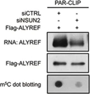
5-methylcytosin dot blot using Anti-5-methylcytosine (5-mC) antibody [33D3] ab10805. Publication image and figure legend from Yang, X., Yang, Y., et al., 2017,Cell Res, PubMed 28418038.
NSUN2 regulates nuclear-cytoplasmic shuttling and RNA-binding ability of ALYREF. (A) Immunofluorescence staining of ALYREF (red color) and ASF (green color) upon NSUN2 knockdown (top); line scan graphs (middle) and peak density quantification of line scan graphs (bottom) for ALYREF are also shown. Scale bar, 10 μm. Error bars indicate SEM (n = 120). (B) Western blotting (left) and quantification (right) of nuclear and cytoplasmic distribution of ALYREF in control and NSUN2-knockdown HeLa cells. The protein loading for the cytoplasmic fraction is about 2.5-fold higher than that for the nuclear fraction. PARP1 and TUBULIN serve as nuclear and cytoplasmic markers, respectively. Error bars indicate SEM (n = 3). (C) Immunofluorescence staining of ALYREF (red color) in NSUN2-knockdown HeLa cells transfected with control EGFP (EGFP-EV), EGFP-tagged siNSUN2-insensitive wild-type NSUN2 (EGFP-WT-Ins) or mutant (EGFP-DM-Ins) plasmids (top); line scan graphs (middle) and peak density quantification of line scan graphs (bottom) for ALYREF are also shown. Scale bar, 10 μm. Error bars indicate SEM (n = 120). (D) Western blotting (top) and quantification (bottom) of nuclear and cytoplasmic distribution of ALYREF in NSUN2-knockdown HeLa cells transfected with empty Myc expression vector (Myc-EV), Myc-tagged siNSUN2-insensitive wild-type NSUN2 (Myc-WT-Ins), or Mutant (Myc-DM-Ins). The protein loading for the cytoplasmic fraction is about 2.5-fold higher than that for the nuclear fraction. PARP1 and TUBULIN serve as nuclear and cytoplasmic markers, respectively. Error bars indicate SEM (n = 3). (E) PAR-CLIP assay (left) and quantification (right) of RNA pulled down by Flag-ALYREF upon NSUN2 knockdown. RNA labeled with biotin at 3' end of RNA (End Biotinylation Kit, Thermo) was visualized by the chemiluminescent nucleic acid detection module. m5C-modified RNAs were visualized by dot blotting using m5C antibody. Error bars indicate SEM (n = 3). (F) Rescue PAR-CLIP assay (left) and quantification (right) of RNA pulled down by Flag-ALYREF in NSUN2-knockdown HeLa cells transfected with Myc-EV, Myc-WT-Ins, or Myc-DM-Ins. RNA labeled with biotin at 3' end of RNA (End Biotinylation Kit, Thermo) was visualized by the Chemiluminescent nucleic acid detection module. Error bars indicate SEM (n = 3). P values were calculated by Student's t-test.
- IHC
CiteAb
Immunohistochemistry - Anti-5-methylcytosine (5-mC) antibody [33D3] (AB10805)
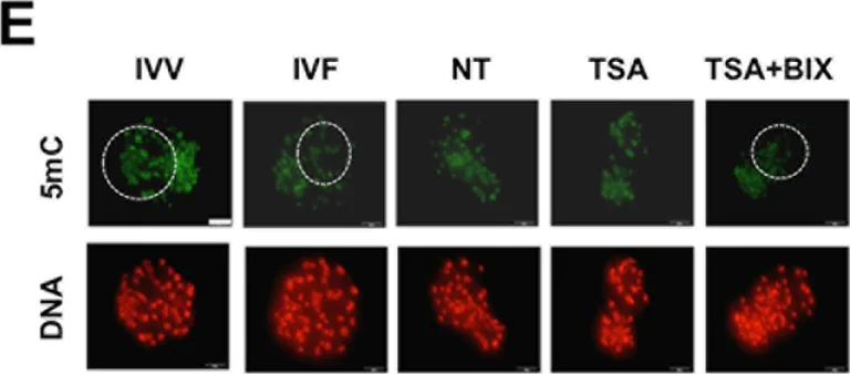
5-methylcytosine immunohistochemistry-immunofluorescence using Anti-5-methylcytosine (5-mC) antibody [33D3] ab10805. Publication image and figure legend from Cao, Z., Hong, R., et al., 2017, PLoS One, Pubmed 28114389.
TSA and BIX-01294 treatment combined restores H3K9me2 and 5mC levels in SCNT embryos.SCNT embryos at the pronuclear stage were cultured in PZM-3 supplemented with 50 nM TSA or 50 nM TSA and 1 μm BIX-01294 for 24 h and SCNT embryos cultured in PZM-3 containing DMSO were served as NT control. Then DMSO, TSA and BIX-01294 in different groups were washed out and embryos were cultured in fresh PZM-3 medium until day7. We isolated and fixed the 4-cell and blastocysts from different groups including NT control, TSA treatment, TSA and BIX-01294 combination treatment for subsequent experiments. At the same time, in vivo fertilized (IVV) and in vitro fertilized (IVF) 4-cell and blastocysts were also collected and fixed. Finally, 4-cell embryos (A) and blastocysts (C) were stained for H3K9me2 (green) and DNA (propidium iodide, red). Bottom panels showed the merged images (yellow) between H3K9me2 signals (green) and DNA staining (red). (E) Blastocysts from all groups were stained for 5mC (green) and DNA (propidium iodide, red). Shown in A, C and E are representative Z-sections obtained by confocal microscopy from one experiment. White dash circle denotes ICM area in C and TE region in E, respectively. Scale bars = 50μm. (B, D) Quantification of the H3K9me2 fluorescence intensity in nucleus of 4-cell and trophectoderm cells. (F) Quantification of the 5mC fluorescence intensity in nucleus of trophectoderm cells. Data in B, D and F are shown as mean ± S.E.M. All experiments above were repeated three times with 20 embryos per group. Values with different superscripts across groups indicate significant differences (p < < 0.05) and *p < < 0.05.
代替製品


組合せ製品


Primary Antibodies
AB172730
Rabbit IgG, monoclonal [EPR25A] - Isotype Control
KD Validated|RabMAb|Advanced Validation|Recombinant

- 0
- 0
Reactivity data
製品の詳細
Anti-5-methylcytosine (5-mC) antibody [33D3] (ab10805) has been cited over 177 times in peer reviewed journals and is trusted by the scientific community.
Abcam's high quality validation processes ensure Anti-5-methylcytosine (5-mC) antibody [33D3] (ab10805) has high sensitivity and specificity.
Anti-5-methylcytosine (5-mC) antibody [33D3] (ab10805) has 11 independent reviews from customers.
Anti-5-methylcytosine (5-mC) antibody [33D3] (ab10805) specifically detects 5-methylcytosine (5-mC) (UniProt ID: ; Molecular weight: kDa) and is sold in 50 µg selling sizes.
Storage in frost-free freezers is not recommended. Should this product contain a precipitate microcentrifugation before use.While older lots have performed well in ICC, we have received inconsistent results with the latest lots. Unfortunately, we can no longer guarantee this antibody for use in ICC.
出荷温度及び保存条件
製品の状態
精製方法
バッファー組成
出荷温度
短期保存期間
短期保存温度
長期保存温度
分注に関する情報
保管に関する情報
補足情報
This supplementary information is collated from multiple sources and compiled automatically.
Biological function summary
Cytosine methylation affects DNA-protein interactions leading to changes in chromatin structure and the regulation of gene activity. 5-methylcytosine is an important player in the chromatin modification complex where it collaborates with other epigenetic marks to control gene expression profiles. This modified nucleotide helps to stabilize gene silencing maintaining the closed chromatin state which inhibits the transcription machinery's access to specific genomic regions. Such silencing is vital during processes like X-chromosome inactivation and imprinting.
Pathways
Researchers have identified 5-mC within two critical pathways: DNA methylation and demethylation cycle and histone modification pathway. In the DNA methylation pathway 5-mC interacts closely with proteins like DNA methyltransferases (DNMTs) which directly add methyl groups to cytosine bases. In the context of histone modification 5-mC influences the binding of methyl-CpG-binding domain (MBD) proteins which read and interpret methylated DNA marks affecting histone modification and DNA accessibility.
製品プロトコール
- Visit the General protocols
- Visit the Troubleshooting
文献 (228)
Recent publications for all applications. Explore the full list and refine your search
Clinical epigenetics 17:151 PubMed40993735
2025
Applications
Unspecified application
Species
Unspecified reactive species
Biological & pharmaceutical bulletin 48:1343-1350 PubMed40930835
2025
Applications
Unspecified application
Species
Unspecified reactive species
Oncogene 44:3609-3624 PubMed40764753
2025
Applications
Unspecified application
Species
Unspecified reactive species
Frontiers in molecular neuroscience 18:1628860 PubMed40747242
2025
Applications
Unspecified application
Species
Unspecified reactive species
Journal of ovarian research 18:162 PubMed40707993
2025
Applications
Unspecified application
Species
Unspecified reactive species
Molecular cancer 24:201 PubMed40691844
2025
Applications
Unspecified application
Species
Unspecified reactive species
Animal bioscience 38:1973-1983 PubMed40241588
2025
Applications
Unspecified application
Species
Unspecified reactive species
Journal of experimental & clinical cancer research : CR 44:121 PubMed40234937
2025
Applications
Unspecified application
Species
Unspecified reactive species
Cancer communications (London, England) 45:846-879 PubMed40227950
2025
Applications
Unspecified application
Species
Unspecified reactive species
Journal of translational medicine 23:385 PubMed40170133
2025
Applications
Unspecified application
Species
Unspecified reactive species
Abcam product promise
Please note: All products are 'FOR RESEARCH USE ONLY. NOT FOR USE IN DIAGNOSTIC OR THERAPEUTIC PROCEDURES'.
For licensing inquiries, please contact partnerships@abcam.com