Human VAPA knockout HeLa cell line
Human VAPA knockout HeLa cell line
Be the first to review this product! Submit a review
|
(0 Publication)

- NGS
Lab
Next Generation Sequencing - Human VAPA knockout HeLa cell line (AB275831)
1 bp deletion after Pro55 of the WT protein
- WB
Lab
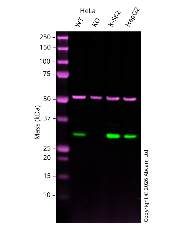
Western blot - Human VAPA knockout HeLa cell line (AB275831)
Western blot : Anti-VAPA antibody [EPR13589(B)] - N-terminal ab181067 staining at 1/20000 dilution, shown in green; Mouse anti alpha Tubulin ab7291 loading control staining at 1/20000 dilution, shown in magenta. A band was observed at 28 kDa in Wild-type HeLa cell lysates with no signal observed at this size in VAPA knockout HeLa cell line. To generate this image, samples were run on an SDS-PAGE gel then transferred onto a nitrocellulose membrane. Membranes were blocked in 5% Milk in TBS-0.1 % Tween® 20 (TBS-T) before incubation with primary antibodies overnight at 4 °C. Blots were washed four times in TBS-T, incubated with secondary antibodies for 1 h at room temperature, washed again four times then imaged. Secondary antibodies used were Goat anti-Rabbit 800CW and Goat anti-Mouse 680RD at 1/20,000 dilution.
All lanes:
Western blot - Anti-VAPA antibody [EPR13589(B)] - N-terminal (<a href='/products/primary-antibodies/vapa-antibody-epr13589b-n-terminal-ab181067'>ab181067</a>) at 1/20000 dilution
Lane 1:
Western blot - Human wild-type HeLa cell line (ab271142) at 20 µg
Lane 2:
Western blot - Human VAPA knockout HeLa cell line (ab275831) at 20 µg
Lane 3:
K562 at 20 µg
Lane 4:
HepG2 at 20 µg
Secondary
All lanes:
Goat anti-Rabbit 800CW & Goat anti-Mouse 680RD at 1/20000 dilution
Predicted band size: 28 kDa
Observed band size: 28 kDa
false
組合せ製品


製品の詳細
Recommended control: Human wild-type HeLa cell line (ab271142). Please note a wild-type cell line is not automatically included with a knockout cell line order, if required please add recommended wild-type cell line at no additional cost using the code WILDTYPE-TMTK1.
We will provide viable cells that proliferate on revival.
This product is subject to limited use licenses from The Broad Institute and ERS Genomics Limited, and is developed with patented technology. For full details of the limited use licenses and relevant patents please refer to our limited use license and patent pages.
製品内容
出荷温度及び保存条件
遺伝子名
遺伝子編集のタイプ
遺伝子編集の方法
接合型
出荷温度
短期保存期間
短期保存温度
長期保存温度
補足情報
This supplementary information is collated from multiple sources and compiled automatically.
Biological function summary
In the cellular environment VAPA plays an important role in maintaining homeostasis by regulating lipid transport and metabolism. It forms a complex with several other proteins such as OSBP (oxysterol-binding protein) and CERT (ceramide transport protein) which facilitates the exchange of lipids between the ER and the Golgi apparatus. This interaction underlines VAPA's role in lipid droplet dynamics and the synthesis of phosphatidylinositols which are critical for cell signaling and membrane integrity.
Pathways
The involvement of VAPA is significant in lipid metabolism and membrane trafficking pathways. It interacts with proteins like VAMP (vesicle-associated membrane proteins) and SNARE (soluble N-ethylmaleimide-sensitive factor attachment protein receptor) complexes which are important for exocytosis and endocytosis processes. VAPA's interaction in these pathways supports the movement of materials across cellular membranes and its regulation of lipid exchange impacts the signaling pathways influencing cell growth and differentiation.
Cell culture
Biosafety level
EU: 2 US: 2
Adherent/suspension
Adherent
Gender
Female
初回取り扱いガイドライン
Upon arrival, the vial should be stored in liquid nitrogen vapor phase and not at -80°C. Storage at -80°C may result in loss of viability. <p>1. Thaw the vial in 37°C water bath for approximately 1-2 minutes.<br>2. Transfer the cell suspension (0.8 mL) to a 15 mL/50 mL conical sterile polypropylene centrifuge tube containing 8.4 mL pre-warmed culture medium, wash vial with an additional 0.8 mL culture medium (total volume 10 mL) to collect remaining cells, and centrifuge at 201 x g (rcf) for 5 minutes at room temperature. 10 mL represents minimum recommended dilution. 20 mL represents maximum recommended dilution.<br>3. Resuspend the cell pellet in 5 mL pre-warmed culture medium and count using a haemocytometer or alternative cell counting method seed all remaining cells into a T25.<br>4. Incubate the culture at 37°C incubator with 5% CO<sub>2</sub>. Check the culture one day after revival and continue to check until 80% confluent. Media change can be given if needed.<br>5. Once confluent passage into an appropriate flask at a density of 2x10<sup>4</sup> cells/cm<sup>2</sup>. Seeding density is given as a guide only and should be scaled to align with individual lab schedules. Cultures should be monitored daily.</p>
継代培養ガイドライン
- All seeding densities should be based on cell counts gained by established methods.
- A guide seeding density of 2x10⁴ cells/cm2 is recommended.
- Cells should be passaged when they have achieved 80-90% confluence.
培養培地
DMEM (High Glucose) + 10% FBS
凍結保存培地
Cell Freezing Medium-DMSO Serum free media, contains 8.7% DMSO in MEM supplemented with methyl cellulose.
Abcam product promise
Please note: All products are 'FOR RESEARCH USE ONLY. NOT FOR USE IN DIAGNOSTIC OR THERAPEUTIC PROCEDURES'.
For licensing inquiries, please contact partnerships@abcam.com