Human MYOF (Myoferlin) knockout HeLa cell line
Human MYOF (Myoferlin) knockout HeLa cell line
- Advanced Validation
- 詳細を見る
Be the first to review this product! Submit a review
|
(0 Publication)
- WB
Lab
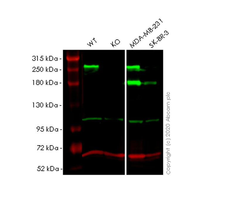
Western blot - Human MYOF (Myoferlin) knockout HeLa cell line (AB265782)
Lanes 1-4 : Merged signal (red and green). Green - ab178386 observed at 250180 kDa. Red - loading control ab7291 observed at 50 kDa.
ab178386 Anti-Myoferlin antibody [EPR18887] was shown to specifically react with Myoferlin in wild-type HeLa cells. Loss of signal was observed when knockout cell line ab265782 (knockout cell lysate ab257547) was used. Wild-type and Myoferlin knockout samples were subjected to SDS-PAGE. ab178386 and Anti-alpha Tubulin antibody [DM1A] - Loading Control (ab7291) were incubated overnight at 4°C at 1 in 1000 dilution and 1 in 20000 dilution respectively. Blots were developed with Goat anti-Rabbit IgG H&L (IRDye® 800CW) preadsorbed (ab216773) and Goat anti-Mouse IgG H&L (IRDye® 680RD) preadsorbed (ab216776) secondary antibodies at 1 in 20000 dilution for 1 hour at room temperature before imaging.
All lanes:
Western blot - Anti-Myoferlin antibody [EPR18887] (<a href='/products/primary-antibodies/myoferlin-antibody-epr18887-ab178386'>ab178386</a>) at 1/1000 dilution
Lane 1:
Wild-type HeLa cell lysate at 20 µg
Lane 2:
MYOF knockout HeLa cell lysate at 20 µg
Lane 2:
Western blot - Human MYOF (Myoferlin) knockout HeLa cell line (ab265782)
Lane 3:
MDA-MB-231 cell lysate at 20 µg
Lane 4:
SK-BR-3 cell lysate at 20 µg
Secondary
All lanes:
Western blot - Goat anti-Rabbit IgG H&L (IRDye® 800CW) preadsorbed (<a href='/products/secondary-antibodies/goat-rabbit-igg-h-l-irdye-800cw-preadsorbed-ab216773'>ab216773</a>) at 1/10000 dilution
Predicted band size: 235 kDa
Observed band size: 180 kDa,250 kDa
false

- Sanger seq
Unknown
Sanger Sequencing - Human MYOF (Myoferlin) knockout HeLa cell line (AB265782)
Homozygous : 10 bp deletion in exon 6.
組合せ製品
Primary Antibodies
AB252126
Alexa Fluor® 488 Anti-CD133 antibody [EPR20980-104]
RabMAb|Recombinant
- 0
- 0

Cell Lines & Lysates
AB276116
Human CASP1 (Caspase-1) knockout THP-1 cell line
Advanced Validation
- 0
- 0
Reactivity data
製品の詳細
We will provide viable cells that proliferate on revival.
This product is subject to limited use licenses from The Broad Institute and ERS Genomics Limited, and is developed with patented technology. For full details of the limited use licenses and relevant patents please refer to our limited use license and patent pages.
製品内容
出荷温度及び保存条件
遺伝子名
遺伝子編集のタイプ
遺伝子編集の方法
ノックアウト検証方法
接合型
出荷温度
短期保存温度
長期保存温度
補足情報
This supplementary information is collated from multiple sources and compiled automatically.
Biological function summary
The protein supports membrane dynamics and stabilization. Myoferlin is not part of a large complex but interacts with other proteins at the cellular membrane. It regulates cellular processes such as proliferation differentiation and endocytosis. In muscle cells it assists in the organization and function of the cytoskeleton maintaining proper cellular architecture and signaling.
Pathways
Myoferlin engages in critical roles in pathways related to cell membrane repair and trafficking. It associates with pathways like the myogenesis pathway and integrin signaling. In these pathways it interacts with other proteins such as dysferlin and caveolin-3 which facilitate membrane repair and receptor localization respectively. Myoferlin ensures efficient cellular response to membrane damage maintaining tissue integrity.
Quality control
STR analysis
CSF1PO, D13S317, D7S820, D5S818, TH01, D16S539, TPOX
Cell culture
Biosafety level
EU: 2 US: 2
Adherent/suspension
Adherent
Gender
Female
初回取り扱いガイドライン
Upon arrival, the vial should be stored in liquid nitrogen vapor phase and not at -80°C. Storage at -80°C may result in loss of viability. <p>1. Thaw the vial in 37°C water bath for approximately 1-2 minutes.<br>2. Transfer the cell suspension (0.8 mL) to a 15 mL/50 mL conical sterile polypropylene centrifuge tube containing 8.4 mL pre-warmed culture medium, wash vial with an additional 0.8 mL culture medium (total volume 10 mL) to collect remaining cells, and centrifuge at 201 x g (rcf) for 5 minutes at room temperature. 10 mL represents minimum recommended dilution. 20 mL represents maximum recommended dilution.<br>3. Resuspend the cell pellet in 5 mL pre-warmed culture medium and count using a haemocytometer or alternative cell counting method seed all remaining cells into a T25.<br>4. Incubate the culture at 37°C incubator with 5% CO<sub>2</sub>. Check the culture one day after revival and continue to check until 80% confluent. Media change can be given if needed.<br>5. Once confluent passage into an appropriate flask at a density of 2x10<sup>4</sup> cells/cm<sup>2</sup>. Seeding density is given as a guide only and should be scaled to align with individual lab schedules. Cultures should be monitored daily.</p>
継代培養ガイドライン
- All seeding densities should be based on cell counts gained by established methods.
- A guide seeding density of 2x10⁴ cells/cm2 is recommended.
- Cells should be passaged when they have achieved 80-90% confluence.
培養培地
DMEM (High Glucose) + 10% FBS
凍結保存培地
Cell Freezing Medium-DMSO Serum free media, contains 8.7% DMSO in MEM supplemented with methyl cellulose.
Abcam product promise
Please note: All products are 'FOR RESEARCH USE ONLY. NOT FOR USE IN DIAGNOSTIC OR THERAPEUTIC PROCEDURES'.
For licensing inquiries, please contact partnerships@abcam.com