アブカムでは最適な動作のために Google Chrome など最新ブラウザでの閲覧を推奨します。

私たちはウェブサイトをできるだけ使いやすくするために、クッキーを使用しています。

クッキー・ポリシーでは、使用するクッキーをオプトアウトする方法について説明しています。

クッキーの設定を変更しないままでいる場合、このポリシーに同意しているとみなされます。

続ける 続ける

お問い合わせは電話 +81-(0)3-6231-0940 または メールでどうぞ

  • アカウント
  • ログアウト
ログイン または 新規登録

Welcome

ログイン または

すでにアカウントをお持ちですか?

新規登録
カート
クイック・オーダー
Abcam homepage

  • 研究用製品
    製品タイプ別
    一次抗体
    二次抗体
    イムノアッセイキット・試薬
    細胞・組織イメージング
    細胞・バイオケミカルアッセイ
    タンパク質・ペプチド
    製品タイプ別
    プロテオミクス
    アゴニスト・アクチベータ-・アンタゴニスト・インヒビター
    細胞株・ライゼート
    miRNA マルチプレックス・アッセイキット
    マルチプレックス・アッセイ
    研究分野別
    癌
    循環器系
    細胞生物学
    エピジェネティクス
    メタボリズム
    発生生物学
    研究分野別
    免疫学
    微生物学
    脳神経科学
    シグナル伝達
    ステムセル
  • 診断と治療のソリューション
    カスタム・ソリューション & パートナーシップ

    あなたの診断法や治療法を発展させるための、カスタム抗体開発およびコマーシャル・パートナーシップ

    私たちと共にカスタム・ソリューションを創造する

    私たちとパートナーになる

  • サポート
    サポートハブ

    研究のためのサポートとアドバイス

    サポートハブを表示

    プロトコール

    実験のステップごとの詳細

    プロトコールを表示

  • イベント
    • イベント・カレンダー
    • 癌
    • 循環器
    • エピジェネティクス&核内情報
    • 免疫学
    • ニューロサイエンス
    • ステムセル
    • 展示会
    • ウェビナー
    最新のイベント情報

    世界中でアブカムが主催する研究会やセミナーの日程、内容、演者など

    イベント・カレンダー

  • パスウェイ
    細胞シグナル伝達パスウェイ

    すべてのパスウェイを見る

    インタラクティブ・パスウェイを見る

  • ブログ
    アブカム・ブログ

    最新の情報はこちら.

    • キャンペーン
    • 製品情報
    • 技術情報
  1. Link

    pericentrin-antibody-centrosome-marker-ab4448.pdf

  1. Send me a copy of this email
    I agree to the terms and conditions.
Cell Biology Cell Cycle Cell Division Spindle
Share by email

Anti-Pericentrin 抗体 - Centrosome Marker (ab4448)

  • Datasheet
  • SDS
Reviews (32)Q&A (5)References (360)

Product price, shipping and contact information

Currently unavailable

申し訳ございませんが、現在表示できません。

こちらにお問い合わせいただくか、またはしばらく経ってから改めてお試しください


Loading size & price…


出荷および受注について

Abpromise

保証された製品品質、優れたカスタマー・サポート。 詳細はこちら。

Immunocytochemistry/ Immunofluorescence - Anti-Pericentrin antibody - Centrosome Marker (ab4448)
  • Immunocytochemistry/ Immunofluorescence - Anti-Pericentrin antibody - Centrosome Marker (ab4448)
  • Immunocytochemistry/ Immunofluorescence - Anti-Pericentrin antibody - Centrosome Marker (ab4448)
  • Immunocytochemistry/ Immunofluorescence - Anti-Pericentrin antibody - Centrosome Marker (ab4448)
  • Immunocytochemistry/ Immunofluorescence - Anti-Pericentrin antibody - Centrosome Marker (ab4448)
  • Immunocytochemistry/ Immunofluorescence - Anti-Pericentrin antibody - Centrosome Marker (ab4448)
  • Immunocytochemistry/ Immunofluorescence - Anti-Pericentrin antibody - Centrosome Marker (ab4448)

Key features and details

  • Rabbit polyclonal to Pericentrin - Centrosome Marker
  • Suitable for: ICC/IF
  • Reacts with: Mouse, Human
  • Isotype: IgG

リコンビナント抗体で、ロット間での高い再現性を実現

Product image
Anti-Pericentrin antibody [EPR21987] (ab220784)
  • 異なるロット間での安定した再現性
  • 容易なスケールアップ
  • 評価試験による特異性の確認済み
  • 倫理基準に準拠 - アニマル・フリーの生産

製品の概要

  • 製品名

    Anti-Pericentrin antibody - Centrosome Marker
    Pericentrin 一次抗体 製品一覧
  • 製品の詳細

    Rabbit polyclonal to Pericentrin - Centrosome Marker
  • 由来種

    Rabbit
  • 特異性

    This antibody should recognise both Pericentrin and Kendrin (also known as Pericentrin-2).
  • アプリケーション

    適用あり: ICC/IFmore details
  • 種交差性

    交差種: Mouse, Human
    交差が予測される動物種: Rat, Rabbit, African green monkey
  • 免疫原

    Fusion protein corresponding to Mouse Pericentrin aa 100-600.

  • ポジティブ・コントロール

    • ICC: Human coronary artery endothelial, MCF7, HeLa and NIH/3T3 cells.
  • 特記事項

    Reproducibility is key to advancing scientific discovery and accelerating scientists’ next breakthrough.

    Abcam is leading the way with our range of recombinant antibodies, knockout-validated antibodies and knockout cell lines, all of which support improved reproducibility.

    We are also planning to innovate the way in which we present recommended applications and species on our product datasheets, so that only applications & species that have been tested in our own labs, our suppliers or by selected trusted collaborators are covered by our Abpromise™ guarantee.

    In preparation for this, we have started to update the applications & species that this product is Abpromise guaranteed for.

    We are also updating the applications & species that this product has been “predicted to work with,” however this information is not covered by our Abpromise guarantee.

    Applications & species from publications and Abreviews that have not been tested in our own labs or in those of our suppliers are not covered by the Abpromise guarantee.

    Please check that this product meets your needs before purchasing. If you have any questions, special requirements or concerns, please send us an inquiry and/or contact our Support team ahead of purchase. Recommended alternatives for this product can be found below, as well as customer reviews and Q&As.

製品の特性

  • 製品の状態

    Liquid
  • 保存方法

    Shipped at 4°C. Store at +4°C short term (1-2 weeks). Upon delivery aliquot. Store at -20°C or -80°C. Avoid freeze / thaw cycle.
  • バッファー

    pH: 7.40
    Preservative: 0.02% Sodium azide
    Constituent: PBS

    Batches of this product that have a concentration < 1mg/ml may have BSA added as a stabilising agent. If you would like information about the formulation of a specific lot, please contact our scientific support team who will be happy to help.
  • Concentration information loading...
  • 精製度

    Protein G purified
  • ポリ/モノ

    ポリクローナル
  • アイソタイプ

    IgG
  • 研究分野

    • Cell Biology
    • Cell Cycle
    • Cell Division
    • Spindle
    • Tags & Cell Markers
    • Subcellular Markers
    • Organelles
    • Centrosome

関連製品

  • Compatible Secondaries

    • Goat Anti-Rabbit IgG H&L (Alexa Fluor® 488) (ab150077)
    • Goat Anti-Rabbit IgG H&L (HRP) (ab205718)
  • Isotype control

    • Rabbit IgG, polyclonal - Isotype Control (ChIP Grade) (ab171870)

アプリケーション

Our Abpromise guarantee covers the use of ab4448 in the following tested applications.

The application notes include recommended starting dilutions; optimal dilutions/concentrations should be determined by the end user.

アプリケーション Abreviews 特記事項
ICC/IF Use a concentration of 0.1 - 0.5 µg/ml.

ターゲット情報

  • 機能

    Integral component of the filamentous matrix of the centrosome involved in the initial establishment of organized microtubule arrays in both mitosis and meiosis. Plays a role, together with DISC1, in the microtubule network formation. Is an integral component of the pericentriolar material (PCM). May play an important role in preventing premature centrosome splitting during interphase by inhibiting NEK2 kinase activity at the centrosome.
  • 組織特異性

    Expressed in all tissues tested, including placenta, liver, kidney and thymus.
  • 関連疾患

    Microcephalic osteodysplastic primordial dwarfism 2
  • ドメイン

    Composed of a coiled-coil central region flanked by non-helical N- and C-terminals.
  • 細胞内局在

    Cytoplasm > cytoskeleton > microtubule organizing center > centrosome. Centrosomal at all stages of the cell cycle. Remains associated with centrosomes following microtubule depolymerization. Colocalized with DISC1 at the centrosome.
  • Target information above from: UniProt accession O95613 The UniProt Consortium
    The Universal Protein Resource (UniProt) in 2010
    Nucleic Acids Res. 38:D142-D148 (2010) .

    Information by UniProt
  • 参照データベース

    • Entrez Gene: 5116 Human
    • Entrez Gene: 18541 Mouse
    • Omim: 605925 Human
    • SwissProt: O95613 Human
    • SwissProt: P48725 Mouse
    • Unigene: 474069 Human
    • Unigene: 251794 Mouse
    • 別名

      • Centrosome Marker antibody
      • Ken antibody
      • Kendrin antibody
      • KIAA0402 antibody
      • MOPD2 antibody
      • PCN antibody
      • PCNT 2 antibody
      • PCNT antibody
      • PCNT B antibody
      • PCNT_HUMAN antibody
      • PCNT1 antibody
      • PCNT2 antibody
      • PCNTB antibody
      • PCTN2 antibody
      • Pericentrin 1 antibody
      • Pericentrin 2 antibody
      • Pericentrin 380 antibody
      • Pericentrin antibody
      • Pericentrin B antibody
      • Pericentrin-B antibody
      • SCKL4 antibody
      see all

    画像

    • Immunocytochemistry/ Immunofluorescence - Anti-Pericentrin antibody - Centrosome Marker (ab4448)
      Immunocytochemistry/ Immunofluorescence - Anti-Pericentrin antibody - Centrosome Marker (ab4448)Image courtesy of an anonymous abreview

      Human coronary artery endothelial cells (HCAEC) were labeled for Pericentrin using ab4448 at 1/100 dilution in ICC/IF. Primary incubation was for 1 hour at room temperature (green). The seciondary antibody was a Goat anti-rabbit IgG AlexaFluor® conjugate.

      Fixation: Formaldehyde. Permeabilisation: 0.1% Trition X-100 in HBSS.

    • Immunocytochemistry/ Immunofluorescence - Anti-Pericentrin antibody - Centrosome Marker (ab4448)
      Immunocytochemistry/ Immunofluorescence - Anti-Pericentrin antibody - Centrosome Marker (ab4448)Ruppenthal et al PLoS One. 2018 Jan 25;13(1):e0191734. doi: 10.1371/journal.pone.0191734. eCollection 2018. Fig 2. Reproduced under the Creative Commons license http://creativecommons.org/licenses/by/4.0/

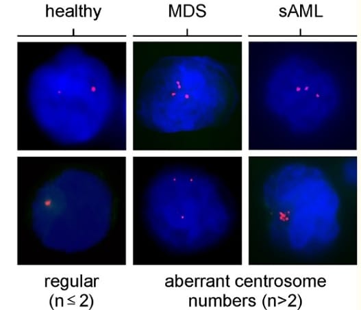
      A representative panel of indirect immunofluorescence microscopic images shows normal (regular, n ≤ 2) and aberrant centrosome numbers (n > 2) in interphase cells.

      Centrosomes were stained using anti-pericentrin antibody ab4448 (magenta), nuclear DNA is shown in blue (DAPI).

      Statistical methods: Kruskal-Wallis test. Mann-Whitney U tests followed by Bonferroni-Holm p-value correction were made as post-hoc tests in order to compare the MDS and sAML patients with the control group.

    • Immunocytochemistry/ Immunofluorescence - Anti-Pericentrin antibody - Centrosome Marker (ab4448)
      Immunocytochemistry/ Immunofluorescence - Anti-Pericentrin antibody - Centrosome Marker (ab4448)This image is courtesy of Gordon Chan, University of Alberta

      IF staining of pericentrin in MCF7 (Human breast adenocarcinoma cell line) cells.

      The top panel is an interphase cell showing centrosome staining. 

      The bottom panel shows a mitotic cell with spindle pole staining. 

      ab4448 was used at 1/500, but also works at higher dilutions (1/1000-1/2000).

      Top panel - 630X magnification; Bottom panel -1000X magnification.

      The secondary antibody was Alexa-Fluor®488 anti-rabbit.

    • Immunocytochemistry/ Immunofluorescence - Anti-Pericentrin antibody - Centrosome Marker (ab4448)
      Immunocytochemistry/ Immunofluorescence - Anti-Pericentrin antibody - Centrosome Marker (ab4448)This image is courtesy of Roberto Giambruno, Marilena Ciciarello and Patrizia Lavia

      NIH/3T3 (Mouse embryo fibroblast cell line) cells were fixed in 100% methanol for 6 minutes at -20°C, washed 3 times in PBS then incubated with ab4448 (1/2000) for 1 hour at room temperature.

      The panel of images shows the nuclei stained with DAPI (blue), ab4448 staining is shown in green. 100x magnification.


    • Immunocytochemistry/ Immunofluorescence - Anti-Pericentrin antibody - Centrosome Marker (ab4448)
      Immunocytochemistry/ Immunofluorescence - Anti-Pericentrin antibody - Centrosome Marker (ab4448)

      ICC/IF image of ab4448 stained HeLa (Human epithelial cell line from cervix adenocarcinoma) cells.

      The cells were fixed in methanol (5 minutes) and incubated with the antibody (ab4448, 1 µg/ml) for 1 hour at room temperature. The secondary antibody (green) was Alexa Fluor® 488 goat anti-rabbit IgG (H+L) used at a 1/1000 dilution for 1 hour. Image-iTTM FX Signal Enhancer was used as the primary blocking agent, 5% BSA (in TBS-T) was used for all other blocking steps. DAPI was used to stain the cell nuclei (blue). Alexa Fluor® 594 WGA was used to label plasma membranes (red).

    • Immunocytochemistry/ Immunofluorescence - Anti-Pericentrin antibody - Centrosome Marker (ab4448)
      Immunocytochemistry/ Immunofluorescence - Anti-Pericentrin antibody - Centrosome Marker (ab4448)

      ab4448 stained NIH/3T3 (Mouse embryo fibroblast cell line) cells.

      The cells were fixed in 100% methanol for 5 minutes at -20°C and then incubated in 1% BSA / 10% normal goat serum / 0.3M glycine in 0.1% PBS-Tween for 1 hour at room temperature to permeabilize the cells and block non-specific protein-protein interactions. The cells were then incubated with the antibody (ab4448 at 5 µg/ml) overnight at +4°C. The secondary antibody (pseudo-colored green) was Goat Anti-Rabbit IgG H&L (Alexa Fluor® 488) preadsorbed (ab150081) used at a 1/1000 dilution for 1 hour at room temperature. Alexa Fluor® 594 WGA was used to label plasma membranes (pseudo-colored red) at a 1/200 dilution for 1 hour at room temperature.

      DAPI was used to stain the cell nuclei (pseudo-colored blue) at a concentration of 1.43µM for 1 hour at room temperature.

    • Immunocytochemistry/ Immunofluorescence - Anti-Pericentrin antibody - Centrosome Marker (ab4448)
      Immunocytochemistry/ Immunofluorescence - Anti-Pericentrin antibody - Centrosome Marker (ab4448)

      ICC/IF image of ab4448 stained HeLa (Human epithelial cell line from cervix adenocarcinoma) cells.

      The cells were fixed in 100% methanol (5 minutes) and then incubated in 1% BSA / 10% normal goat serum / 0.3M glycine in 0.1% PBS-Tween for 1 hour to permeabilize the cells and block non-specific protein-protein interactions. The cells were then incubated with the antibody (ab4448, 1 µg/ml) overnight at +4°C. The secondary antibody (green) was DyLight® 488 goat anti-rabbit IgG - H&L, pre-adsorbed (ab96899) used at a 1/250 dilution for 1 hour. Alexa Fluor® 594 WGA was used to label plasma membranes (red) at a 1/200 dilution for 1 hour.

      DAPI was used to stain the cell nuclei (blue) at a concentration of 1.43µM.

    プロトコール

    • Recommended ICC/IF protocol with ab4448

    Click here to view the general protocols

    データシートおよび資料

    • Datasheet
    • SDS
  • 参考文献 (360)

    ab4448 を使用した論文を発表された方は、こちらまでお知らせください。データシートに掲載させていただきます。

    ab4448 は 360 報の論文で使用されています。

    • Huhn O  et al. Distinctive phenotypes and functions of innate lymphoid cells in human decidua during early pregnancy. Nat Commun 11:381 (2020). PubMed: 31959757
    • Denu RA  et al. Centrosome Amplification in Cancer Disrupts Autophagy and Sensitizes to Autophagy Inhibition. Mol Cancer Res 18:33-45 (2020). PubMed: 31604847
    • Naso FD  et al. Excess TPX2 Interferes with Microtubule Disassembly and Nuclei Reformation at Mitotic Exit. Cells 9:N/A (2020). PubMed: 32041138
    • Mikulenkova E  et al. NANOG/NANOGP8 Localizes at the Centrosome and is Spatiotemporally Associated with Centriole Maturation. Cells 9:N/A (2020). PubMed: 32168958
    • Wu D & Dean J EXOSC10 sculpts the transcriptome during the growth-to-maturation transition in mouse oocytes. Nucleic Acids Res 48:5349-5365 (2020). PubMed: 32313933
    View all Publications for this product

    レビューと Q&A

    Show All レビュー Q&A
    レビューを送る 質問を送る

    1-10 of 37 Abreviews or Q&A

    Western blot abreview for Anti-Pericentrin antibody - Centrosome Marker

    Inconclusive
    Abreviews
    Abreviews
    abreview image
    Application
    Western blot
    Sample
    Human Cell lysate - whole cell (Dopaminergic Neurons, HEK293T and HeLa)
    Gel Running Conditions
    Reduced Denaturing (NuPage 4-12% MES buffer)
    Loading amount
    30 µg
    Specification
    Dopaminergic Neurons, HEK293T and HeLa
    Blocking step
    Milk as blocking agent for 1 hour(s) and 0 minute(s) · Concentration: 5% · Temperature: 20°C
    Read More

    Abcam user community

    Verified customer

    投稿 Nov 20 2020

    Immunocytochemistry/ Immunofluorescence abreview for Anti-Pericentrin antibody - Centrosome Marker

    Excellent
    Abreviews
    Abreviews
    abreview image
    Application
    Immunocytochemistry/ Immunofluorescence
    Sample
    Human Cell (Human coronary artery endothelial cells HCAEC)
    Permeabilization
    Yes - 0.1% Trition X-100 in HBSS
    Specification
    Human coronary artery endothelial cells HCAEC
    Blocking step
    Serum as blocking agent for 30 minute(s) · Concentration: 2% · Temperature: 21°C
    Fixative
    Formaldehyde
    Read More

    Abcam user community

    Verified customer

    投稿 Feb 19 2020

    Immunocytochemistry/ Immunofluorescence abreview for Anti-Pericentrin antibody - Centrosome Marker

    Excellent
    Abreviews
    Abreviews
    abreview image
    Application
    Immunocytochemistry/ Immunofluorescence
    Sample
    Human Cell (HAP1)
    Permeabilization
    Yes - 0.5% Triton 5 minutes
    Specification
    HAP1
    Blocking step
    Serum as blocking agent for 30 minute(s) · Concentration: 5% · Temperature: 25°C
    Fixative
    Paraformaldehyde
    Read More

    Abcam user community

    Verified customer

    投稿 Dec 14 2018

    Immunocytochemistry/ Immunofluorescence abreview for Anti-Pericentrin antibody - Centrosome Marker

    Excellent
    Abreviews
    Abreviews
    Application
    Immunocytochemistry/ Immunofluorescence
    Sample
    Human Cell (colon carcinoma cells)
    Permeabilization
    Yes - Triton
    Specification
    colon carcinoma cells
    Fixative
    Paraformaldehyde
    Read More

    Abcam user community

    Verified customer

    投稿 Sep 11 2018

    Immunocytochemistry/ Immunofluorescence abreview for Anti-Pericentrin antibody - Centrosome Marker

    Good
    Abreviews
    Abreviews
    abreview image
    Application
    Immunocytochemistry/ Immunofluorescence
    Sample
    African green monkey Cell (BS-C-1 cells)
    Permeabilization
    Yes - 0.2% Triton X
    Specification
    BS-C-1 cells
    Blocking step
    Serum as blocking agent for 30 minute(s) · Concentration: 5% · Temperature: RT°C
    Fixative
    Paraformaldehyde
    Read More

    Mr. J. Y. Seo

    Verified customer

    投稿 Apr 09 2018

    Immunocytochemistry/ Immunofluorescence abreview for Anti-Pericentrin antibody - Centrosome Marker

    Good
    Abreviews
    Abreviews
    abreview image
    Application
    Immunocytochemistry/ Immunofluorescence
    Sample
    Mouse Cell (brain section)
    Specification
    brain section
    Fixative
    Formaldehyde
    Read More

    Abcam user community

    Verified customer

    投稿 Dec 18 2017

    Western blot abreview for Anti-Pericentrin antibody - Centrosome Marker

    Good
    Abreviews
    Abreviews
    abreview image
    Application
    Western blot
    Sample
    Human Cell lysate - whole cell (293T)
    Gel Running Conditions
    Reduced Denaturing (4-12 Bis tris)
    Loading amount
    5e+006 cells
    Specification
    293T
    Blocking step
    Milk as blocking agent for 12 hour(s) and 0 minute(s) · Concentration: 5% · Temperature: 4°C
    Read More

    Abcam user community

    Verified customer

    投稿 Mar 18 2016

    Immunocytochemistry/ Immunofluorescence abreview for Anti-Pericentrin antibody - Centrosome Marker

    Excellent
    Abreviews
    Abreviews
    abreview image
    Application
    Immunocytochemistry/ Immunofluorescence
    Sample
    Human Cell (HT1080)
    Permeabilization
    Yes - 0.5% Triton X100
    Specification
    HT1080
    Blocking step
    BSA as blocking agent for 1 hour(s) and 0 minute(s) · Concentration: 3% · Temperature: 18°C
    Fixative
    Paraformaldehyde
    Read More

    Abcam user community

    Verified customer

    投稿 Mar 18 2016

    Immunocytochemistry/ Immunofluorescence abreview for Anti-Pericentrin antibody

    Poor
    Abreviews
    Abreviews
    Application
    Immunocytochemistry/ Immunofluorescence
    Sample
    Mouse Cell (mouse prostate and mammary epithelial)
    Specification
    mouse prostate and mammary epithelial
    Blocking step
    BSA as blocking agent for 1 hour(s) and 0 minute(s) · Concentration: 5µg/mL · Temperature: 25°C
    Fixative
    try both PFA and methanol
    Read More

    Abcam user community

    Verified customer

    投稿 Aug 13 2014

    Immunocytochemistry/ Immunofluorescence abreview for Anti-Pericentrin antibody

    Excellent
    Abreviews
    Abreviews
    abreview image
    Application
    Immunocytochemistry/ Immunofluorescence
    Blocking step
    BSA as blocking agent for 1 hour(s) and 0 minute(s) · Concentration: 5% · Temperature: RT°C
    Sample
    Human Cell (MCF10A Breast Cancer Cell Line)
    Specification
    MCF10A Breast Cancer Cell Line
    Permeabilization
    No
    Fixative
    1% Gluteraldehyde
    Read More

    Abcam user community

    Verified customer

    投稿 May 13 2014

    1-10 of 37 Abreviews or Q&A

    •  戻る
    • 1
    • 2
    • 3
    • 4
    • 次へ 

    Please note: All products are "FOR RESEARCH USE ONLY. NOT FOR USE IN DIAGNOSTIC PROCEDURES"
    For licensing inquiries, please contact partnerships@abcam.com

    技術情報や製品情報をメールで受け取る 登録
    製品・研究分野別
    • 癌
    • 循環器
    • 細胞生物学
    • 発生
    • エピジェネティクス&核内情報
    • 免疫学
    • メタボリズム
    • 感染症
    • ニューロサイエンス
    • シグナル伝達
    • ステムセル
    製品・タイプ別
    • 一次抗体
    • 二次抗体
    • バイオケミカルズ
    • アイソタイプ・コントロール
    • マルチカラー・セレクター
    • キット
    • ローディング・コントロール
    • ライゼート
    • ペプチド
    • タンパク質
    • スライド
    • タグ抗体・細胞マーカー
    • その他試薬
    サービス&サポート
    • サポート
    • よくあるお問い合わせ
    • プロトコール&トラブルシュート
    • ご購入に関して
    • お問い合わせフォーム
    • トレーニング
    アブカムについて
    • 企業ウェブサイト
    • 投資家の皆様へ
    • 企業情報
    • 採用情報
    • アブカムについて
    イベント
    • 展示会
    • カンファレンス
    ブログ

    ブログを読む

    International websites
    • abcam.com
    • abcam.cn

    Join with us

    • LinkedIn
    • facebook
    • Twitter
    • YouTube
    • Terms of sale
    • Website terms of use
    • Cookie policy
    • Privacy policy
    • Legal
    © 1998-2021 Abcam plc. All rights reserved.