Anti-GAPDH 抗体 - Loading Control (ab9485)
")
Key features and details
- Rabbit polyclonal to GAPDH - Loading Control
- Suitable for: IHC-P, WB, ICC/IF
- Reacts with: Mouse, Human
- Isotype: IgG
リコンビナント抗体で、ロット間での高い再現性を実現


- 異なるロット間での安定した再現性
- 容易なスケールアップ
- 評価試験による特異性の確認済み
- 倫理基準に準拠 - アニマル・フリーの生産
製品の概要
-
製品名
Anti-GAPDH antibody - Loading Control
GAPDH 一次抗体 製品一覧 -
製品の詳細
Rabbit polyclonal to GAPDH - Loading Control -
由来種
Rabbit -
特異性
From Mar 2024, QC testing of replenishment batches of this polyclonal changed. All tested and expected application and reactive species combinations are still covered by our Abcam product promise. However, we no longer test all applications. For more information on a specific batch, please contact our Scientific Support who will be happy to help. You may also be interested in our alternative recombinant antibody, ab313650.
-
アプリケーション
適用あり: IHC-P, WB, ICC/IFmore details -
種交差性
交差種: Mouse, Human
交差が予測される動物種: Rat, Chicken, Dog, Saccharomyces cerevisiae, Xenopus laevis, Schizosaccharomyces pombe, African green monkey
-
免疫原
Full length native protein (purified) corresponding to Human GAPDH.
-
特記事項
The Life Science industry has been in the grips of a reproducibility crisis for a number of years. Abcam is leading the way in addressing this with our range of recombinant monoclonal antibodies and knockout edited cell lines for gold-standard validation. Please check that this product meets your needs before purchasing.
If you have any questions, special requirements or concerns, please send us an inquiry and/or contact our Support team ahead of purchase. Recommended alternatives for this product can be found below, along with publications, customer reviews and Q&As
製品の特性
-
製品の状態
Liquid -
保存方法
Shipped at 4°C. Store at +4°C short term (1-2 weeks). Upon delivery aliquot. Store at -20°C or -80°C. Avoid freeze / thaw cycle. -
バッファー
Preservative: 0.02% Sodium azide
Constituents: 98.98% PBS, 1% BSA
Batches of this product that have a concentration < 1mg/ml may have BSA added as a stabilising agent. If you would like information about the formulation of a specific lot, please contact our scientific support team who will be happy to help. -
 Concentration information loading...
 -
精製度
Protein A purified -
ポリ/モノ
ポリクローナル -
アイソタイプ
IgG -
研究分野
関連製品
-
Alternative Versions
-
Compatible Secondaries
-
Conjugation kits
-
Isotype control
-
Recombinant Protein
-
Related Products
- Prestained Protein Ladder - Broad molecular weight (10 - 245 kDa) (ab116028)
- Anti-SDHB antibody [21A11AE7] (ab14714)
- Anti-SDHA antibody [2E3GC12FB2AE2] (ab14715)
- Anti-alpha Tubulin antibody [DM1A] - Loading Control (ab7291)
- Anti-beta Actin antibody [mAbcam 8224] - Loading Control (ab8224)
- Anti-GAPDH antibody [mAbcam 9484] - Loading Control (ab9484)
アプリケーション
The Abpromise guarantee
Abpromise保証は、 次のテスト済みアプリケーションにおけるab9485の使用に適用されます
アプリケーションノートには、推奨の開始希釈率がありますが、適切な希釈率につきましてはご検討ください。
| アプリケーション | Abreviews | 特記事項 |
|---|---|---|
| IHC-P |
Use a concentration of 5 µg/ml. Perform heat mediated antigen retrieval with citrate buffer pH 6 before commencing with IHC staining protocol.
|
|
| WB | (74) |
1/2500. Detects a band of approximately 40 kDa (predicted molecular weight: 37 kDa).
Some customers have experienced that milk significantly decreases the signal in WB compared to BSA. In-house we use BSA. We recommend Goat Anti-Rabbit IgG H&L (Alexa Fluor® 790) (ab175781) secondary antibody. |
| ICC/IF | (6) |
Use a concentration of 5 µg/ml.
We recommend Goat Anti-Rabbit IgG H&L (Alexa Fluor® 488) preadsorbed (ab150081) secondary antibody. |
| 特記事項 |
|---|
|
IHC-P
Use a concentration of 5 µg/ml. Perform heat mediated antigen retrieval with citrate buffer pH 6 before commencing with IHC staining protocol. |
|
WB
1/2500. Detects a band of approximately 40 kDa (predicted molecular weight: 37 kDa). Some customers have experienced that milk significantly decreases the signal in WB compared to BSA. In-house we use BSA. We recommend Goat Anti-Rabbit IgG H&L (Alexa Fluor® 790) (ab175781) secondary antibody. |
|
ICC/IF
Use a concentration of 5 µg/ml. We recommend Goat Anti-Rabbit IgG H&L (Alexa Fluor® 488) preadsorbed (ab150081) secondary antibody. |
ターゲット情報
-
機能
Has both glyceraldehyde-3-phosphate dehydrogenase and nitrosylase activities, thereby playing a role in glycolysis and nuclear functions, respectively. Participates in nuclear events including transcription, RNA transport, DNA replication and apoptosis. Nuclear functions are probably due to the nitrosylase activity that mediates cysteine S-nitrosylation of nuclear target proteins such as SIRT1, HDAC2 and PRKDC (By similarity). Glyceraldehyde-3-phosphate dehydrogenase is a key enzyme in glycolysis that catalyzes the first step of the pathway by converting D-glyceraldehyde 3-phosphate (G3P) into 3-phospho-D-glyceroyl phosphate. -
パスウェイ
Carbohydrate degradation; glycolysis; pyruvate from D-glyceraldehyde 3-phosphate: step 1/5. -
配列類似性
Belongs to the glyceraldehyde-3-phosphate dehydrogenase family. -
翻訳後修飾
S-nitrosylation of Cys-152 leads to interaction with SIAH1, followed by translocation to the nucleus.
ISGylated. -
細胞内局在
Cytoplasm > cytosol. Nucleus. Cytoplasm > perinuclear region. Membrane. Translocates to the nucleus following S-nitrosylation and interaction with SIAH1, which contains a nuclear localization signal (By similarity). Postnuclear and Perinuclear regions. - Information by UniProt
-
参照データベース
- Entrez Gene: 374193 Chicken
- Entrez Gene: 403755 Dog
- Entrez Gene: 2597 Human
- Entrez Gene: 100042025 Mouse
- Entrez Gene: 14433 Mouse
- Entrez Gene: 24383 Rat
- Entrez Gene: 685186 Rat
- Entrez Gene: 380259 Xenopus laevis
see all -
別名
- 38 kDa BFA-dependent ADP-ribosylation substrate antibody
- aging associated gene 9 protein antibody
- Aging-associated gene 9 protein antibody
see all
画像
-
Western blot - Anti-GAPDH antibody - Loading Control (ab9485)All lanes : Anti-GAPDH antibody - Loading Control (ab9485) at 1/2500 dilution
Lane 1 : HeLa (Human epithelial carcinoma cell line) Whole Cell Lysate
Lane 2 : A431 (Human epithelial carcinoma cell line) Whole Cell Lysate
Lane 3 : A549 (Human lung adenocarcinoma epithelial cell line) Whole Cell Lysate
Lysates/proteins at 20 µg per lane.
Secondary
All lanes : Goat Anti-Rabbit IgG H&L (Alexa Fluor® 790) (ab175781) secondary antibody at 1/10000 dilution
Predicted band size: 37 kDa
Observed band size: 37 kDaThis blot was produced using a 4-12% Bis-tris gel under the MOPS buffer system. The gel was run at 200V for 50 minutes before being transferred onto a Nitrocellulose membrane at 30V for 70 minutes. The membrane was then blocked for an hour using Licor blocking buffer before being incubated with ab9485 overnight at 4°C. Antibody binding was detected using Goat Anti-Rabbit IgG H&L (Alexa Fluor® 790) (ab175781) secondary antibody at a 1:10,000 dilution for 1hr at room temperature and then imaged using the Licor Odyssey CLx.
-
 Western blot - Anti-GAPDH antibody - Loading Control (ab9485)Image from Wu T et al., PLoS One, 14(4), Fig 3.; doi: 10.1371/journal.pone.0216042. Reproduced under the Creative Commons license http://creativecommons.org/licenses/by/4.0/.
HEK293 cells stably transfected with pINDUCER10-shNF90/NF110 (D2) or pINDUCER10-shNF45 (D5) were treated without or with doxycycline for 96 h, then serum starved for 12 h and treated with PMA (20 ng/mL) for 2 h. Protein lysates (20 µg/lane) were separated by SDS-PAGE and transferred to PVDF membranes.
Loading control: Rabbit polyclonal to GAPDH (ab9485) at 1/1000 dilution.
Secondary antibodies (HRP) were used at 1/10,000 dilution.
-
 Immunohistochemistry (Formalin/PFA-fixed paraffin-embedded sections) - Anti-GAPDH antibody - Loading Control (ab9485)
IHC image of ab9485 staining GAPDH in human pancreas formalin fixed paraffin embedded tissue sections*, performed on a Leica Bond. The section was pre-treated using heat mediated antigen retrieval with sodium citrate buffer (pH6, epitope retrieval solution 1) for 20 mins. The section was then incubated with ab9485, 5μg/ml working concentration, for 15 mins at room temperature and detected using an HRP conjugated compact polymer system. DAB was used as the chromogen. The section was then counterstained with haematoxylin and mounted with DPX. No primary antibody was used in the secondary only control (shown on the inset).
For other IHC staining systems (automated and non-automated) customers should optimize variable parameters such as antigen retrieval conditions, primary antibody concentration and antibody incubation times.
*Tissue obtained from the Human Research Tissue Bank, supported by the NIHR Cambridge Biomedical Research Centre -
 Western blot - Anti-GAPDH antibody - Loading Control (ab9485)All lanes : Anti-GAPDH antibody - Loading Control (ab9485) at 1 µg/ml
Lane 1 : HeLa cell lysate
Lane 2 : Jurkat cell lysate
Lane 3 : A431 cell lysate
Lane 4 : HEK-293 cell lysate
Lane 5 : HepG2 cell lysate
Lysates/proteins at 20 µg per lane.
Predicted band size: 37 kDaWestern blot image using 4-20% Optiblot gel with the Prism Ultra Protein Ladder (ab116028) 5µl used. We recommend using our ECL substrate kit (ab65623).
20ug of Lysate per lane and detection using ab9485 diluted to 1ug/ml.
Lane 1: HeLa cell lysate
Lane 2: Jurkat cell lysate
Lane 3: A431 cell lysate
Lane 4: HEK-293 cell lysate
Lane 5: HepG2 cell lysate. -
 Immunocytochemistry/ Immunofluorescence - Anti-GAPDH antibody - Loading Control (ab9485)
ab9485 staining GAPDH in HeLa cells. The cells were fixed with 100% methanol (5min) and then blocked in 1% BSA/10% normal goat serum/0.3M glycine in 0.1%PBS-Tween for 1h. The cells were then incubated with ab9485 at 5μg/ml and ab7291 at 1µg/ml overnight at +4°C, followed by a further incubation at room temperature for 1h with Goat Anti-Rabbit IgG H&L (Alexa Fluor® 488) preadsorbed (ab150081) secondary antibody at 2 μg/ml (shown in green) and Goat Anti-Mouse IgG H&L (Alexa Fluor® 594) preadsorbed (ab150120) secondary antibody at 2 μg/ml (shown in pseudo color red). Nuclear DNA was labelled in blue with DAPI.
Negative controls: 1– Rabbit primary antibody and anti-mouse secondary antibody; 2 – Mouse primary antibody and anti-rabbit secondary antibody. Controls 1 and 2 indicate that there is no unspecific reaction between primary and secondary antibodies used.
-
 Western blot - Anti-GAPDH antibody - Loading Control (ab9485)This image is courtesy of an anonymous AbreviewAll lanes : Anti-GAPDH antibody - Loading Control (ab9485) at 1/1000 dilution
Lane 1 : Mouse hepatocytes - untreated
Lane 2 : Mouse hepatocytes - treated with LPS (100 ng/mL) for 1 hour
Lane 3 : Mouse hepatocytes - treated with LPS (100 ng/mL) for 12 hours
Lysates/proteins at 20 µg per lane.
Secondary
All lanes : Goat anti-rabbit secondary antibody (HRP) at 1/10000 dilution
Developed using the ECL technique.
Performed under reducing conditions.
Predicted band size: 37 kDa
Observed band size: 37 kDa
Exposure time: 1 minutePrimary incubation: 16 hours at 4°C
Blocking: 5% milk for 1 hour at room temperature
-
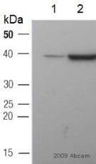 Western blot - Anti-GAPDH antibody - Loading Control (ab9485)This image is a courtesy of Anonymous AbreviewAll lanes : Anti-GAPDH antibody - Loading Control (ab9485) at 1/2500 dilution
Lane 1 : Lysate prepared from human Huh-7 cells at 2 µg
Lane 2 : Lysate prepared from human Huh-7 cells at 20 µg
Secondary
All lanes : HRP-conjugated sheep polyclonal to rabbit IgG at 1/20000 dilution
Performed under reducing conditions.
Predicted band size: 37 kDa
Observed band size: 40 kDa why is the actual band size different from the predicted?
Exposure time: 5 minutes
-
 Immunocytochemistry/ Immunofluorescence - Anti-GAPDH antibody - Loading Control (ab9485)
ab9485 staining GAPDH in NIH3T3 cells. The cells were fixed with 4% formaldehyde (10min), permeabilized with 0.1% Triton X-100 for 5 minutes and then blocked in 1% BSA/10% normal goat serum/0.3M glycine in 0.1%PBS-Tween for 1h. The cells were then incubated with ab9485 at 5μg/ml and ab195889 at 1/250 overnight at +4°C, followed by a further incubation at room temperature for 1h with Goat Anti-Rabbit IgG H&L (Alexa Fluor® 488) preadsorbed (ab150081) secondary antibody at 2 μg/ml (shown in green). Nuclear DNA was labelled in blue with DAPI. Image was taken with a confocal microscope (Leica-Microsystems, TCS SP8).
-
 Western blot - Anti-GAPDH antibody - Loading Control (ab9485)Anti-GAPDH antibody - Loading Control (ab9485) at 1/1000 dilution + Mouse Embryonic lung whole tissue lysate at 30 µg
Developed using the ECL technique.
Performed under reducing conditions.
Predicted band size: 37 kDa
Exposure time: 15 seconds
プロトコール
データシートおよび資料
-
SDS download
-
Datasheet download
参考文献 (2835)
ab9485 は 2835 報の論文で使用されています。
- Aoki H et al. Thymidine Kinase 2 and Mitochondrial Protein COX I in the Cerebellum of Patients with Spinocerebellar Ataxia Type 31 Caused by Penta-nucleotide Repeats (TTCCA)n. Cerebellum 22:70-84 (2023). PubMed: 35084690
- Huang L et al. TBX3 stimulates proliferation and stem cell self-renewal in bladder carcinoma. Histol Histopathol 38:65-72 (2023). PubMed: 35856500
- Liu CL et al. Aberrant Expression of Solute Carrier Family 35 Member A2 Correlates With Tumor Progression in Breast Cancer. In Vivo 37:262-269 (2023). PubMed: 36593004
- Miskiewicz EI et al. Phosphoserine-86-HSPB1 (pS86-HSPB1) is cytoplasmic and highly induced in rat myometrium at labour. Histochem Cell Biol 159:149-162 (2023). PubMed: 36260112
- Shi CJ et al. TGFβR-1/ALK5 inhibitor RepSox induces enteric glia-to-neuron transition and influences gastrointestinal mobility in adult mice. Acta Pharmacol Sin 44:92-104 (2023). PubMed: 35794374