Donkey Anti-Rabbit IgG H&L (HRP) (ab205722)
 (ab205722)")
Key features and details
- Donkey Anti-Rabbit IgG H&L (HRP)
- Conjugation: HRP
- Host species: Donkey
- Isotype: IgG
- Suitable for: IHC-P, IP, WB, ELISA
 Related conjugates and formulations
製品の概要
-
製品名
Donkey Anti-Rabbit IgG H&L (HRP)
IgG 二次抗体 製品一覧 -
由来種
Donkey -
ターゲット生物種
Rabbit -
特異性
The antibody used for conjugation reacts with rabbit immunoglobulins of all classes. Cross-reactions as determined by ELISA for the unconjugated antibody (ab182020): Human IgG, mouse IgG, rat IgG, goat IgG and chicken IgY, less than 2%. -
アプリケーション
適用あり: IHC-P, IP, WB, ELISAmore details -
免疫原
The details of the immunogen for this antibody are not available.
-
標識
HRP
製品の特性
-
製品の状態
Liquid -
保存方法
Shipped at 4°C. Store at +4°C short term (1-2 weeks). Upon delivery aliquot. Store at -20°C. Avoid freeze / thaw cycle. Store In the Dark. -
バッファー
pH: 7.40
Preservative: 0.1% Proclin 300 Solution
Constituents: PBS, 1% BSA, 30% Glycerol (glycerin, glycerine) -
 Concentration information loading...
 -
精製度
Immunogen affinity purified -
特記事項(精製)
This antibody was isolated by affinity chromatography using antigen coupled to agarose beads and conjugated to Horse Radish Peroxidase (HRP). -
ポリ/モノ
ポリクローナル -
アイソタイプ
IgG -
研究分野
関連製品
-
Alternative Versions
- Donkey Anti-Rabbit IgG H&L (Alexa Fluor® 488) (ab150073)
- Donkey Anti-Rabbit IgG H&L (Alexa Fluor® 555) (ab150074)
- Donkey Anti-Rabbit IgG H&L (Alexa Fluor® 647) (ab150075)
- Donkey Anti-Rabbit IgG H&L (Alexa Fluor® 594) (ab150076)
- Donkey Anti-Rabbit IgG H&L (Alexa Fluor® 568) (ab175470)
- Donkey Anti-Rabbit IgG H&L (Alexa Fluor® 405) (ab175651)
- Donkey Anti-Rabbit IgG H&L (Alexa Fluor® 750) (ab175731)
- Donkey Anti-Rabbit IgG H&L (Alexa Fluor® 680) (ab175772)
- Donkey Anti-Rabbit IgG H&L (Alexa Fluor® 790) (ab175780)
- Donkey Anti-Rabbit IgG H&L (ab182020)
- Donkey Anti-Rabbit IgG H&L (Biotin) (ab207999)
アプリケーション
The Abpromise guarantee
Abpromise保証は、 次のテスト済みアプリケーションにおけるab205722の使用に適用されます
アプリケーションノートには、推奨の開始希釈率がありますが、適切な希釈率につきましてはご検討ください。
| アプリケーション | Abreviews | 特記事項 |
|---|---|---|
| IHC-P |
1/2000 - 1/20000.
|
|
| IP |
Use at an assay dependent concentration.
|
|
| WB | (2) |
1/2000 - 1/50000.
|
| ELISA |
Use at an assay dependent concentration.
|
| 特記事項 |
|---|
|
IHC-P
1/2000 - 1/20000. |
|
IP
Use at an assay dependent concentration. |
|
WB
1/2000 - 1/50000. |
|
ELISA
Use at an assay dependent concentration. |
画像
-
Western blot - Donkey Anti-Rabbit IgG H&L (HRP) (ab205722)All lanes : Anti-beta Actin antibody (ab8227) at 1 µg/ml
Lane 1 : Liver (Human) Tissue Lysate
Lane 2 : Liver (Mouse) Tissue Lysate
Lane 3 : Liver (Rat) Tissue Lysate
Lane 4 : HeLa (Human epithelial carcinoma cell line) Whole Cell Lysate
Lane 5 : NIH 3T3 (Mouse embryonic fibroblast cell line) Whole Cell Lysate
Lane 6 : PC12 (Rat adrenal pheochromocytoma cell line) Whole Cell Lysate
Lysates/proteins at 10 µg per lane.
Secondary
All lanes : Donkey Anti-Rabbit IgG H&L (HRP) (ab205722) at 1/10000 dilution
Developed using the ECL technique.
Performed under reducing conditions.
Observed band size: 42 kDa why is the actual band size different from the predicted?
Exposure time: 10 secondsThis blot was produced using a 4-12% Bis-tris gel under the MOPS buffer system. The gel was run at 200V for 50 minutes before being transferred onto a Nitrocellulose membrane at 30V for 70 minutes. The membrane was then blocked for an hour using 2% Bovine Serum Albumin before being incubated with ab8227 overnight at 4°C. Antibody binding was detected using ab205722, and visualised using ECL development solution ab133406.
-
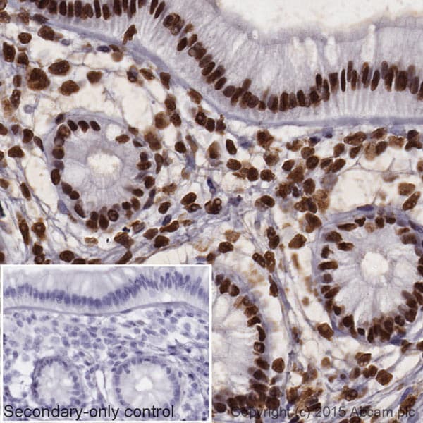 Immunohistochemistry (Formalin/PFA-fixed paraffin-embedded sections) - Donkey Anti-Rabbit IgG H&L (HRP) (ab205722)
IHC image of Histone H4 staining in a section of formalin-fixed paraffin-embedded normal human colon tissue*. The section was pre-treated using pressure cooker heat mediated antigen retrieval with sodium citrate buffer (pH6) for 30mins, and incubated overnight at +4°C with ab177840 at 1ug/ml. An HRP-conjugated secondary (Ab205722, 1/10000 dilution) was used to detect the primary for 1hr at room temperature. DAB was used as the chromogen (ab103723), diluted 1/100 and incubated for 10min at room temperature. The section was counterstained with haematoxylin and mounted with DPX. The inset negative control image is taken from an identical assay without primary antibody.
For other IHC staining systems (automated and non-automated) customers should optimize variable parameters such as antigen retrieval conditions, primary antibody concentration and antibody incubation times.
*Tissue obtained from the Human Research Tissue Bank, supported by the NIHR Cambridge Biomedical Research Centre
-
 Western blot - Donkey Anti-Rabbit IgG H&L (HRP) (ab205722)All lanes : Anti-beta Actin antibody (ab8227) at 1 µg/ml
Lane 1 : Liver (Mouse) Tissue Lysate
Lane 2 : Liver (Rat) Tissue Lysate
Lysates/proteins at 10 µg per lane.
Secondary
All lanes : ab205722 (Left Image) at 1/10,000 and a competitor secondary (Right Image) at 1/10,000. Notice the decreased signal of the competitor product.
Performed under reducing conditions.
Observed band size: 42 kDa why is the actual band size different from the predicted?
Exposure time: 10 secondsThis blot was produced using a 4-12% Bis-tris gel under the MOPS buffer system. The gel was run at 200V for 50 minutes before being transferred onto a Nitrocellulose membrane at 30V for 70 minutes. The membrane was then blocked for an hour using 2% Bovine Serum Albumin before being incubated with ab8227 overnight at 4°C. Antibody binding was detected using ab205722 (Left Image) and a competitor secondary (Right Image), and visualised using ECL development solution ab133406.
-
 Immunohistochemistry (Formalin/PFA-fixed paraffin-embedded sections) - Donkey Anti-Rabbit IgG H&L (HRP) (ab205722)
IHC image of beta tubulin staining in a section of formalin-fixed paraffin-embedded normal human colon tissue*. The section was pre-treated using pressure cooker heat mediated antigen retrieval with sodium citrate buffer (pH6) for 30mins, and incubated overnight at +4°C with ab6046 at 5ug/ml. An HRP-conjugated secondary (Ab205722, 1/10000 dilution) was used to detect the primary for 1hr at room temperature. DAB was used as the chromogen (ab103723), diluted 1/100 and incubated for 10min at room temperature. The section was counterstained with haematoxylin and mounted with DPX. The inset negative control image is taken from an identical assay without primary antibody.
For other IHC staining systems (automated and non-automated) customers should optimize variable parameters such as antigen retrieval conditions, primary antibody concentration and antibody incubation times.
*Tissue obtained from the Human Research Tissue Bank, supported by the NIHR Cambridge Biomedical Research Centre
-
 Western blot - Donkey Anti-Rabbit IgG H&L (HRP) (ab205722)All lanes : No Primary Antibody
Lane 1 : Liver (Mouse) Tissue Lysate
Lane 2 : Liver (Rat) Tissue Lysate
Lysates/proteins at 10 µg per lane.
Secondary
All lanes : ab205722 (Left Image) 1/2000 and a competitor secondary (Right Image) 1/2000. Notice the increased background of the competitor product.
Performed under reducing conditions.
Exposure time: 10 secondsThis blot was produced using a 4-12% Bis-tris gel under the MOPS buffer system. The gel was run at 200V for 50 minutes before being transferred onto a Nitrocellulose membrane at 30V for 70 minutes. The membrane was incubated overnight with 2% Bovine Serum Albumin at 4°C. Any non-specific background binding was assessed by incubating the membrane with ab205722 (Left Image) and a competitor secondary (Right Image), and visualised using ECL development solution ab133406.
-
 Western blot - Donkey Anti-Rabbit IgG H&L (HRP) (ab205722)All lanes : Anti-STAT3 antibody [EPR787Y] (ab68153) at 1/2000 dilution
Lane 1 : A431 (Human epithelial carcinoma cell line) Whole Cell Lysate
Lane 2 : Heart (Mouse) Tissue Lysate
Lane 3 : Heart (Rat) Tissue Lysate
Lysates/proteins at 10 µg per lane.
Secondary
All lanes : Donkey Anti-Rabbit IgG H&L (HRP) (ab205722) at 1/2000 dilution
Developed using the ECL technique.
Performed under reducing conditions.
Observed band size: 88 kDa why is the actual band size different from the predicted?
Exposure time: 20 minutesThis blot was produced using a 4-12% Bis-tris gel under the MOPS buffer system. The gel was run at 200V for 50 minutes before being transferred onto a Nitrocellulose membrane at 30V for 70 minutes. The membrane was then blocked for an hour using 3% milk before being incubated with ab68153 overnight at 4°C. Antibody binding was detected using ab205722, and visualised using ECL development solution ab133406.
-
 Western blot - Donkey Anti-Rabbit IgG H&L (HRP) (ab205722)All lanes : No Primary Antibody
Lane 1 : Liver (Human) Tissue Lysate
Lane 2 : Liver (Mouse) Tissue Lysate
Lane 3 : Liver (Rat) Tissue Lysate
Lane 4 : HeLa (Human epithelial carcinoma cell line) Whole Cell Lysate
Lane 5 : NIH 3T3 (Mouse embryonic fibroblast cell line) Whole Cell Lysate
Lane 6 : PC12 (Rat adrenal pheochromocytoma cell line) Whole Cell Lysate
Lysates/proteins at 10 µg per lane.
Secondary
All lanes : Donkey Anti-Rabbit IgG H&L (HRP) (ab205722) at 1/2000 dilution
Performed under reducing conditions.
Exposure time: 10 secondsThis blot was produced using a 4-12% Bis-tris gel under the MOPS buffer system. The gel was run at 200V for 50 minutes before being transferred onto a Nitrocellulose membrane at 30V for 70 minutes. The membrane was incubated overnight with 2% Bovine Serum Albumin at 4°C. Any non-specific background binding was assessed by incubating the membrane with only the secondary antibody (ab205722), and visualised using ECL development solution ab133406.
-
 ELISA - Donkey Anti-Rabbit IgG H&L (HRP) (ab205722)
Cross-reactivity of the polyclonal secondary antibody ab182020 was tested using a sandwich ELISA approach. The wells were coated with the indicated IgG standards at 1 µg/ml (50 µl/well) and incubated overnight at 4°C, followed by a 5% BSA blocking step for 2h at RT. ab182020 was then added starting at 1 µg/ml and gradually diluted 1/4 (50 µl/well), followed by incubation for 2h. For the detection Goat anti-Donkey IgG H&L (HRP) (ab6988) was used at 1/20,000 dilution (50 µl/well), followed by incubation for 1h at RT.
For the batch tested, ab182020 showed a cross-reactivity below 2% towards human IgG, mouse IgG, rat IgG, goat IgG and chicken IgY.
This data was developed using the unconjugated antibody (ab182020).
プロトコール
To our knowledge, customised protocols are not required for this product. Please try the standard protocols listed below and let us know how you get on.
データシートおよび資料
-
SDS download
-
Datasheet download
参考文献 (19)
ab205722 は 19 報の論文で使用されています。
- Nishida Y et al. Oxidative stress induces MUC5AC expression through mitochondrial damage-dependent STING signaling in human bronchial epithelial cells. FASEB Bioadv 5:171-181 (2023). PubMed: 37020748
- Backe MB et al. PICK1-Deficient Mice Maintain Their Glucose Tolerance During Diet-Induced Obesity. J Endocr Soc 7:bvad057 (2023). PubMed: 37200849
- Scharmacher J et al. The pro-inflammatory signature of lipopolysaccharide in spontaneous contracting embryoid bodies differentiated from mouse embryonic stem cells. J Cell Mol Med 27:2045-2058 (2023). PubMed: 37315183
- Luo T et al. Curcumin inhibits esophageal squamous cell carcinoma progression through down-regulating the circNRIP1/miR-532-3p/AKT pathway. Environ Toxicol 38:2705-2716 (2023). PubMed: 37471645
- Grabowska A et al. Activation-induced chromatin reorganization in neurons depends on HDAC1 activity. Cell Rep 38:110352 (2022). PubMed: 35172152